Original Article
Effect of TSLP on the function of platelets and IL-25 in
chronic obstructive pulmonary disease
Lingyun Wu1*, Li Fang1*, Xiuping Xu1, Dingbang Pei1, Wei Zhou1, Haijun Wan2
1Department of Geriarics, The Third Hospital of Quzhou, Quzhou, Zhejiang, China; 2Department of Cardiothoracic Surgery, Quzhou People’s Hospital, Quzhou, Zhejiang, China. *Equal contributors and co-first authors.
Received December 4, 2018; Accepted April 9, 2019; Epub May 15, 2019; Published May 30, 2019
Abstract: Objective: To investigate the effect of thymic stromal lymphopoietin (TSLP) on the function of platelets and IL-25 in patients with chronic obstructive pulmonary disease (COPD) and its monitoring value. Methods: 126 patients with COPD and 119 healthy subjects were selected for retrospective analysis. TSLP and IL-25 levels in the serum were measured using enzyme-linked immunosorbent assay (ELISA). The platelet level was detected using the automatic blood cell analyzer, while platelet aggregation ability was detected using the platelet aggregation analyzer. The correlation between TSLP and the partial pressure of oxygen (PaO2) and partial pressure of carbon dioxide (PaCO2) was analyzed. Results: The TSLP in the COPD group was significantly higher than that in the control group (P<0.001). IL-25, platelet count, and platelet aggregation rate in the high-TSLP group were significantly higher
than those in the low-TSLP group (P<0.001). The PaO2 in the high-TSLP group was significantly lower than that in
the low-TSLP group (P<0.001), while the PaCO2 in the high-TSLP group was significantly higher than that in the
low-TSLP group (P<0.001). Linear correlation analysis showed that low-TSLP was negatively correlated with PaO2 (r=-0.880, P<0.001) but positively correlated with PaCO2 (r=0.878, P<0.001). Conclusion: The expression of TSLP can be used as an effective indicator for monitoring the severity of COPD in patients, and is considered as a potential target for the treatment of COPD.
Keywords: TSLP, platelets, IL-25, chronic obstructive pulmonary disease
Introduction
Chronic obstructive pulmonary disease (COPD)
is a disease characterized by persistent airflow
limitation. It is a common disease in the respi-ratory system. The majority of patients affected by the disease are mainly middle-aged and older men [1].
According to statistics, in 2016, the number of new patients with COPD in the world exceeded 5 million, which is about 10 times higher than the number of patients ten years ago [2]. More statistical reports show that about 8.2% of peo-ple over the age of 40 will have COPD [3]. Ad- ditionally, the mortality rate of COPD is
extreme-ly high, and it is now one of the top five diseas -es that endanger human life [4]. According to
statistics, the five-year survival rate of patients
with COPD is only 28.4% [5]. Several studies report that COPD is expected to have the third highest mortality rate, after gastric and lung
cancer, in 2030 [6]. COPD mainly manifests as cough, expectoration, and dyspnea. Damage to
the lungs can also cause inflammation of sur -rounding organs and accelerate the deteriora-tion of the disease [7]. The pathogenesis of
COPD has not yet been clarified, but it is
believed that smoking is the leading cause of COPD [8]. Through advancements in research, several studies have shown that infection, air, protease-antiprotease imbalance, and oxida-tive stress may cause COPD [9-11]. Efforts have been made to effectively improve the diagnosis
and treatment of COPD, but no significant
br-eakthrough has been made yet. In clinical
prac-tice, COPD is classified as an incurable chronic
disease similar to diabetes and hypertension. To reduce the effects of the disease on patients, it is necessary to take large quantities of medi-cation for a long time [12, 13].
discov-find a potential target for future
treatment of COPD.
Materials and methods
General information
From February 2016 to March 2017, One hundred twenty-six pa- tients with COPD who were admit-ted to our hospital and 119 he- althy subjects (a total of 243 par-ticipants) were selected for retro-spective analysis. 124 patients with COPD were assigned to the COPD group while 119 healthy su- bjects were assigned to the co- ntrol group. The experiment was approved by the Ethics Committ- ee of Quzhou People’s Hospital, and all the above participants signed an informed consent form.
ery of thymic stromal lymphopoietin (TSLP), an interleukin-7 (IL-7)-like cytokine. Its role in acti-vating various cells (such as monocytes and T cells) has been shown to contribute to the de- velopment of various lung diseases and tumors [14-16].
At present, the role of TSLP is not clear. This study investigated the expression of TSLP and its effects on IL-25 and platelet function in patients with COPD. The aim of the study was to
Inclusion and exclusion criteria
Inclusion criteria: Only patients who were diag-nosed with COPD according to the 2014 COPD diagnosis guideline [17], aged between 30-70 years, and had undergone surgery and treat-ment with antibiotics were included in the study. Additionally, only complete cases and patients who cooperated with our hospital staff were included. Exclusion criteria: Patients with tumors, long-term use of glucocorticoids, car-diovascular and cerebrovascular diseases, im- munological diseases, infectious diseases, pn- eumonia, organ failures, mental disorders, and physical disabilities were excluded from the study. Additionally, pregnant patients and long-term bedridden patients were excluded.
Methods
[image:2.612.88.317.95.512.2]Sample collection: Patients in the COPD group were treated with targeted therapy after admis-sion. Using anticoagulation and coagulation tubes, 4 ml of venous blood was taken from each patient. Platelet levels in the anticoagula-tion tubes were measured by an automatic blood cell analyzer (DxH800 Analyzer, Beckman Coultry Technology, Inc.) while platelet aggrega-tion was measured using a platelet aggregaaggrega-tion analyzer (Chrono-log 700 Platelet Aggregation Analyzer, Beckman Coultry Technology, Inc.). The coagulation tube was kept at 20°C for 20
Table 1. Comparison of clinical data between the COPD and control groups [n (%)]
COPD group
(n=124) Control group (n=119) X2 or t P
Age 52.84±8.84 53.67±9.16 0.719 0.473
Body weight (KG) 66.13±12.34 64.81±13.47 0.426 0.797 Body temperature (°C) 36.28±0.84 36.12±0.70 1.610 0.109
Sex 0.065 0.799
Male 107 (86.29) 104 (87.39) Female 17 (13.71) 15 (12.61)
Living Environment 0.699 0.403
Town 77 (62.10) 80 (67.23)
Rural 47 (37.90) 39 (32.77)
Smoking 0.934 0.334
Yes 114 (91.94) 105 (88.24)
No 10 (8.06) 14 (11.76)
Sports habit 0.266 0.606
Yes 24 (19.35) 20 (16.81)
No 100 (80.65) 99 (83.19)
Figure 1. Expression levels of TSLP in the COPD and control groups. The TSLP in the COPD group was
[image:2.612.86.361.96.324.2]min, centrifuged for 10 min (4000 rpm/min), and the supernatant was divided into two parts.
Pressure of oxygen (PaO2) and carbon dioxide (PaCO2): Blood gas analyzer (Kangli BG-800 blood gas analyzer, Jinan Zhengrong Medical Instrument Co., Ltd.) was used for partial pres-sure of oxygen (PaO2) and partial pressure of carbon dioxide (PaCO2).
ELISA: Enzyme-linked immunosorbent assay (ELISA) was used to detect the serum TSLP and IL-25 (the kit was purchased from R&D sys-tems, USA, 300-62, MAB13992), and the de- tection process was performed in strict accor-dance with the manufacturer’s instructions.
Observation indicators: According to the ex- pression level of TSLP, patients with COPD we- re divided into a high-TSLP group and a low-TSLP group. Parameters of interest included the clinical information of both COPD and con-trol groups, differential expression of TSLP in both groups, effect of TSLP on IL-25 and plate-lets, IL-25 and platelet count, aggregation abil-ity, and correlation between TSLP and PaO2 and PaCO2.
Statistical method: The data were analyzed and processed using the SPSS22.0 statistical soft-ware. Data on sex and smoking status in the clinical information of patients were expressed
ment, and smoking and exercise status in the clinical data of the two groups. This demon-strated that the two groups were comparable (Table 1).
TSLP is increased in the COPD group
The TSLP in the COPD group was 207.63±37.48
ng/L. This was significantly higher than that in
the control group which was 61.83±8.24 ng/L (P<0.001) (Figure 1).
Clinical data between the high-TSLP and low-TSLP groups has no difference
According to the median expression of TSLP, 66 patients with COPD were assigned to the high-TSLP group (high-TSLP>207.63 ng/L), and the rem- aining 58 were assigned to the low-TSLP group
(TSLP≤207.63 ng/L). No significant difference
(P>0.050) was observed after comparing the age, weight, body temperature, sex, living envi-ronment, and smoking and exercise status in the clinical data of the high-TSLP and low-TSLP groups. This showed that the two groups were comparable (Table 2).
IL-25 expression is higher in the high-TSLP group
IL-25 in the high-TSLP group was 23.66±4.58
[image:3.612.91.366.94.332.2]pg/mL. This was significantly higher than that
Table 2. Comparison of clinical data between the high TSLP and low TSLP groups [n (%)]
High TSLP
group (n=66) group (n=58) XLow TSLP 2 or t P
Age 53.16±8.25 54.08±7.44 0.649 0.518
Body weight (KG) 64.27±11.54 62.37±12.36 0.885 0.378 Body temperature (°C ) 37.38±0.62 37.25±0.74 1.064 0.289 course of disease (d) 8.68±2.16 9.04±2.25 0.908 0.366
Sex 0.301 0.583
Male 58 (87.88) 49 (84.48)
Female 8 (12.12) 9 (15.52)
Living Environment 0.133 0.715
Town 40 (60.61) 37 (63.79)
Rural 26 (39.39) 21 (36.21)
Smoking 0.764 0.382
Yes 64 (93.94) 52 (89.66)
No 4 (6.06) 6 (10.34)
Sports habit 0.653 0.419
Yes 11 (16.67) 13 (22.41)
No 55 (83.33) 45 (77.59)
as rates. Comparison between groups was performed using the chi-square test. Data on the ex- pression of TSLP and IL-25 were presented as mean ± standard deviation. The t-test was used for comparison between groups. Correlation was analyzed using linear correlation. Statistical
sig-nificance was set at P<0.050.
Results
General information of the COPD and control groups
These 243 participants comp- rised 211 males and 32 fem- ales, aged 42-64 years, with an average age of 54.27±8.66 ye-
ars. No significant difference
environ-in the low-TSLP group which was 16.34±5.07 pg/mL (P<0.001) (Figure 2).
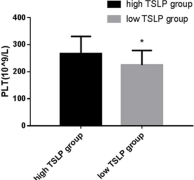
Platelet aggregation rate is higher in the high-TSLP group
The platelet count in the high-TSLP group was 267.57±62.87×10⁹/L, which was
significant-ly higher than that in the low-TSLP group (224.39±54.21×10⁹/L) (P<0.001). The platelet aggregation rate in the high-TSLP group was
36.84±8.57. This was significantly higher
th-an that in the low-TSLP group which was 21.86±6.94 (P<0.001) (Figures 3 and 4).
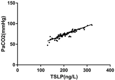
Blood gas indicator PaO2 is lower while PaCO2 is higher in the high-TSLP group
The PaO2 in the high-TSLP group was 52.96±
6.97 mmHg, which was significantly lower
th-an that in the low-TSLP group (59.84±5.71 mmHg) (P<0.001). The PaCO2 in the high-TSLP
group (78.56±8.14 mmHg) was significantly
higher than that in the low-TSLP group (72.17±6.39 mmHg) (P<0.001). Linear correla-tion analysis showed that the TSLP was nega-tively correlated with PaO2 (r=-0.880, P<0.001) but was positively correlated with PaCO2 (r=0.878, P<0.001) (Table 3, Figures 5 and 6).
Discussion
[image:4.612.323.519.70.266.2]COPD is a common respiratory disease, which is prevalent in middle-aged and elderly people. Recently, the incidence and mortality rates of COPD have been observed to increase every year [18]. Currently, there are limited treatment options for COPD in clinics. Some patients still
Figure 2. IL-25 expression levels in the high-TSLP and
low-TSLP groups. The level of IL-25 was significantly
higher in the high-TSLP group than that in the low-TSLP group. *represents P<0.001 for a comparison with the IL-25 level in the high-TSLP group.
Figure 3. Platelet counts in the high-TSLP and low-TSLP groups. Platelet counts in the high-low-TSLP group
were significantly higher than those in the low-TSLP
[image:4.612.92.288.71.270.2]group. *represents P<0.001 for the platelet count compared with that of the high-TSLP group.
Figure 4. Platelet aggregation rate in the high-TSLP and low-TSLP groups. The platelet aggregation rate
in the high-TSLP group was significantly higher than
[image:4.612.93.287.359.539.2]attention now being focused on TSLP. As an IL-7-like cytokine, it has some effect on human epithelial, thymic (Th) stromal, and vascular endothelial cells [21]. In recent years, studies have also shown that these cells inhibit and
pro-mote both the expression of inflamma -tory factors and the differentiation of
experience no fundamental changes in their pathological condition after treatment. Trea- tments may even worsen the lung function [19]. Some patients have poor autoimmune func-tion, which is more likely to cause various car-diopulmonary complications, threatening the life and health of patients [20]. Therefore, there
is an urgent need to find a method for effective
treatment of COPD. Research has begun to explore targeted treatments for COPD with
subgroups of Th cells through the TSLP recep-tor. These cells participate in the development
of some forms of inflammation, autoimmune
diseases, and tumors [22]. IL-25, a cytokine produced by Th2 cells, enhances Th2
cell-medi-ated inflammatory response. It promotes air
-way hyper-responsiveness and eosinophil infil -tration in lung tissue [23]. In the pathogenesis of COPD, platelet adhesion and aggregation also play an important role. Platelet membrane surface receptors and platelet agonists can cause molecular structure deformation, which leads to platelet aggregation and release [24]. Currently, the expression of TSLP in patients with COPD is still poorly understood. This study investigated the expression of TSLP and its effects on IL-25 and platelet function in patients
with COPD. The aim of the study was to find a
potential target for future treatment of COPD. The results of this study showed that the expression of TSLP in the COPD group was
sig-nificantly higher than that in the control group,
suggesting that TSLP may be involved in the development of COPD. Studies have shown that there is a strong correlation between TSLP and
inflammatory infections [25]. Therefore, it is
suspected that when the bronchial epithelial barrier is destroyed in patients with COPD, TSLP activates CD4+ T cells, which promotes high expression of TSLP. The level of IL-25 in the
high-TSLP group was significantly higher than
that in the low-TSLP group, suggesting that TSLP can promote IL-25 expression. TSLP can trigger the abnormal reaction of eosinophils by activating the extracellular signal p38 and causing mitosis in proteases [26]. IL-25 not only induces the release of chemokines from eosin-ophils, but also delays the apoptosis of eosino-phils [27]. In patients with COPD, high expres-sion of TSLP activates eosinophils. Additionally, eosinophils secrete IL-25 thus resulting in a
partial inflammatory response in the patient’s
[image:5.612.87.306.91.313.2]body via the modulation of L-selectin. This leads to an increase in the proliferation of eosinophils, and promotes the activation of
Table 3. Comparison of blood gas indices between high TSLP group and low TSLP group
High TSLP
[image:5.612.91.288.384.521.2]group (n=66) group (n=58)Low TSLP t P PaO2 (mmHg) 52.96±6.97 59.84±5.71 5.962 <0.001 PaCO2 (mmHg) 78.56±8.14 72.17±6.39 4.815 <0.001
Figure 5. Correlation analysis between TSLP and PaO2. Linear correlation analysis showed that TSLP was negatively correlated with PaO2 (r=-0.880, P<0.001).
TSLP and the production of inflammatory cyto -kines by Th2 cells. The patient’s condition is then aggravated through a vicious cycle. The platelet aggregation ability of the high-TSLP
group was significantly higher than that of the
low-TSLP group, suggesting that TSLP can pro-mote platelet aggregation. This is because TSLP induces the expression of platelet ago-nists. TSLP promotes platelet activation thr- ough receptors and accelerates the secretion
of α-particles and dense particles. TSLP rapidly
aggregates into platelet membranes, which causes the activation of integrin and leads to the aggregation of a large number of platelets. These platelets are released into the blood, which subsequently block the normal blood
flow in the lung tissue, thus aggravating the
condition of the patient. The role of TSLP and its effects on platelet function require further investigation. A comparison of the blood gas function between the high-TSLP and low-TSLP groups revealed that the PaCO2 in the
high-TSLP group was significantly lower than that in
the low-TSLP group, and the PaCO2 in the
high-TSLP group was significantly higher than that in
the low-TSLP group. Correlation analysis sho- wed that TSLP was positively correlated with PaCO2. This suggests that TSLP can be used as an effective indicator to monitor the severity of COPD in patients.
A limitation of this study is its small sample size. Another limitation is that it is a single-cen-ter study. We will conduct a long-single-cen-term follow-up survey of this study, and further explore the effect of TSLP on IL-25 and platelet function in future studies to improve our experiments. In summary, TSLP is highly expressed in pati- ents with COPD and can promote the expres-sion of IL-25 and platelet aggregation. TSLP can be used as an effective indicator for monitoring the severity of COPD in patients and is consid-ered as a potential target for the treatment of COPD.
Disclosure of conflict of interest
None.
Address correspondence to: Haijun Wan, Depart- ment of Cardiothoracic Surgery, Quzhou People’s Hospital, No.2, Bell Tower Road, Quzhou 324000, Zhejiang, China. Tel: +86 0570 3012329; E-mail: [email protected]
References
[1] Barnes PJ. Inflammatory mechanisms in pa -tients with chronic obstructive pulmonary dis-ease. J Allergy Clin Immunol 2016; 138: 16-27. [2] Vestbo J, Anderson JA, Brook RD, Calverley PM, Celli BR, Crim C, Martinez F, Yates J, Newby DE, SUMMIT Investigators. Fluticasone furoate and vilanterol and survival in chronic obstructive pulmonary disease with heightened cardiovas-cular risk (SUMMIT): a double-blind random- ised controlled trial. Lancet 2016; 387: 1817-1826.
[3] Singh D, Papi A, Corradi M, Pavlišová I, Mo- ntagna I, Francisco C, Cohuet G, Vezzoli S, Scuri M and Vestbo J. Single inhaler triple ther-apy versus inhaled corticosteroid plus
long-acting β2-agonist therapy for chronic obstruc -tive pulmonary disease (TRILOGY): a double-blind, parallel group, randomised controlled trial. Lancet 2016; 388: 963-973.
[4] Martinez FD. Early-life origins of chronic ob-structive pulmonary disease. N Engl J Med 2016; 375: 871-878.
[5] Vedel-Krogh S, Nielsen SF, Lange P, Vestbo J and Nordestgaard BG. Blood eosinophils and exacerbations in chronic obstructive pulmo-nary disease. The copenhagen general popula-tion study. Am J Respir Crit Care Med 2016; 193: 965-974.
[6] Bhatt SP, Soler X, Wang X, Murray S, Anzueto AR, Beaty TH, Boriek AM, Casaburi R, Criner GJ and Diaz AA. Association between functional small airway disease and FEV1 decline in chronic obstructive pulmonary disease. Am J Respir Crit Care Med 2016; 194: 178-184. [7] George L and Brightling CE. Eosinophilic airway
inflammation: role in asthma and chronic ob -structive pulmonary disease. Ther Adv Chronic Dis 2016; 7: 34-51.
[8] Wang Z, Bafadhel M, Haldar K, Spivak A, Ma- yhew D, Miller BE, Tal-Singer R, Johnston SL, Ramsheh MY and Barer MR. Lung microbiome dynamics in chronic obstructive pulmonary disease exacerbations. Eur Respir J 2016; 47: 1082-92.
[9] Siu AL, Bibbins-Domingo K, Grossman DC, Davidson KW, Epling JW, García FA, Gillman M, Kemper AR, Krist AH and Kurth AE. Screening for chronic obstructive pulmonary disease: US preventive services task force recommenda-tion statement. JAMA 2016; 315: 1372-1377. [10] Vestbo J, Papi A, Corradi M, Blazhko V, Mon-
tagna I, Francisco C, Cohuet G, Vezzoli S, Scuri
M and Singh D. Single inhaler extrafine triple
[20] Ehteshami-Afshar S, FitzGerald JM, Doyle-Wa- ters MM, Sadatsafavi M. The global economic burden of asthma and chronic obstructive pul-monary disease. Int J Tuberc Lung Dis 2016; 20: 11-23.
[21] Demehri S, Cunningham TJ, Manivasagam S, Ngo KH, Tuchayi SM, Reddy R, Meyers MA, DeNardo DG and Yokoyama WM. Thymic stro-mal lymphopoietin blocks early stages of br- east carcinogenesis. J Clin Invest 2016; 126: 1458-1470.
[22] Kim J, Kim BE, Lee J, Han Y, Jun HY, Kim H, Choi J, Leung DY and Ahn K. Epidermal thymic stromal lymphopoietin predicts the develop-ment of atopic dermatitis during infancy. J Allergy Clin Immunol 2016; 137: 1282-1285, e1284.
[23] Lee JB, Chen CY, Liu B, Mugge L, Angkasekwinai P, Facchinetti V, Dong C, Liu YJ, Rothenberg ME and Hogan SP. IL-25 and CD4+ TH2 cells en-hance type 2 innate lymphoid cell–derived IL-13 production, which promotes IgE-mediated experimental food allergy. J Allergy Clin Im- munol 2016; 137: 1216-1225, e1215. [24] von Moltke J, Ji M, Liang HE and Locksley RM.
Tuft-cell-derived IL-25 regulates an intestinal ILC2–epithelial response circuit. Nature 2016; 529: 221.
[25] Oyoshi MK, Venturelli N and Geha RS. Thymic stromal lymphopoietin and IL-33 promote skin
inflammation and vaccinia virus replication in
a mouse model of atopic dermatitis. J Allergy Clin Immunol 2016; 138: 283-286.
[26] Dong H, Hu Y, Liu L, Zou M, Huang C, Luo L, Yu C, Wan X, Zhao H and Chen J. Distinct roles of short and long thymic stromal lymphopoietin isoforms in house dust mite-induced asthmat-ic airway epithelial barrier disruption. Sci Rep 2016; 6: 39559.
[27] Lam EP, Kariyawasam HH, Rana BM, Durham SR, McKenzie AN, Powell N, Orban N, Lennartz-Walker M, Hopkins C and Ying S. IL-25/IL-33-responsive TH2 cells characterize nasal polyps with a default TH17 signature in nasal muco-sa. J Allergy Clin Immunol 2016; 137: 1514-1524.
[11] Lipson DA, Barnacle H, Birk R, Brealey N, Locantore N, Lomas DA, Ludwig-Sengpiel A, Mohindra R, Tabberer M and Zhu CQ. FULFIL trial: once-daily triple therapy for patients with chronic obstructive pulmonary disease. Am J Respir Crit Care Med 2017; 196: 438-446. [12] To T, Zhu J, Larsen K, Simatovic J, Feldman L,
Ryckman K, Gershon A, Lougheed MD, Licskai C and Chen H. Progression from asthma to chronic obstructive pulmonary disease. Is air pollution a risk factor? Am J Respir Crit Care Med 2016; 194: 429-438.
[13] Portegies ML, Lahousse L, Joos GF, Hofman A, Koudstaal PJ, Stricker BH, Brusselle GG and Ikram MA. Chronic obstructive pulmonary dis-ease and the risk of stroke. The Rotterdam Study. Am J Respir Crit Care Med 2016; 193: 251-258.
[14] Buchheit KM, Cahill KN, Katz HR, Murphy KC, Feng C, Lee-Sarwar K, Lai J, Bhattacharyya N, Israel E and Boyce JA. Thymic stromal lympho-poietin controls prostaglandin D2 generation in patients with aspirin-exacerbated respirato-ry disease. J Allergy Clin Immunol 2016; 137: 1566-1576, e1565.
[15] Stier MT, Bloodworth MH, Toki S, Newcomb DC, Goleniewska K, Boyd KL, Quitalig M, Ho- tard AL, Moore ML and Hartert TV. Respiratory syncytial virus infection activates IL-13–pro-ducing group 2 innate lymphoid cells through thymic stromal lymphopoietin. J Allergy Clin Immunol 2016; 138: 814-824, e811.
[16] Mehta AK, Duan W, Doerner AM, Traves SL, Broide DH, Proud D, Zuraw BL and Croft M. Rhinovirus infection interferes with induction of tolerance to aeroantigens through OX40 li-gand, thymic stromal lymphopoietin, and IL-33. J Allergy Clin Immunol 2016; 137: 278-288, e276.
[17] Wurst KE, Kelly-Reif K, Bushnell GA, Pascoe S and Barnes N. Understanding asthma-chronic obstructive pulmonary disease overlap syn-drome. Respir Med 2016; 110: 1-11.
[18] Vogelmeier CF, Criner GJ, Martinez FJ, Anzueto A, Barnes PJ, Bourbeau J, Celli BR, Chen R, Decramer M and Fabbri LM. Global strategy for the diagnosis, management, and prevention of chronic obstructive lung disease 2017 report. GOLD executive summary. Am J Respir Crit Care Med 2017; 195: 557-582.
![Table 1. Comparison of clinical data between the COPD and control groups [n (%)]](https://thumb-us.123doks.com/thumbv2/123dok_us/1263024.654090/2.612.86.361.96.324/table-comparison-clinical-data-copd-control-groups-n.webp)
![Table 2. Comparison of clinical data between the high TSLP and low TSLP groups [n (%)]](https://thumb-us.123doks.com/thumbv2/123dok_us/1263024.654090/3.612.91.366.94.332/table-comparison-clinical-data-high-tslp-tslp-groups.webp)