Original Article
Effects of miR-149-3p-mediated PI3K/AKT signaling
pathways on proliferation and apoptosis of endothelial
cells from hemangioma in children
Xiaoyun Gao1, Junping Wen2, Yu Yang3, Chengyong Huang4
Departments of 1Pediatric Surgery, 2Endocrinology, 3Plastic Surgery, 4Clinical Laboratory, Provincial Clinical
College of Fujian Medical University, Fujian Provincial Hospital, Fuzhou 350001, Fujian Province, China Received February 14, 2019; Accepted March 12, 2019; Epub July 15, 2019; Published July 30, 2019
Abstract: Objective: The aim of the current study was investigate the effects of miR-149-3p-mediated phosphatidy-lositol-3-kinase/protein kinase B (PI3K/AKT) signaling pathways on proliferation and apoptosis of endothelial cells from hemangiomas in children. Methods: Hemangioma tissues and normal tissues adjacent to cancer from 59 children were collected, detecting expression of protein kinase B (AKT) using immunohistochemistry. Endothelial cells of normal tissues adjacent to cancer (Normal group) and hemangioma tissues were extracted. The latter were divided into the Model group (without any treatment), NC group (transfection of negative plasmid), miR-149-3p mimic group (transfection of miR-149-3p mimic plasmid), miR-149-3p inhibitor group (transfection of miR-149-3p inhibitor plasmid), LY294002 group (inhibitor of PI3K/AKT signaling pathways), and miR-149-3p mimic + LY294002 group. Dual-luciferase report gene assay was used to verify targeted relationships between miR-149-3p and AKT. Quantitative Real-time Polymerase Chain Reaction (qRT-PCR) was used to detect mRNA expression of miR-149-3p, PI3K, AKT, and myc. Western blot was used to detect protein expression of PI3K, p-PI3K, AKT, p-AKT, and c-myc. MTT (3-(4,5-dimetyl-2-thiazolyl)-2,5-diphenyl-2-H-tetrazolium bromide, Thiazolyl Blue Tetrazolium Bromide) was used to detect proliferation of cells. Flow cytometry with Annexin V staining was used to detect apoptosis. Results: Positive expression rates of AKT in hemangioma tissues were significantly higher than those in normal tissues (P<0.05). According to dual-luciferase report gene assay, AKT was the target gene of miR-149-3p. According to cell experiments, compared with the Normal group, in Model, NC, miR-149-3p mimic, miR-149-3p inhibitor, LY294002, and miR-149-3p mimic + LY294002 groups, mRNA expression of miR-149-3p was decreased. Moreover, mRNA expression of c-myc, PI3K, and AKT, as well as phosphorylation protein expression of PI3K and AKT, was increased. Proliferation was increased but apoptosis was decreased (all P<0.05). There were no statistically significant differ-ences in indicators between Model and NC groups (all P>0.05). Conclusion: Results suggest that miR-149 inhibits activation of PI3K/AKT signaling pathways in a targeted way, promoting apoptosis and inhibiting proliferation of endothelial cells from hemangiomas in children.
Keywords: miR-149, PI3K/AKT signaling pathway, hemangiomas in children, endothelial cells, proliferation, apop-tosis
Introduction
Hemangiomas, a common angiogenic disease in children, are benign tumors of vascular endo-thelial cells [1]. Some hemangiomas involute by themselves. However, large and fast-growing hemangiomas are prone to ruptures, causing infections, disfigurement, and even coagulation disorders [2]. Currently, pathological mecha-nisms of proliferation and involution remain unclear in clinical practice. Thus, effective tr- eatments are lacking. Overactivity of blood
ves-sels and excessive proliferation of endothelial cells are the biggest pathological characteris-tics of hemangiomas in the proliferative phase. Therefore, key points in the treatment of hem-angiomas and regression of hemhem-angiomas are the reduction of proliferation and promotion of apoptosis of endothelial cells [3].
messenger and further activates AKT, a down-stream molecule of PI3K [4]. According to a study, PI3K and AKT regulate growth, prolifera-tion, apoptosis, and the glycol-metabolism of cells [5]. According to a study by Pan WK et al., protein expression of PI3K and p-AKT in tissues of children with hemangiomas is significantly higher than that in normal children [6]. Acc- ording to a study by Zheng N and others, AKT signaling pathways in hemangiomas are over-activated. Inhibiting the activation of this path-way in a targeted path-way significantly inhibits pro -liferation of endothelial cells from hemangio-masand growth of hemangiomas[7]. Micro- RNAs are endogenous, micromolecular, and non-coding RNAs, which regulate expression of genes and contribute to differentiation, devel-opment, and metabolism of cells [8]. In recent years, more and more studies have shown that miR-149-3p is lowly expressed in tumors. For example, Ke Y explored epithelial-mesenchy-mal transition of non-sepithelial-mesenchy-mall cell lung cancer (NSCLC) cells. They found that miR-149-3p tar-geted to inhibit expression of FOXM1, acting as a tumor suppressor RNA in NSCLC [9]. According to a study by Xue L and others, low expression of miR-149-3p activates AKT signaling path-ways to accelerate the progression of gliomas. This is related to poor prognosis of patients [10]. Moreover, miR-149-3p has been confir-med to act together with AKT, regulating cancer progression. However, it remains unclear whe- ther miR-149-3p is associated with hemangio-mas in children. Therefore, based on previous studies, miR-149-3p was hypothesized to medi-ate PI3K/AKT signaling pathways, regulating progression of hemangiomas in children. The- refore, related experiments were carried out.
Materials and methods
Research objects
Hemangioma tissues and normal tissues adja-cent to cancer specimens were collected from 59 children pathologically diagnosed and un- dergoing surgical resections in Fujian Provincial Hospital, Provincial Clinical Medical College of Fujian Medical University, from January 2013 to December 2017. According to Mulliken classifi -cation criteria, 21 cases were in the prolifera-tive phase and 38 cases were in the extinction phase [11]. Parts of hemangioma tissue and normal tissue specimens were fixed with 10%
neutral formaldehyde solution (Cannice Bio- medical Technology Co., Ltd., China) for subse-quent experiments. The remaining specimens were washed with PBS solution (Dycent Biotech (Shanghai) Co., Ltd.) at 4°C and stored in liquid nitrogen at -80°C. Inclusion criteria: (1) Children with complete data, diagnosed with hemangio-mas by cytology or histology; (2) Children that underwent surgical resections, with biopsy specimens sectioned through clinical evalua-tion; and (3) Children without other genetic dis-eases or history of cancer. Exclusion criteria: (1) Children with severe complications; (2) Children with endocrine diseases; (3) Children with in- complete treatment data; and (4) Children with degenerative biopsy specimens during preser-vation. The use of specimens was agreed upon by the children and their families. This study was approved by the Ethics Committee of Fujian Provincial Hospital, Provincial Clinical Medical College of Fujian Medical University.
Immunohistochemistry
cells: positive cells/all cells * 100% >20% con -sidered as positive (+), and positive cells/all cells * 100% ≤20% as negative (-).
Culturing, morphological observations, and
identification of endothelial cells from heman -giomas
Cell culturing: Fresh hemangioma and normal tissues adjacent to cancer tissues were washed with PBS over 5 minutes for a total of 3 times. They were trimmed into tissue masses with 1 mm3, added with 2 mL of 0.25 trypsin (Procell Life Science & Technology Co., Ltd., Wuhan, China), and digested in a water bath at 37°C for 15 minutes. The trypsin was discarded, adding 4 mL of culture solution containing 10% fetal bovine serum (FBS) (Thermo Fisher Scientific, USA) to terminate digestion. The digested tu- mor was inoculated in the bottom of the culture flask with 3-5 mm in intervals. It was added with approximately 1 mL of complete endothe-lial cell culture medium (Sciencell, USA). The flask was inverted into the incubator, incubated at 37°C for 24 hours, and added with 2 mL of complete endothelial cell culture solution. The solution was changed once every two days. The tissue mass was removed one week later. When cell density reached 80%, the cells were passed on. Cultured cells were purified through mechanical curettage, enzyme digestion, and the repeated adherence method.
Morphological observation of endothelial cells from hemangiomas: Morphological changes of the cultured cells were observed under a micro-scope (Nikon, Japan).
Detection of factor VIII-related antigen by immunohistochemistry (SP method): Endothe- lial cells were inoculated in a 24-well plate, placed in a round coverslip, cultured for 48 hours, washed with PBS over 5 minutes for a total of 3 times, fixed with 95% ethanol at room temperature for 15 minutes, incubated with 0.3% H2O2 at room temperature for 10 minutes, washed with PBS for 3 minutes, and sealed with 100 μL of 5% BSA for 10 minutes. The cells were then added dropwise with rabbit human antibody of factor VIII-related anti-gen (Shanghai Yuping Biotechnology Co., Ltd., China) at 1:100, stayed at 4°C overnight, and washed with PBS. Afterward, they were added dropwise with HRP-labeled goat anti-rabbit sec-ondary antibody IgG (1:2,000, Shanghai Baiyan
Biotechnology Co., Ltd., China), incubated at room temperature for 10 minutes, washed with PBS, added dropwise with streptomycin anti-biotin-peroxidase solution, incubated at room temperature for 10 minutes, washed with PBS, colored with DAB (Abcam, UK), re-stained with hematoxylin, and sealed. Under the micro-scope, five visual fields (200×) were randomly selected for observation and photographing, calculating the percentage of positive cells: positive cells/all cells * 100%.
Cell transfection and grouping
Endothelial cells in adjacent normal tissues were placed in the Normal group. Endothelial cells from hemangiomas were divided into the Model group (without any treatment), NC group (transfection of negative plasmid), miR-149-3p mimic group (transfection of miR-149-3p mimic plasmid), mir-149-3p inhibitor group (transfec-tion of mir-149-3p inhibitor plasmid), LY294002 group (inhibitor of PI3K/AKT signaling path-ways), and miR-149-3p mimic + LY294002 group. Cells were inoculated into a 24-well plate 24 hours before transfection. They were transfected according to the instructions of Lipofectamine 2000 (Shanghai Huiying Bio- logical Technology Co., Ltd., China) when cell density reached 30-50%. Moreover, 100 pmol of negative plasmid, miR-149-3p mimic plas-mid, miR-149-3p inhibitor plasplas-mid, and the inhibitor of PI3K/AKT signaling pathways were diluted with 250 µL of serum-free medium Opti-MEM (model: 31985070, Gibco, USA). They were gently mixed and incubated at room tem-perature for 5 minutes. Next, 5 µL of Lipo- fectamine 2000 was diluted with 250 µL of serum-free medium Opti-MEM, gently mixed, and incubated at room temperature for 5 min-utes. The two materials mentioned above were mixed evenly, incubated at room temperature for 20 minutes, added into wells, and cultured with 5% CO2 at 37°C. After 6-8 hours, the medi-um was replaced with a complete medimedi-um. After 24-48 hours, subsequent experiments were carried out.
Dual-luciferase report gene assay
extract plasmid and co- nduct PCR. This was id- entified with enzyme di-gestion and sent to In- vitrogen USA for sequ- encing. The plasmid co- ntaining the correct ta- rget sequence was na- med as a wild type. With the wild type as a tem-plate, miR-149-3p and AKT1 3’-UTR area were [12]. Recognition sites of restriction enzymes
SpeI and HindIII were introduced into upstream and downstream primers. Te complementary sequence mutation site of the seed sequence was designed on AKT wild type. After restriction enzyme digestion, T4 DNA ligase was used to insert target fragments into the pMIR-reporter. Correctly sequenced luciferase report plasmids WT and MUT were, respectively, co-transfected with miR-149-3p into HEK-293T (Wuhan Yipu Biotechnology Co., Ltd., China). After 48 hours, the cells were collected, lysed, and centrifuged at 2,000 r/min for 5 minutes. The supernatant was taken to detect the fluorescence intensity using a Fluor tester (Promega, USA).
Plasmid construction: The template was the whole blood genome DNA of healthy people, while polymerase chain reaction (PCR) amplifi -cation contained AKT1 3’-UTR area of miR-149-3p binding sites. Primers were designed and synthesized according to AKT1 gene 3’UTR sequence and recognition sites of SpeI and HindⅢ were introduced into upstream and downstream primers. PCR primers were syn-thesized by Shanghai GenePharma Co., Ltd. See Table 1 for sequences. After enzyme diges-tion, purificadiges-tion, and recycling, purified and recovered products ligated the inserted frag-ment at 16°C overnight. These were trans-formed into DH5α competent cells. After plates were coated, positive mono-clones were select-ed and culturselect-ed in a shake-flask overnight to
bound to the base and mutated into a meaning-less sequence. Site-directed mutant primers were designed (See Table 1), with specific oper -ations in accord with instructions. The success-ful mutant plasmid was named as a mutant. It was sequenced after PCR, identifying with enzyme digestion. The above plasmids were purchased from Promega, USA [13].
qRT-PCR
After 48 hours, total RNA and tissues were extracted, according to manufacturer instruc-tions of TRIzol (Thermo Fisher Scientific, USA). Concentrations and purities of RNA were detected using an ultraviolet spectrophotome-ter (Beijing Mairuibo Biotechnology Co., Ltd., China). Samples with a purity of A260/A280 = 1.8-2.0 were adjusted to 50 ng/μL. PrimeSc-riptTM RT reagent Kit (Takara, Japan) was used to reversely transcribe RNA into cDNA (50 ng/ μL), which was frozen at -80°C for use. With ref -erence to GenBank database, primers were designed using the Primer 5.0 computer soft-ware and synthesized by Annoron Biotechnology Co., Ltd. (See Table 2). According to ABI 7900HT Quantitative Real-time Polymerase Chain Re- action (qRT-PCR), GAPDH and U6 served as internal references. Reaction conditions were pre-denaturation at 95°C for 30 seconds, degeneration at 95°C for 5 seconds, annealing at 58°C for 30 seconds, and extension at 72°C for 15 seconds, for 40 cycles. Relative
expres-Table 1. Primer sequences of PCR
Primer Sequence
Wild type AKT13’-UTR upstream 5’-CTGACTGTCCCACCGGGAGCCTCCCCCTCAGATGATCTCTCCACGGTAGCACTTGACCTTTTCGACC-3’
Wild type AKT13’-UTR downstream 5’-TCGAGGTCGAAAAGGTCAAGTGCTACCGTGGAGAGATCATCTGAGGGGGAGGCTCCCGGTGGGACAGTCAGAGCT-3’ Mutant AKT13’-UTR upstream 5’-CTGACTGTCCCACCGGGAGCCTCCCCCTCAGATGAAGAGAGGTCGGTAGCACTTGACCTTTTCGACC-3’
Mutant AKT13’-UTR downstream 5’-TCGAGGTCGAAAAGGTCAAGTGCTACCGTGGAGAGATCATCTGAGGGGGAGGCTCCCGGTGGGACAGTCAGAGCT-3’
[image:4.612.89.530.86.152.2]Note: PCR, polymerase chain reaction.
Table 2. Primer sequences of qRT-PCR
Gene Upstream sequence Downstream sequence
miR-149-3p 5’-TTTAGGGAGGGACGGGG-3’ 5’-GTGCGTGTCGTGGAGTCG-3’
PI3K 5’-CATCACTTCCTCCTGCTCTAT-3’ 5’-CAGTTGTTGGCAATCTTCTTC-3’ AKT 5’-GGACAACCGCCATCCAGACT-3’ 5’-GCCAGGGACACTCCATCTC-3’ c-myc 5’-TGCTGCATGAAGAGACACCG-3’ 5’-TTTCAACTGTTCTCGCCGTT-3’ GAPDH 5’-AGCCACATCGCTCAGACACC-3’ 5’-GTACTCAGCGGCCAGCATCG-3’
U6 5’-CTCGGCTTCGGCAGCACA-3’ 5’-AACGCTTCACGAATTTGCGT-3’
[image:4.612.88.407.189.282.2]sion of miR-149, PI3K, AKT, and c-myc mRNA was calculated using 2-ΔΔCt and the formulas
were: ΔCt = Cttarget gene - Ctinternal reference gene, ΔΔCt = ΔCttreatment group - ΔCtcontrol group. Three same wells were set for each gene in the samples.
Western blotting
After 48 hours, total proteins were extracted using the protein extraction kit (Bestbio, Sh- anghai). Phosphorylated proteins were extra- ced using phosphorylated protein extraction kit (Beijing Biolab Technology Co., Ltd., China). Concentrations of protein samples were deter-mined using the BCA kit (Pierce, USA), with 10% SDS-PAGE gel (Beyotime Institute of Biotech- nology, China) prepared. A total of 50 μg of pro -tein samples was added to each well, separat-ed by electrophoresis, transferrseparat-ed to PVDF membranes, and sealed with TBST buffer con-taining 5% skim milk powder for 2 hours. After washing with TBST, the membrane was added dropwise with primary antibodies rabbit anti-human PI3K (1:1,000, abcam, UK), p-PI3K (1:1,000, abcam, UK), AKT (1:500, abcam, UK), p-AKT (1:500), c-myc (1:1,000 abcam, UK), and GAPDH (1:10,000, abcam, UK), all of which were purchased from Abcam, UK. The mem-brane was incubated at 4°C overnight, rinsed with PBST over 10 minutes for 3 times, added with peroxidase-labeled goat anti-rabbit IgG (1:2,000, Shanghai Baiyan Biotechnology Co., Ltd., China) as a secondary antibody, incubated at room temperature for 1 hour, washed with TBST over 10 minutes for 3 times, colored in ECL reaction solution (Millipore, USA), exposed in a cassette, and developed. With GAPDH as an internal reference, relative expression of proteins = the gray value of the target protein/ the gray value of GAPDH. The gray value of the target band was analyzed using Western blot analysis software Image J (National Institutes of Health, USA).
MTT
After 48 hours, cells were inoculated in a 96-well plate with 3*103 - 6*103 cells and 0.1 mL per well for 6 wells. They were incubated in an incubator for 24, 48, and 72 hours, with the following experiments carried out. A total of 20 μL of prepared MTT solution (Beijing Solarbio Science & Technology Co., Ltd., China) at 5 mg/ mL was added to each well and incubated at 37°C for 2 hours. Afterward, the supernatant
was aspirated and discarded and 150 μL of DMSO was added to each well to measure the OD value at 490 nm using an ELISA detector (Beijing Nuoyawei Instruments Co., Ltd., China). Flow cytometry
After 48 hours, the cells were digested with trypsin without EDTA, collected in a flow tube, and centrifuged. The supernatant was discard-ed. They were washed with cold PBS over 5 minutes for 3 times and centrifuged at 2,000 r/ min, with the supernatant discarded. According to instructions for the Annexin-V-FITC cell apop-tosis detection kit (Dalian Melonepharma Te- chnology Co., Ltd., China), Annexin-V-FITC, PI and HEPES buffer were prepared into Annexin-V-FITC/PI dye liquor at 1:2:50. A total of 1*106 cells were resuspended with 100 μL of dye liquor, mixed evenly by shaking, incubated at room temperature for 15 minutes, added with 1 mL of HEPES buffer, and mixed evenly by shaking. Flow cytometer (BD, USA) excited band-pass filters with 525 nm and 620 nm at 488 nm were used to detect FITC and PI fluo -rescence, respectively, as well as apoptosis. Statistical analysis
Statistical data were processed and analyzed using SPSS statistical software, version 21.0. Measurement data are expressed by mean ± standard deviation (_x ± sd). Multiple groups of data were compared by analysis of variance (ANOVA) and tested by homogeneity of vari-ance. When ANOVA was significantly different, q-test was used for pairwise comparisons. When the variance was unequal, a nonpara-metric rank test was used. P<0.05 indicates statistically significant differences.
Results
General information
2.5 cm*4 cm. Based on Mulliken classification, 21 cases were in the proliferative phase and 38 cases in the involuting phase. See Table 3
for more details.
Immunohistochemical detection of positive expression of AKT protein in hemangiomas and normal tissues
According to immunohistochemistry (Figure 1), AKT was positively expressed in the cytoplasm and the positive particles were brownish yellow. The positive expression rate of AKT protein was (14.11±2.32)% in normal tissues adjacent to cancer tissues, significantly lower than the (53.16±6.13)% found in hemangioma tissues, with statistically significant differences (P< 0.05).
Morphological observation and identification
of endothelial cells from hemangiomas
After inoculation for 3 to 5 days, endothelial cells from hemangiomas crawled out of the tis-sue mass and swam out from the edge of it. They were round, oval, or polygonal, with intact cytomembranes. There was a round or oval nucleus under the microscope. After culturing for 2-3 weeks, endothelial cells spread all over the bottom of the culture flask and grew in a vortex way, arranging in a “pebble shape”. Factor VIII-related antigen was mainly expre-
ssed in the cytoplasm and cytomembranes of endothelial cells from hemangiomas, with obvi-ous brownish yellow particles and a positive expression rate of more than 95%. According to the morphological characteristics of cells under the microscope and results of immunohisto-chemical staining, the cultured cells had endo-thelial cell specificity. They were endoendo-thelial cells from hemangiomas. See Figure 2.
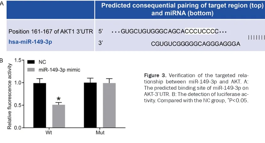
Dual luciferase reporter gene assay
According to online analysis software, there was a specific binding domain between AKT and miR-149-3p sequences. AKT was identified as the target gene of miR-149-3p (Figure 3A). According to luciferase reporter gene assay, the luciferase signal of the Wt-miR-149-3p/AKT co-transfection group in the miR-149-3p mimic group was lower than that in the NC group (P<0.05), without significant differences in the luciferase activity of Mut-3’UTR between the two groups (P>0.05). Results indicate that miR-149-3p can specifically bind to AKT, the target gene of miR-149-3p.
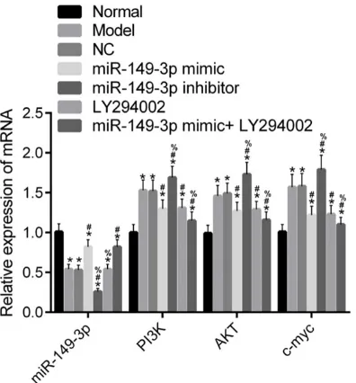
Detection of mRNA expression of miR-149-3p, PI3K, AKT, and c-myc by qRT-PCR
[image:6.612.90.287.82.322.2]According to qRT-PCR (Figure 4), compared with the Normal group, mRNA expression of miR-149-3p was decreased. However, that of c-myc, PI3K, and AKT was increased in Model, NC, miR-149-3p mimic, miR-149-3p inhibitor, LY294002, and miR-149-3p mimic + LY294002 groups (all P<0.05). Compared with the Model group, mRNA expression of miR-149-3p was increased. However, that of c-myc, PI3K, and AKT was decreased in the miR-149-3p mimic and miR-149-3p mimic + LY294002 groups (all P<0.05). Moreover, mRNA expression of c-myc, PI3K, and AKT in the LY294002 group was decreased (all P<0.05), without statistically sig-nificant differences, compared to that of miR-149-3p (P>0.05). Also, mRNA expression of miR-149-3p in the miR-149-3p inhibitor group was decreased. However, that of c-myc, PI3K, and AKT was increased in the miR-149-3p inhibitor group (all P<0.05). There were no sig-nificant differences in expression of genes between the Model and NC groups (all P>0.05). Compared with the miR-149-3p mimic group, mRNA expression of miR-149-3p was decr- eased in the LY294002 group (P<0.05), with-out significant differences compared to that of
Table 3. General information
Item Case (n)
Gender
Male 32
Female 27
Age 3 months - 4 years old
Hemangioma sites
Lip and cheek 24
Nose 6
Limbs 12
Back 8
Chest 4
Abdomen 5
Hemangioma area
Smallest 1.5 cm*2 cm
Largest 2.5 cm*4 cm
Mulliken classification
Figure 1. Immunohistochemical staining. Para-cancerous tissue was the normal tissue adjacent to cancer tissue and cancer tissue was hemangioma tissue. A: Immunohistochemical staining (200×). B: Histogram of the positive expression rate of AKT protein. Compared with the normal tissue adjacent to cancer, ***P<0.001.
Figure 2. Morphological observation and immunohistochemistry of endothelial cells from hemangioma. A: Cells covered with the culture flask after culture for 3 weeks (100×). B: The staining of factor VIII-related antigen of hem-angioma cells (immunohistochemistry, 400×).
[image:7.612.92.514.494.722.2]other genes (all P>0.05). Furthermore, mRNA expression of c-myc, PI3K, and AKT in the miR-149-3p mimic + LY294002 group was decr- eased (all P<0.05), without significant differ -ences compared to that of miR-149-3p (P> 0.05). Finally, mRNA expression of miR-149-3p in the miR-149-3p inhibitor group was decr- eased. However, that of c-myc, PI3K, and AKT was increased (all P<0.05).
Detection of protein expression of PI3K, p-PI3K, AKT, p-AKT, and c-myc by Western blot-ting
According to Western blotting (Figure 5), com-pared with the Normal group, protein expres-sion of c-myc, PI3K, p-PI3K, AKT, and p-AKT was increased in Model, NC, miR-149-3p mi- mic, 149-3p inhibitor, LY294002, and miR-149-3p mimic + LY294002 groups (all P<0.05). Compared with the Model group, protein expression in 3p mimic and miR-149-3p mimic + LY294002 groups was decreased (all P<0.05). Protein expression in the LY29- 4002 group was decreased (all P<0.05). Protein expression in the miR-149-3p inhibitor group was increased (all P<0.05). There were no sig-nificant differences in protein expression
be-tween Model and NC groups (all P>0.05). Co- mpared with the miR-149-3p mimic group, pro-tein expression in the LY294002 group was not significantly different (all P>0.05). Protein ex-pression in the miR-149-3p mimic + LY294002 group was decreased (all P<0.05). Protein expression in the miR-149-3p inhibitor group was increased (all P<0.05).
Detection of proliferation of cells by MTT
According to MTT (Figure 6), there were no sig-nificant differences in OD values between the groups at 24 hours (all P>0.05). At 48 and 72 hours, compared with the Normal group, OD values in the Model, NC, miR-149-3p mimic, 3p inhibitor, LY294002, and miR-149-3p mimic + LY294002 groups were increased (all P<0.05). At 48 and 72 hours, compared with the Model group, OD values in the miR-149-3p inhibitor group were increased (both P<0.05). Levels were decreased in the miR-149-3p mimic, LY294002, and miR-miR-149-3p mimic + LY294002 groups (all P<0.05). There were no significant differences between Model and NC groups at each time point (both P>0.05). At 48 and 72 hours, compared with the miR-149-3p mimic group, OD values in the LY29- 4002 group were not significantly different (all P>0.05), but were decreased in the miR-149-3p mimic + LY294002 group (all P<0.05).
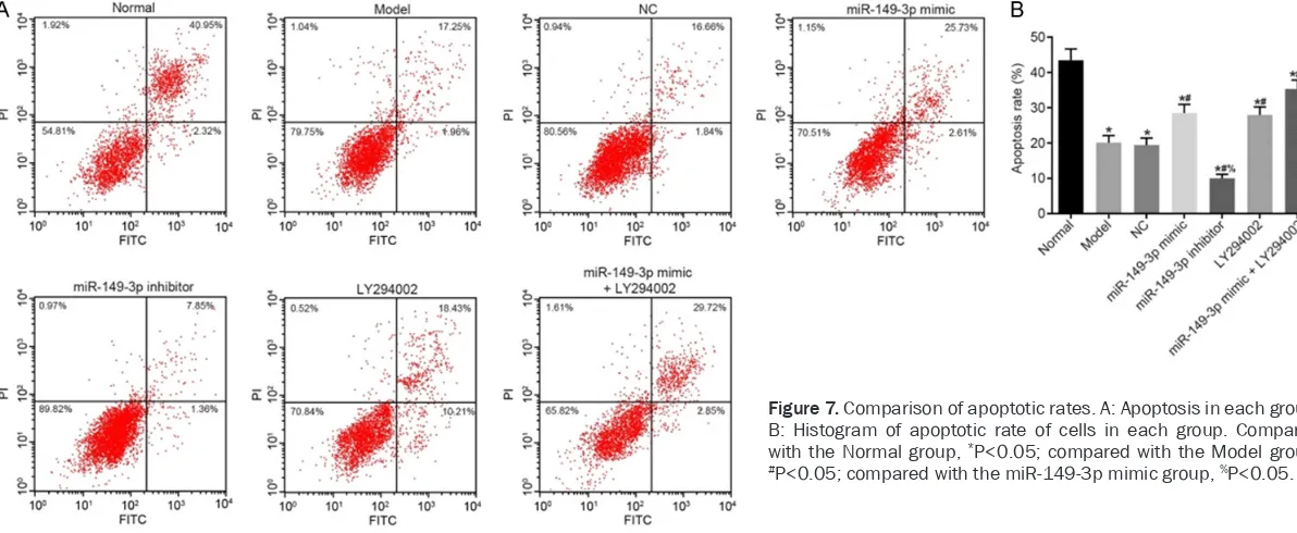
Detection of apoptosis by flow cytometry
[image:8.612.92.290.69.282.2]According to flow cytometry (Figure 7), com-pared with the Normal group, apoptosis rates in Model, NC, miR-149-3p mimic, miR-149-3p inhibitor, LY294002, and miR-149-3p mimic + LY294002 groups were decreased significantly (all P<0.05). Compared with the Model group, apoptosis rates in the miR-149-3p inhibitor group were decreased significantly (P<0.05), but were increased significantly in the miR-149-3p mimic, LY294002, and miR-149-miR-149-3p mimic + LY294002 groups (all P<0.05). There were no statistically significant differences between Model and NC groups (P>0.05). Compared with the miR-149-3p mimic group, apoptosis rates in the miR-149-3p inhibitor group were dec- reased significantly (P<0.05), but were inc-reased significantly in the miR-149-3p mimic + LY294002 group (P<0.05). There were no sig-nificant differences between miR-149-3p mimic and LY294002 groups (P>0.05).
Discussion
Recent studies have confirmed that PI3K/AKT signaling pathways play an important role in cell metabolism, cell cycle control, angiogene-sis, and chemotherapy resistance [14-16]. AKT, a downstream molecule of PI3K, is an impor-tant check point of PI3K/AKT signaling path-ways. Its phosphorylation is an important mark-er for activation of this pathway [17, 18]. Li and others have explored the relationship between
hemangiomas in children and PI3K/AKT signal-ing pathways, findsignal-ing that phosphorylation of AKT can activate its downstream mTOR protein, accelerate cell progression from G1 to S phas-es, and promote proliferation of hemangioma cells [19]. Immunohistochemistry was used in this study to observe positive expression of AKT in hemangioma and normal tissues. Po- sitive expression of AKT found in hemangioma tissues was significantly increased, confirming that AKT might be related to the pathogenesis of hemangiomas.
[image:9.612.92.521.69.299.2]Moreover, miR-149-3p has been proven to play a protective role in cancers. For example, Cao found that glycyrrhetinic acid inhibits the pro-gression of gastric cancer through activating miR-149-3p-Wnt signaling pathways [20]. St- udying the biological characteristics of bladder cancer cells, Yang confirmed that miR-149-3p inhibits S100A4 in a targeted way, thereby inhibiting proliferation, migration, and invasion of bladder cancer cells [21]. Additionally, there is a study confirming that miR-149 inhibits the growth and metastasis of NSCLC by inhibiting the FOXM1/cyclin D1/MMP2 axis [22]. These studies have confirmed the protective role of miR-149 in cancer progression. The current study verified the targeted relationship between miR-149 and AKT through dual luciferase report Figure 5. Comparison of protein expression of PI3K, p-PI3K, AKT, p-AKT, and c-myc. A: Protein band; B: Protein expression histogram. Compared with the Normal group, *P<0.05; compared with the Model group, #P<0.05; com-pared with the miR-149-3p mimic group, %P<0.05. PI3K, phosphatidylositol-3-kinase; AKT, protein kinase B.
[image:9.612.89.285.362.512.2]gene assay. Additionally, qRT-PCR was used to observe expression of miR-149. Compared with the Normal group, expression of miR-149-3p was significantly decreased. However, mRNA and protein expression of PI3K, p-PI3K, AKT, and p-AKT was significantly increased in other groups, proving that PI3K/AKT signaling path-ways are significantly activated in hemangioma cells of children. Compared with the Model group, expression of PI3K, p-PI3K, AKT, and p-AKT in the miR-149-3p inhibitor group was increased, indicating that miR-149-3p knock-out can promote activation of PI3K/AKT signal-ing pathways.
As a proto-oncogene, c-myc has been proven to be involved in the gene control of cells, acceler-ating the transformation of cells into malignant phenotypes [23, 24]. Studies have found that expression of c-myc is abnormally increased in lung cancer, liver cancer, breast cancer, and thyroid cancer [25-28]. Expression of c-myc in other groups was significantly higher than that in the Normal group. Overexpression of miR-149-3p or inhibiting activation of PI3K/AKT sig-naling pathways reduced its expression, further confirming the protective roles of miR-149 over -expression and inhibiting activation of PI3K/ AKT signaling pathways in hemangiomas in children. MTT and flow cytometry were used to detect proliferation and apoptosis of cells. Compared with the normal group, proliferation was significantly enhanced but apoptosis was weakened in other groups. Overexpression of miR-149-3p or inhibiting activation of PI3K/AKT signaling pathways can inhibit proliferation and promote apoptosis of endothelial cells from hemangiomas. The combination of the two is more effective. This study explored the relation-ship between miR-149-3p, PI3K/AKT signaling pathways, and the biological characteristics of endothelial cells from hemangiomas in chil-dren. However, it needs to be further verified whether miR-149-3p and this pathway are affected by related target genes. In addition, experimental conditions may have affected the results of this study.
In summary, miR-149-3p can inhibit expression of AKT in a targeted way and mediate PI3K/AKT signaling pathways, thus promoting apoptosis and inhibiting proliferation of endothelial cells from hemangiomas. This plays a protective role in children with hemangiomas. Moreover, miR-149-3p is expected to become a potential tar-get for treatment of hemangiomas in children.
Acknowledgements
This work was supported by Fujian Provincial Medical Innovation Course for the influence of
PI3K/AKT/mTOR/4EBP1 signaling pathway on occurrence and development of pediatric hem-angioma (2016-CX-6).
Disclosure of conflict of interest
None.
Address correspondence to: Junping Wen, Depart- ment of Endocrinology, Provincial Clinical College of Fujian Medical University, Fujian Provincial Hos- pital, No.134 East Street, Fuzhou 350001, Fujian Province, China. Tel: +86-13559925729; E-mail: wenjunping52@163.com
References
[1] Prey S, Voisard JJ, Delarue A, Lebbe G, Taieb A, Leaute-Labreze C and Ezzedine K. Safety of propranolol therapy for severe infantile hem-angioma. JAMA 2016; 315: 413-415.
[2] Munabi NC, England RW, Edwards AK, Kita- jewski AA, Tan QK, Weinstein A, Kung JE, Wilcox M, Kitajewski JK, Shawber CJ and Wu JK. Propranolol targets hemangioma stem cells via cAMP and mitogen-activated protein kinase regulation. Stem Cells Transl Med 2016; 5: 45-55.
[3] Zuccolo E, Bottino C, Diofano F, Poletto V, Codazzi AC, Mannarino S, Campanelli R, Fois G, Marseglia GL, Guerra G, Montagna D, Laforenza U, Rosti V, Massa M and Moccia F. Constitutive store-operated Ca(2+) entry leads to enhanced nitric oxide production and prolif-eration in infantile hemangioma-derived endo-thelial colony-forming cells. Stem Cells Dev 2016; 25: 301-319.
[4] Dolly SO, Wagner AJ, Bendell JC, Kindler HL, Krug LM, Seiwert TY, Zauderer MG, Lolkema MP, Apt D, Yeh RF, Fredrickson JO, Spoerke JM, Koeppen H, Ware JA, Lauchle JO, Burris HA 3rd and de Bono JS. Phase I study of apitolisib (GDC-0980), dual phosphatidylinositol-3-kina- se and mammalian target of rapamycin kinase inhibitor, in patients with advanced solid tu-mors. Clin Cancer Res 2016; 22: 2874-2884. [5] Yang W, Hosford SR, Dillon LM, Shee K, Liu SC,
[6] Pan WK, Li P, Guo ZT, Huang Q and Gao Y. Propranolol induces regression of hemangio-ma cells via the down-regulation of the PI3K/ Akt/eNOS/VEGF pathway. Pediatr Blood Can- cer 2015; 62: 1414-1420.
[7] Zheng N, Ding X, Sun A and Jahan R. PDK1 ac-tivity regulates proliferation, invasion and gr- owth of hemangiomas. Cell Physiol Biochem 2015; 36: 1903-1910.
[8] Huang C, Huang J, Ma P and Yu G. microR-NA-143 acts as a suppressor of hemangioma growth by targeting Bcl-2. Gene 2017; 628: 211-217.
[9] Ke Y, Zhao W, Xiong J and Cao R. miR-149 Inhibits Non-Small-Cell Lung Cancer Cells EMT by Targeting FOXM1. Biochem Res Int 2013; 2013: 506731.
[10] Xue L, Wang Y, Yue S and Zhang J. Low MiR-149 expression is associated with unfavorable prognosis and enhanced Akt/mTOR signaling in glioma. Int J Clin Exp Pathol 2015; 8: 11178-11184.
[11] Mulliken JB and Glowacki J. Hemangiomas and vascular malformations in infants and chil-dren: a classification based on endothelial characteristics. Plast Reconstr Surg 1982; 69: 412-422.
[12] Li XY, Luo QF, Wei CK, Li DF, Li J and Fang L. MiRNA-107 inhibits proliferation and migration by targeting CDK8 in breast cancer. Int J Clin Exp Med 2014; 7: 32-40.
[13] Collin SP. Topographic organization of the gan-glion cell layer and intraocular vascularization in the retinae of two reef teleosts. Vision Res 1989; 29: 765-775.
[14] Yang SX, Polley E and Lipkowitz S. New insights on PI3K/AKT pathway alterations and clinical outcomes in breast cancer. Cancer Treat Rev 2016; 45: 87-96.
[15] Zhang D, Sun G, Zhang H, Tian J and Li Y. Long non-coding RNA ANRIL indicates a poor prog-nosis of cervical cancer and promotes carcino-genesis via PI3K/Akt pathways. Biomed Phar- macother 2017; 85: 511-516.
[16] Erdogan S, Doganlar O, Doganlar ZB, Serttas R, Turkekul K, Dibirdik I and Bilir A. The flavo-noid apigenin reduces prostate cancer CD- 44(+) stem cell survival and migration through PI3K/Akt/NF-kappaB signaling. Life Sci 2016; 162: 77-86.
[17] Dey N, De P and Leyland-Jones B. PI3K-AKT-mTOR inhibitors in breast cancers: from tumor cell signaling to clinical trials. Pharmacol Ther 2017; 175: 91-106.
[18] Baek SH, Ko JH, Lee JH, Kim C, Lee H, Nam D, Lee J, Lee SG, Yang WM, Um JY, Sethi G and Ahn KS. Ginkgolic acid inhibits invasion and migration and TGF-beta-induced EMT of lung cancer cells through PI3K/Akt/mTOR inactiva-tion. J Cell Physiol 2017; 232: 346-354.
[19] Li D, Li P, Guo Z, Wang H and Pan W. Downregulation of miR-382 by propranolol in-hibits the progression of infantile hemangioma via the PTEN-mediated AKT/mTOR pathway. Int J Mol Med 2017; 39: 757-763.
[20] Cao D, Jia Z, You L, Wu Y, Hou Z, Suo Y, Zhang H, Wen S, Tsukamoto T, Oshima M, Jiang J and Cao X. 18beta-glycyrrhetinic acid suppresses gastric cancer by activation of miR-149-3p-Wnt-1 signaling. Oncotarget 2016; 7: 71960-71973.
[21] Yang D, Du G, Xu A, Xi X and Li D. Expression of miR-149-3p inhibits proliferation, migration, and invasion of bladder cancer by targeting S100A4. Am J Cancer Res 2017; 7: 2209-2219.
[22] Zhao L, Liu L, Dong Z and Xiong J. miR-149 suppresses human non-small cell lung cancer growth and metastasis by inhibiting the FO- XM1/cyclin D1/MMP2 axis. Oncol Rep 2017; 38: 3522-3530.
[23] Qiu MK,Wang SQ, Pan C. ROCK inhibition as a potential therapeutic target involved in apopto-sis in hemangioma. Oncol Rep 2017; 37: 2987-2993.
[24] Zhang HF, Wu C, Alshareef A, Gupta N, Zhao Q, Xu XE, Jiao JW, Li EM, Xu LY and Lai R. The PI3K/AKT/c-MYC axis promotes the acquisi-tion of cancer stem-like features in esophageal squamous cell carcinoma. Stem Cells 2016; 34: 2040-2051.
[25] Ritorto MS, Rhode H, Vogel A, Borlak J. Regu- lation of glycosylphosphatidylinositol-anchor- ed proteins and GPI-phospholipase D in a c-Myc transgenic mouse model of hepatocellular carcinoma and human HCC. Biol Chem 2016; 397: 1147-1162.
[26] Fan W, Yang H, Liu T, Wang J, Li TW, Mavila N, Tang Y, Yang J, Peng H, Tu J, Annamalai A, Noureddin M, Krishnan A, Gores GJ, Martinez-Chantar ML, Mato JM and Lu SC. Prohibitin 1 suppresses liver cancer tumorigenesis in mice and human hepatocellular and cholangiocarci-noma cells. Hepatology 2017; 65: 1249-1266. [27] Pourteimoor V, Paryan M and Mohammadi-Yeganeh S. microRNA as a systemic interven-tion in the specific breast cancer subtypes with C-MYC impacts; introducing subtype-based ap-praisal tool. J Cell Physiol 2018; 233: 5655-5669.