Original Article
miR-144 inhibits development of meningitis
in rats by down-regulating TLR2
Shuhong Ma
Weinan Vocational and Technical College, Weinan High Tech Zone, Science and Technology Park of West Section of Shengli Road, Weinan City 714026, Shaanxi Province, China
Received July 3, 2018; Accepted September 8, 2018; Epub April 15, 2019; Published April 30, 2019
Abstract: Purpose: This article was to investigate the effect of miR-144 on meningitis in rats and its mechanism. Methods: A rat meningitis model was constructed and divided into the Meningitis group, the Meningitis + agomir
group, and the Meningitis + antagomir group. A sham group was served as control. Loeffler scores, white blood cell count as well as IL-1β and TNF-α level in the cerebrospinal fluid were measured. TLR2 protein expression in brain
tissues was detected by immunohistochemistry and Western blot. miR-144 and TLR2 mRNA expression in brain tissues was determined by qRT-PCR. Luciferase reporter assay was performed to verify the targeting relationship between miR-144 and TLR2. Results: Compared with the sham group, the meningitis rats had much higher body
temperature, lower Loeffler score, higher white blood cell count, IL-1β and TNF-α level, markedly lower miR-144 ex -pression, and much higher TLR2 mRNA and protein expression (all P < 0.05). Compared with the Meningitis group,
infusion of miR-144 agonists decreased white blood cell count as well as IL-1β and TNF-α level, and down-regulated
TLR2 expression (P < 0.05 or P < 0.01). miR-144 inhibited TLR2 expression. Conclusions: miR-144 inhibited devel-opment of meningitis in rats by down-regulating TLR2.
Keywords: Meningitis, miR-144, TLR2, IL-1β, TNF-α
Introduction
Bacterial meningitis is a common infectious disease of the central nervous system in infants and young children. It has a high mortality rate and severely affects the life and health of chil-dren. In addition, meningitis also causes severe sequelae, such as paralysis, hydrocephalus, epilepsy, deaf, blindness, and intellectual dam-age [1, 2]. Therefore, early prevention and diag-nosis of meningitis are very important for im- proving the prognosis of patients.
In recent years, the treatment of diseases at the genetic level has received increasing atten-tion. miRNA are also deeply studied in multiple diseases. As a miRNA, miR-144 has also been reported to be involved in many diseases, espe-cially in tumors, and is considered to be a tumor suppressor in glioblastoma, laryngeal squam- ous cell carcinoma, as well as gastric cancer [3-5]. Meanwhile, miR-144 has also been fou- nd to participate in and regulate inflammatory responses. Dongmin et al. [6] illustrated that in rats with non-alcoholic steatohepatitis,
down-regulation of miR-144 could stimulate the re- lease of pro-inflammatory cytokines. Liu et al. [7] also found that, in monocyte-derived macro-phages infected with M. tuberculosis, inhibition of miR-144 could lead to accelerated secretion of pro-inflammatory cytokines, including TNF-α, IL-1β, and IL-6. However, expression of miR-144 in meningitis and its effect on the development of meningitis have not been reported before. Therefore, this article established a model of rat meningitis through S. pneumoniae infection. The effect of miR-144 on development of men-ingitis in rats was also researched in depth. This study provides an important theoretical basis for the application of miR-144 in the pre-vention and treatment of meningitis.
Methods
Preparation of streptococcus pneumoniae sus-pension
(Bacterial No. 31003). Streptococcus pneu-moniae was inoculated in blood agar medium and cultured in a 37°C, 5% CO2 incubator for 12 hours. Then they were inoculated into VITAL AER broth medium (Bio Mérieux, France), and were collected in the logarithmic phase for centrifugation. After being rinsed with normal saline, Streptococcus pneumoniae was sub-jected to centrifugation again. They were dilut-ed in normal saline with a density of 3 × 10¹⁰/L.
Construction of rat meningitis model
Forty male Sprague-Dawley rats (weighing 200-250 g, purchased from Shanghai Laboratory Animal Center of Chinese Academy of Scien- ces) were housed in cages at 25°C and 40% relative humidity for one week. Rats were ran -domly divided into four groups with 10 rats in each group: Sham group, Meningitis group, Meningitis + agomir group, Meningitis + an- tagomir group. All rats were subjected to men-ingitis model construction surgery after fasting for 12 hours.
All rats were injected intraperitoneally with 10% chloral hydrate (dose: 5 mL/kg) and fixed on a stereotaxic instrument. Totally 50 μL of cere -brospinal fluid was obtained from the cisterna magna by using a microsyringe. At the same injection point, 30 μL of normal saline was injected into rats of the Sham group, while 30 μL of Streptococcus pneumoniae suspension was injected into rats of the Meningitis group, Meningitis + agomir group and Meningitis + antagomir group. One hour later, 20 μL of nor -mal saline was injected into rats of the Sham group again. However, rats of the Meningitis + agomir group were injected with 20 μL of miR-144 agonist, and rats of the Meningitis + antagomir group were injected with 20 μL of miR-144 antagonist. After injection, rats were kept individually in cages with sufficient water and feed. Body temperature was recorded for 5 days. All rats were then anesthetized with 10% chloral hydrate and 50 μL of cerebrospinal fluid was collected from the cisterna magna. Finally, all rats were sacrificed and intact brain tissues were obtained. The use of animals and proce-dures were approved by Weinan Vocational & Technical College Institutional Animal Care and Use Committee.
Neuroethology assessment by Loeffler’s neu-roethology score
One day after surgery, neuroethology of rats were assessed using Loeffler’s neuroethology
score according to the following criteria: 5 points: rats were able to move normally when they were caught on the back, and they were able to turn over within 5 s; 4 points: rats had reduced exercise, but they were able to turn over within 5 s; 3 points: rats were able to turn over more than 5 s; 2 points, rats were unable to turn over; 1 points, rats could not exercise. A higher Loeffler’s neuroethology score indicated less neurological impairment in rats.
White blood cell counts and levels of IL-1β and TNF-α in rat cerebrospinal fluid
The number of white blood cells in cerebrospi-nal fluid collected before and after surgery in rats was measured using the LH750 automatic hematology analyzer (Beckman Coulter, USA). Furthermore, levels of IL-1β and TNF-α in these cerebrospinal fluid were also detected by enzyme-linked immunosorbent assay (ELISA) kit (Endogen, Woburn, MA). All operations are strictly performed according to the instruc- tions.
TLR2 expression detection by immunohisto-chemistry
Paraffin-embedded rat brain tissues were sub -jected to continuous coronal slices. Five con -secutive sections of brain tissues from each rat were randomly selected for dewaxing, rehydra-tion, and antigen retrieval. After incubation with 3% H2O2 for 15 min at room temperature, these brain tissue sections were blocked with goat serum for 15 minutes again. Rabbit anti-Hu-man TLR2 Antibody (1:100, Santa Cruz) was used to incubate these sections for 12 hours at 4°C. Slices were rinsed 3 times by PBS. The secondary antibody was added for 15 minute incubation at 37°C. After rinsed 3 times by PBS, horseradish peroxidase-labeled strepta- vidin working solution was added to continue incubation for 15 minutes. Brain tissue sec-tions were subjected to DAB chromogenic reac-tion and were counterstain with hematoxylin for 30 seconds. Neutral gel used to seal them. Five areas of each slice were observed under a microscope and the number of TLR2 positive cells was counted. Brown-yellow particles ap- pearing in the cytoplasm and cell membrane were considered as TLR2-positive cells.
Culture and transfection of 293T Cells
CGCTTCGGCAGCACATATACT, U6-R, ACGCTTCA- CGAATTTGCGTGTC; TLR2-F, CCAAAGAGCTCG-TAGCATCC, TLR2-R, AGGGGCTTCACTTCTCTG- CT; GAPDH-F, TGGCAAAGTGGAGATTGTTGCC, GAPDH-R, AAGATGGTGATGGGCTTCCCG. In this research, 2-ΔΔCt method was selected to
calcu-late the fold change of genes to be detected.
Western blot analysis
Total proteins in tissues and cells were collect-ed after they were lyscollect-ed by lysis buffer (Cell Signaling Technology). Proteins were trans-ferred onto polyvinylidene difluoride (PVDF) membranes after they were separated by sodi-um dodecyl sulfate polyacrylamide gel elec- trophoresis (SDS-PAGE). Skim milk (5%) was added to block these membranes for 1 hour at room temperature. Membranes were sequen-tially subjected to primary antibody (mouse anti-human TLR2 antibody, 1:1000, Abcam, Cambridge, UK) incubation for 12 hours at 4°C and secondary antibody (horse radish peroxi-dase conjugated secondary antibody, Beijing Zhongshan Jinqiao Biotechnology Co., Ltd., China) incubation for 1 hour at room tem- perature.
Statistical analysis
All data were processed by SPSS 19.0 and were expressed as mean ± SD. Comparison between two groups was conducted by t-test. One-way ANOVA was selected to perform com-parison among multiple groups. P < 0.05 was considered statistically significant. In this re-search, all experiments were repeated 3 times.
Results
Clinical features of rats
Before surgery, the difference in body tempera-ture, Loeffler scores, white blood cell count, IL-1β, and TNF-α level was not obvious among groups. After surgery, compared to the Sham group, rats of the Meningitis group, the Me- ningitis + agomir group and the Meningitis + antagomir group were with much higher bo- dy temperature (P < 0.05), obviously lower Loeffler’s scores (P < 0.05), and markedly higher white blood cell count, IL-1β and TNF-α level (P < 0.05). Additionally, when com- pared to the Meningitis group, rats of the Meningitis + agomir group had dramatically lower body temperature (P < 0.05), significant-10% fetal bovine serum (FBS) and were placed
in a 5% CO2, 37°C incubator for subculture. 293T cells in logarithmic growth phase were transfected with miR-144 mimics negative con-trol (NC), miR-144 mimics, miR-144 inhibitors NC and miR-144 inhibitors respectively by using Lipfectamine 2000 (Thermo Fisher Scientific, Waltham, MA, USA) according to the instruc-tions. These transfected cells were grouped in turn as follows: mimics NC group, miR-144 mimics group, inhibitors NC group and miR-144 inhibitors group. 293T cells without any treat-ment were served as the control group. Cells of each group were prepared as cell suspen-sions by DMEM (10% FBS) at a density of 1 × 10⁵ cells/Ml, and were inoculated in a 24-well plates with 2 mL of cell suspensions each well. Plates were were placed in a 5% CO2 37°C in- cubator for continued culture.
Luciferase reporter assay
Target scan online prediction software was used to predict the targeted relationship be- tween miR-144 and TLR2. Mutant-type (MT) and wild-type (WT) sequences of TLR2 mRNA containing the binding site were designed ac- cording to the predicted results. These sequ- ences were constructed on vectors to transfect cells of mimics NC group and miR-144 mimics group respectively. After 48 hours, residual liq-uid in each well was discarded and cells were rinsed twice with PBS. Then 100 μL of Passive Lysis Buffer was added into each well for 15 minutes slightly shaking at room temperature. Cell lysates in each well were collected for lu- ciferase activity detection.
Quantitative real-time polymerase chain reac-tion (qRT-PCR)
CT-ly higher Loeffler’s scores (P < 0.05), and obvi-ously lower white blood cell count, IL-1β and TNF-α level (P < 0.05). However, markedly high -er body temp-erature (P < 0.05), much lower Loeffler’s scores (P < 0.05), as well as dramati-cally higher white blood cell count, IL-1β and TNF-α level (P < 0.05) were found in rats of the Meningitis + antagomir group when compared with the Meningitis group (Figure 1A-D, Table 1).
miR-144 expression in rat brain tissues
miR-144 relative expression in rat brain tissues of the Meningitis group, the Meningitis + agomir
group and the Meningitis + antagomir group was all significantly lower than that of the Sham group (P < 0.01). In addition, when compared with the Meningitis group, miR-144 relative expression was dramatically increased in the Meningitis + agomir group (P < 0.01), while that was markedly decreased in the Meningitis + antagomir group (P < 0.01) (Figure 2).
TLR2 expression in rat brain tissues
Significantly higher TLR2 mRNA and protein relative expression was found in rat brain tis-sues of the Meningitis group, the Meningitis + agomir group, and the Meningitis + antagomir group when compared with the Sham group (P
[image:4.612.98.517.69.334.2]< 0.01). Furthermore, when compared with TLR2 mRNA and protein relative expression in rat brain tissues of the Meningitis group, they were significantly down-regulated in the Meni-ngitis + agomir group (P < 0.01) and dramati-cally up-regulated in the Meningitis + antago- mir group (P < 0.01) (Figure 3A, 3B). These results of TLR2 protein expression had also been demonstrated by immunohistochemistry (Figure 3C).
Figure 1. Changes in clinical features of rats in each group. A. Changes in body temperature. B. Changes in
Loef-fler score. C. Levels of IL-1β and TNF-α in the cerebrospinal fluid before surgery. D. Levels of IL-1β and TNF-α in the cerebrospinal fluid after surgery. *P < 0.05 when compared with the other three groups. #P < 0.05 when compared with the Meningitis group.
Table 1. Changes in white blood cell count of rats (Unit: × 106/L)
Group surgeryBefore after surgeryThe 5th day Sham group 84 ± 7 88 ± 6*
Meningitis group 82 ± 9 3365 ± 681 Meningitis + agomir group 85 ± 6 2872 ± 574#
Meningitis + antagomir group 83 ± 5 3834 ± 862#
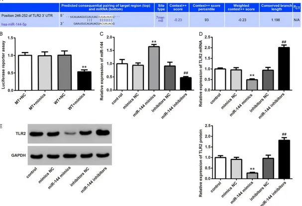
[image:4.612.90.305.431.510.2]TLR2 was the target gene of miR-144
Via Target Scan, we observed that miR-144 was bound to the 3’-UTR region of TLR2 mRNA (Figure 4A). Therefore, reporter gene plasmids with TLR2 3’-UTR (MT or WT) were constructed. After 293T cells of mimics NC group and miR-144 mimics group were transfected by these plasmids, we found that there was no statisti-cally significant difference of luciferase activity intensity between MT + NC group and MT + mimics group. However, when compared with the WT+NC group, the luciferase activiyy inten-sity was significantly decreased in the WT + mimics group (P < 0.05) (Figure 4B).
The role of miR-144 in the regulation of TLR2 was further studied. Results showed that, com-pared with the control and mimics NC groups, cells of the miR-144 mimics group were with much higher miR-144 relative expression and markedly lower TLR2 mRNA and protein relative expression (P < 0.01). At the same time, signifi -cantly increased miR-144 relative expression (P < 0.01) and dramatically increased TLR2 mRNA and protein relative expression (P < 0.01) were found in the miR-144 inhibitors group when compared with the control and inhibitors NC group (Figure 4C-E). The above results further indicated that TLR2 was a target gene of miR-144 and TLR2 was negatively regu-lated by miR-144.
Discussion
Bacterial meningitis is one of the most com-mon diseases in children, which can cause severe neurological sequelae [8]. Streptococcus pneumoniae meningitis is the most common bacteria in infants with intracranial infections [9]. In this study, the rat meningitis model infected by Streptococcus pneumoniae was successfully constructed. Furthermore, this research also investigated the effect of miR-144 on rats with meningitis. The results dem-onstrated that miR-144 was down-regulated in brain tissues of meningitis rats. Up-regulation of miR-144 could inhibit the development of meningitis in rats through suppressing expres-sion of TLR2.
[image:5.612.90.286.72.261.2]miRNAs are a type of noncoding small RNA that is widely spread in eukaryotes [10]. It has been found to play a pivotal role in many major human diseases, especially in multiple cancers [11]. The influence of miRNAs on inflammatory responses has also been studied [12]. For example, it has been reported that miR-221 could regulate endothelial inflammatory res-ponse by targeting AdipoR1 [13]. miR-155 was found to stimulate expression of IL-1β and it could participate in regulation of inflammatory responses in ischemic cerebral tissues [14]. miR-138 could decrease the levels of IL-6 and IL-8 in human coronary artery endothelial cell injury. It relieved human coronary artery endo-thelial cell injury as well as related inflammato -ry reactions [15]. In this study, white blood cell count as well as IL-1β and TNF-α level were increased significantly in cerebrospinal fluid of meningitis rats. IL-1β and TNF-α were two major pro-inflammatory factors. Elevated levels were indicative of aggravated inflammatory respons -es [16]. White blood cells are an important part of the human body’s defense system. Once the human body is invaded by external bacteria, white blood cells could pass through capillary walls through deformation. Then they are con-centrated on the bacteria invading parts in order to surround and devour them [17-20]. Therefore, it is likely that the body develops inflammation if the number of white blood cells exceeds the normal range. We observed from this study that white blood cell count and levels of IL-1β and TNF-α in rats cerebrospinal fluid were dramatically decreased after meningitis rats were injected with miR-144 agonist. While Figure 2. miR-144 relative expression in rat brain
Figure 3. TLR2 relative expression in rat brain tissues of each group. (A) TLR2 mRNA relative expression in rat brain tissues was detected by qRT-PCR. (B) West-ern blot was used to determine the relative expres-sion of TLR2 protein in rat brain tissues. (C) TLR2 protein relative expression in rat brain tissues was
Figure 4. TLR2 is a target gene of miR-144. (A) The binding site of miR-144 and TLR2 was predicted by Target Scan. (B) Luciferase reporter assay was used to detect
the targeting relationship between miR-144 and TLR2. **P < 0.01 when compared with the WT + NC group. (C) miR-144 relative expression in 293T cells of each group was detected by qRT-PCR. (D) TLR2 mRNA relative expression in 293T cells of each group was determined by qRT-PCR. (E) TLR2 protein relative expression
these indicators were all increased after men-ingitis rats were injected with miR-144 antago-nist. It can be concluded from these results that miR-144 might have an inhibitory effect on the inflammatory response in meningitis rats. Liu et al. [7] noticed from their research that, lower miR-144 expression could stimulate the release of inflammatory factors in monocyte-derived macrophages after they were infected by mycobacterium tuberculosis. Their research was consistent with our results. This article first studied the regulation effect of miR-144 on meningitis rats.
TLR2 is an important member of the Toll-like receptor family, which has an irreplaceable role in innate immunity and inflammation [21]. TLR2 could recognize multiple bacterial lipoproteins, which is the initial barrier to bacterial infection [22]. Previous research has suggested that increased TLR2 expression in monocytes could result in systemic inflammatory response syn -drome after liver transplantation [23]. Li et al. [24] demonstrated that, in burn-related cellular inflammatory reactions, application of TLR2 agonists could stimulate the expression of inflammatory factors, such as IL-1β and TNF-α. There have been some studies on the role of TLR2 in meningitis. TLR2 has also been found to be regulated by some miRNAs. Philippe et al. [25] revealed that TLR2 was closely related to rheumatoid arthritis and miR-19a/b regulated the release of IL-6 and matrix metalloprotein-ase 3 through decreasing expression of TLR2 protein. Xu et al. [26] also noted that TLR2 was expressed lower in rats with chronic obstruc-tive pulmonary disease, which was negaobstruc-tively regulated by miR-344b-1-3p. In this article, we found that TLR2 was negatively regulated by miR-144.
In conclusion, the rat meningitis model was successfully constructed through Streptoco- ccus pneumoniae infection. miR-144 agonist significantly inhibited development of meningi -tis. The relevant mechanism involved in this process was that miR-144 could relieve menin-gitis development by suppressing expression of TLR2. miR-144 might serve as one of the poten-tial therapeutic targets for the diagnosis and treatment of meningitis caused by Strepto- coccus pneumoniae.
Disclosure of conflict of interest
None.
Address correspondence to: Shuhong Ma, Weinan
Vocational and Technical College, Weinan High Tech Zone, Science and Technology Park of West Section
of Shengli Road, Weinan City 714026, Shaanxi Province, China. Tel: +86-0913-3033110; Fax: +86-0913-3033110; E-mail: mashuhong256@163.com
References
[1] Haseloff RF, Dithmer S, Winkler L, Wolburg H
and Blasig IE. Transmembrane proteins of the tight junctions at the blood-brain barrier: struc-tural and functional aspects. Semin Cell Dev Biol 2015; 38: 16-25.
[2] Obermeier B, Daneman R and Ransohoff RM. Development, maintenance and disruption of the blood-brain barrier. Nat Med 2013; 19: 1584-96.
[3] Lan F, Yu H, Hu M, Xia T and Yue X. miR-144-3p
exerts anti-tumor effects in glioblastoma by targeting c-Met. J Neurochem 2015; 135: 274-86.
[4] Zhang SY, Lu ZM, Lin YF, Chen LS, Luo XN, Song XH, Chen SH and Wu YL. miR-144-3p, a
tumor suppressive microRNA targeting ETS-1 in laryngeal squamous cell carcinoma. Onco- target 2016; 7: 11637-50.
[5] Ren K, Liu QQ, An ZF, Zhang DP and Chen XH.
MiR-144 functions as tumor suppressor by targeting PIM1 in gastric cancer. Eur Rev Med Pharmacol Sci 2017; 21: 3028-3037.
[6] Dongmin L, Xuan W, Xi L, Yue L, Li L, Jing Y, Jing L, Qingzhu S, Yili W and Hongmin L. Down-regulation of miR-144 elicits proinflammatory cytokine production by targeting toll-like recep -tor 2 in nonalcoholic steatohepatitis of high-fat-diet-induced metabolic syndrome E3 rats. Mol Cell Endocrinol 2015; 402: 1-12.
[7] Liu HY. Down-regulation of miR-144 after
My-cobacterium tuberculosis infection promotes
inflammatory factor secretion from macro -phages through the Tpl2/ERK pathway. Cell Mol Biol (Noisy-le-grand) 2016; 62: 87-93. [8] Gaini S, Karlsen GH, Nandy A, Madsen H,
Christiansen DH and Á Borg S. Culture nega -tive listeria monocytogenes meningitis result-ing in hydrocephalus and severe neurological sequelae in a previously healthy immunocom-petent man with penicillin allergy. Case Rep Neurol Med 2015; 2015: 248302.
[9] Elena P, Fabio F, Maria CA, Francesco I, Donata M and Gianni P. Interferon-γ from brain leuko -cytes enhances meningitis by type
4strepto-coccus pneumoniae. Front Microbiol 2015; 6:
1340.
[10] Patricia B, Caroline YI and Blanca SS.
Genome-wide analysis of polycistronic microRNAs in cultivated and wild rice. Genome Biol Evol 2016; 8: 1104-14.
progres-sion, miR-200: potential cancer therapeutic targets. Curr Pharm Des 2014; 20: 1896-903. [12] O’Connell RM, Rao DS and Baltimore D. mi
-croRNA regulation of inflammatory responses.
Annu Rev Immunol 2012; 30: 295-312. [13] Chen CF, Huang J, Li H, Zhang C, Huang X, Tong
G and Xu YZ. MicroRNA-221 regulates endo
-thelial nitric oxide production and inflammato -ry response by targeting adiponectin receptor 1. Gene 2015; 565: 246-51
[14] Wen Y, Zhang X, Dong L, Zhao J, Zhang C and
Zhu C. Acetylbritannilactone modulates micro-
RNA-155-mediated inflammatory response in
ischemic cerebral tissues. Mol Med 2015; 21: 197-209.
[15] Li JB, Wang HY, Yao Y, Sun QF, Liu ZH, Liu SQ, Zhuang JL, Wang YP and Liu HY. Overexpression
of microRNA-138 alleviates human coronary
artery endothelial cell injury and inflammatory response by inhibiting the PI3K/Akt/eNOS
pathway. J Cell Mol Med 2017; 21: 1482-1491. [16] Ribeiro AB, de Barcellos-Filho PC, Franci CR,
Menescal-De-Oliveira L and Saia RS.
Pro-inflammatory cytokines, IL-1β and TNF-α, pro -duce persistent compromise in tonic immobili-ty defensive behavior in endotoxemic guinea pigs. Acta Physiologica; 218: 123-135. [17] Lyons TW, Cruz AT, Freedman SB, Neuman MI,
Balamuth F, Mistry RD, Mahajan P, Aronson PL,
Thomson JE and Pruitt CM. Interpretation of
cerebrospinal fluid white blood cell counts in
young infants with a traumatic lumbar punc-ture. Ann Emerg Med 2017; 69: 622-631. [18] Lackner J, Schatzl G, Horvath S, Kratzik C and
Marberger M. Value of counting white blood cells (WBC) in semen samples to predict the presence of bacteria. Eur Urol 2006; 49: 148-52.
[19] Purcell K and Fergie J. Lack of usefulness of an
abnormal white blood cell count for predicting a concurrent serious bacterial infection in in-fants and young children hospitalized with re-spiratory syncytial virus lower rere-spiratory tract infection. Pediatr Infect Dis J 2007; 26: 311-5. [20] Honda T, Uehara T, Matsumoto G, Arai S and
Sugano M. Neutrophil left shift and white
blood cell count as markers of bacterial infec -tion. Clin Chim Acta 2016; 457: 46-53. [21] Li J, Chen S, Cai X, Wang H, Xin W and Wei W.
TLR2 expression doesn’t change in ox-LDL me
-diated inflammation in Human umbilical vein
endothelial cells under high glucose culture. Int J Clin Exp Med 2015; 8: 22004-10. [22] Jin X, Yin S, Zhang Y and Chen X. Association
between TLR2 + 2477G/A polymorphism and bacterial meningitis: a meta-analysis. Epide- miol Infect 2018; 146: 642-647.
[23] Hei Z, Chi X, Nan C, Luo G and Li S.
Upregula-tion of TLR2/4 Expression in mononuclear
cells in postoperative systemic inflammatory
response syndrome after liver transplantation.
Mediators Inflamm 2010; 2010: 519589.
[24] Li L, Xu G and Duan C. TLR2 affects CD86
ex-pression and inflammatory response in burn
injury mice through regulation of p38. Bio- chem Cell Biol 2017; 95: 549-555.
[25] Philippe L, Alsaleh G, Suffert G, Meyer A, Georgel P, Sibilia J, Wachsmann D and Pfeffer S. TLR2 expression is regulated by microRNA
miR-19 in rheumatoid fibroblast-like synovio -cytes. J Immunol 2012; 188: 454-61.
[26] Xu H, Wu Y, Li L, Yuan W, Zhang D, Yan Q, Guo Z and Huang W. MiR-344b-1-3p targets TLR2