Original Article
Correlation between serum total bile acid
levels and coronary heart disease
Hong Yue1, Yihong Liu2, Ruihong Yin3, Yang Liu4
1Second Department of Cardiology, Shouguang People’s Hospital, Shouguang, Shandong Province, China; 2Department of Cardiology, The People’s Hospital of Zhangqiu Area, Jinan, Shandong Province, China;
3Department of Gastroenterology, Ji’nan First People’s Hospital, Ji’nan, Shandong Province, China; 4Department
of Child Healthcare, Linyi People’s Hospital, Linyi, Shandong Province, China
Received July 12, 2019; Accepted September 3, 2019; Epub October 15, 2019; Published October 30, 2019
Abstract: Objective: The aim of the current study was to investigate correlation levels between serum total bile acid and occurrence of coronary heart disease. Methods: Patients that underwent coronary angiographies in Linyi people’s Hospital, between December 2015 to December 2016, were selected. Patients with coronary artery dis-ease (diagnosed by coronary angiographies) were included in the observation group (102 cases). Patients with no
significant abnormalities were included in the control group (80 cases). Basic information of the patients was col -lected and levels of total cholesterol (TC), triacylglycerol (TG), high density lipoprotein-cholesterol (HDL-C), low
den-sity lipoprotein-cholesterol (LDL-C), glucose (Glu), and total bile acid (TBA) were measured. Results were analyzed statistically. Results: Levels of TC, TG, LDL-C, TBA, and Glu in the observation group were significantly higher than those in the control group. HDL-C was significantly lower than that in the control group (all P < 0.05). The abnormal rate of TBA (72.55%) was significantly higher than that of TC, HDL-C, and LDL-C in the observation group (all P < 0.05). Pearson’s linear correlation analysis showed that TC, TG, and LDL-C levels were positively correlated with TBA expression levels and negatively correlated with HDL-C expression levels (all P < 0.05). Logistics multivariate analy
-sis showed that smoking, hypertension, diabetes, hyperlipidemia, and TBA are independent risk factors for coronary
heart disease. Conclusion: Serum total bile acid was closely related to occurrence of coronary heart disease and could be a good response to the metabolic statuses of coronary heart disease patients. Thus, it is worthy of promo-tion as an auxiliary indicator of coronary heart disease detecpromo-tion.
Keywords: Serum total bile acid, coronary heart disease, total cholesterol, atherosclerosis
Introduction
Coronary heart disease (CHD) is a type of car-diovascular disease. With an aging population, irregular diets, and other relative factors, soar-ing incidence rates have been observed [1-3]. Occurrence of coronary heart disease depends largely on metabolic abnormalities of patients. Metabolic abnormalities in the body can lead to increased endotoxins, damaging the lipid mo- lecular layer on the cell membrane surface. It can also damage myocardial cells and the vas-cular wall structure, inducing inflammatory re-actions [4]. Local inflammation could also result in an increase in exotoxin secretion from bacte-ria. This will stimulate the blood vessel walls, causing accumulation of platelets and lipid macromolecules. The cumulative effects may
destroy the permeability of blood vessel walls and cause atherosclerosis. This will increase the resistance to blood transport and cause vascular injuries, eventually leading to myocar-dial insufficiency [5, 6].
significantly lower than those in healthy ani -mals [8, 9]. In addition, studies have shown that exogenous increases in chelating ions could significantly reduce reabsorption of bile acids in the intestine, increase expression of cytochrome P450 7A1 (CYP7A1), accelerate cholesterol degradation, and reduce TC and LDL-C levels. Therefore, correlation levels be- tween serum total bile acid and occurrence of coronary heart disease should receive more clinical attention.
The current study aimed to explore the roles of serum total bile acid levels in the diagnosis and pathogenesis of coronary heart disease, pro-viding a theoretical basis for future clinical treatment.
Materials and methods
Patient information
The present study was approved by the Ethics Committee of the Linyi people’s Hospital. Pa- tients undergoing coronary angiography (CAG) procedures were recruited from Linyi people’s Hospital, between December 2015 to Decem- ber 2016. Based on results of clinical diagno -ses of coronary angiograms, patients with clini-cally dominant coronary artery disease were included in the observation group (102 pati- ents) [10]. Patients with no angiographic chan- ges were included in the control group (80 patients). Inclusion criteria: (A) In the observa-tion group, CAG examinaobserva-tions showed arterial vascular stenosis of more than 50%; (B) In the control group, CAG examinations showed ath-erosclerotic lesions < 50% with no history of strokes; and (C) Aged 50-70 years old [10]. Exclusion criteria: (A) Patients with incomplete clinical data and testing items; (B) Patients wi-th heart failure, myocarditis, and owi-ther types of heart diseases; and (C) Patients with other dis-eases. All included patients provided informed consent.
Methods
Data collection
According to admission diagnoses and medical history investigations, relevant case informa-tion was collected, including gender, age, hei- ght, weight, disease history (hypertension, hy- perglycemia, hyperlipidemia, and heart dis-ease), and smoking status.
Biochemical indicators measurement
Biochemical indicators related to this study at the time of first visit were recorded, including levels of TC, triacylglycerol (TG), high density lipoprotein-cholesterol (HDL-C), LDL-C, glucose (Glu), and total bile acid (TBA).
Coronary angiography determination
Coronary angiography procedures were per-formed using the Digital Subtraction Angio- graphy system (DSA, INNOVA3100). Results of the lesions in patients with coronary heart dis-ease were diagnosed by professional physi-cians after joint determination [10].
Outcome measures
Main outcome measures: Before blood bio -chemical testing, patients were required to ha- ve fasting preparations for more than 8 hours. Anticoagulant tubes were used for venous blood collection the next day. Each tube con-tained 2-3 mL of a total of 2 tubes. TC, TG, HDL-C, LDL-HDL-C, Glu, and TBA levels were determined by enzyme colorimetry using an Olympus bio -chemical analyzer (OLYMPUS Optical Co., Ltd., Au400). The number of abnormalities in TC, TG, HDL-C, LDL-C, and TBA, along with their propor -tion in the total number, were analyzed stati-stically.
per day, with a smoking history of more than 5 years.
Statistical analysis
SPSS 21.0 software was used for statistical analysis. Measurement data are expressed as mean ± standard deviation (_x ± sd). Measure- ment data with normal distribution was exam-ined with t-tests. Measurement data without normal distribution was examined with rank-sum tests. Count data are expressed by per-centages and were compared with χ2 and Fisher’s exact tests. Pearson’s correlation was used to analyze correlation levels between indi -cators of the two groups. Multivariate logistic regression was used to analyze correlation lev -els between multiple factors and occurrence of coronary heart disease. P < 0.05 indicates sta -tistically significant differences.
Results
Comparison of baseline data between two groups
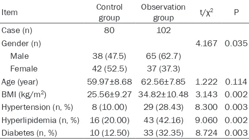
There were no significant differences between the two groups in age (P > 0.05). For the propor-tion of males, BMI, hypertension, hyperglyce -mia, and hyperlipide-mia, results in the observa-tion group were significantly higher than those in the control group (all P < 0.05, Table 1). Comparison of biochemical indicators between two groups
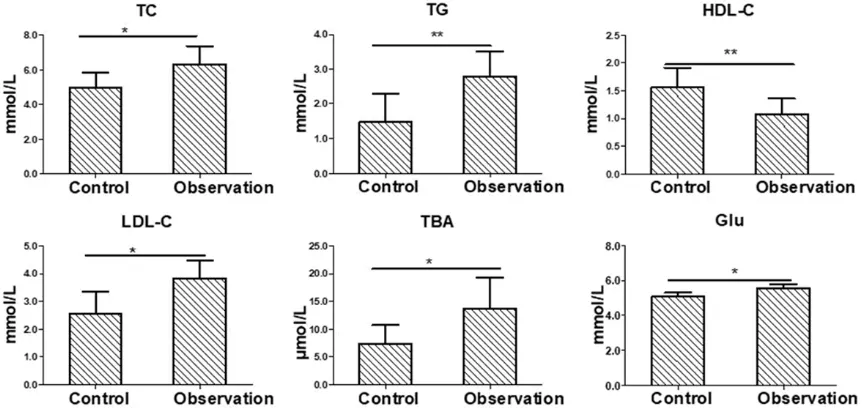
Levels of TC, TG, LDL-C, TBA, and Glu in blood in the observation group were significantly higher than those in the control group, while blood HDL-C levels in the observation group were
sig-Correlation analysis of TBA and influencing
factors of coronary heart disease
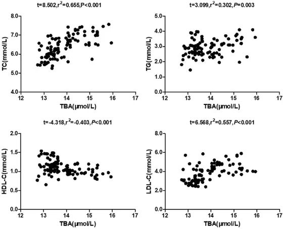
Pearson’s linear correlation analysis showed that TC, TG, and LDL-C levels were positively correlated with TBA expression levels, but neg -atively correlated with HDL-C expression levels (all P < 0.05, Figure 2).
Logistic multivariate regression analysis of coronary heart disease
With or without coronary heart disease was defined as a dependent variable. Smoking, hy-pertension, diabetes, hyperlipidemia, and TBA were used as independent variables. Multiva- riate logistic regression analysis was perform- ed in the previous situation (α level = 0.05). Results showed that all factors mentioned above were included as independent variables of the regression equation as risk factors for occurrence of coronary heart disease (Table 3).
Discussion
In recent years, with an increase in the intake of lipids and bad living habits, the number of patients with coronary heart disease has in- creased significantly. It seriously affects the quality of life of patients, increasing the me- dical burden. Coronary heart disease-related studies have confirmed that the disease is related to genetics, living environment, poor liv-ing habits, and other factors, especially after the intake of a large amounts of lipids [16, 17]. These factors will increase the gastrointestinal glycolysis burden, resulting in excess nutrients. This leads to bile acid and other metabolic dis-orders, elevating blood lipids. Malondialdehy- de, produced by abnormal oxidative
metabo-Table 1. Comparison of clinical baseline data between the two groups
Item Control group Observation group t/χ2 P
Case (n) 80 102
Gender (n) 4.167 0.035
Male 38 (47.5) 65 (62.7)
Female 42 (52.5) 37 (37.3)
Age (year) 59.97±8.68 62.56±7.85 1.222 0.114
BMI (kg/m2) 25.56±9.27 34.82±10.48 3.143 0.002 Hypertension (n, %) 8 (10.00) 29 (28.43) 8.300 0.003
Hyperlipidemia (n, %) 16 (20.00) 43 (42.16) 9.060 0.002
Diabetes (n, %) 10 (12.50) 33 (32.35) 8.724 0.003
nificantly lower than those in the control group (all P < 0.05, Figure 1). Rate of TBA and dyslipidemia in pa-tients with coronary heart disease
[image:3.612.91.347.97.241.2]lism of fat, can easily destroy the lipid bilayer of vascular wall epithelial cells, causing local vas-cular disease [18, 19].
The decomposition of lipid molecules can pro-duce metabolites that adhere to the blood ves-sel walls to form foreign antigens, thereby stim-ulating the body’s immune organs to cause local autoimmune reactions. This damages the integrity of the vessel wall [20]. Therefore, ab- normal lipid metabolism and vascular structur-al damage are the main causes of locstructur-al athe- rosclerotic artery atherosclerosis. Atheroscle- rosis of the arteries will aggravate the agglu- tination of platelets, leading to local vascular insufficiencies. Lesions occurring in the aorta and coronary arteries may induce coronary he- art disease in patients [21, 22].
[image:4.612.92.523.73.277.2]on bile acid content of the intermediate metab-olite. Bile acid is an effective regulating sub -stance in maintaining bile solubility. The struc-ture of cholesterol is stabilized by the dissolu -tion of bile acids. Secre-tion of bile acids in healthy bodies can effectively maintain the dis-solution of cholesterol and other lipids in the blood and bile. When metabolic abnormalities interfere with bile acid secretion, this will indi-rectly lead to hyperlipidemia in the blood, ca- used by high cholesterol. Clinical studies have confirmed that approximately one-third of hu-man TCs are metabolized in the form of bile acids [24]. Therefore, there is a close correla-tion between the metabolism of blood lipids and bile acids. Once metabolic disorders of fat occur, the anabolism of TBA will also be abnormal.
Figure 1. Comparison of biochemical indicators between the two groups (*P < 0.05, **P < 0.01) TC, total choles
-terol; TG, triacylglycerol; HDL-C, high density lipoprotein-choles-terol; LDL-C, low density lipoprotein-choles-terol; Glu,
glucose; TBA, total bile acid.
Table 2. The rate of TBA and dyslipidemia in patients with coro -nary heart disease
Item TG TC HDL-C LDL-C TBA
Abnormal number of cases (n) 75 61 42 48 74
Abnormal rate (%) 73.53 59.80 41.18 47.06 72.55
χ2 0.025 4.861 20.464 12.745
P 0.875 0.028 < 0.001 < 0.001
Note: P value indicates the comparison between abnormal number of abnormal indicators and abnormal number of TBA. TC, total cholesterol; TG, triacylglycerol; HDL-C, high density lipoprotein-cholesterol; LDL-C, low density lipoprotein-choles-terol; TBA, total bile acid.
[image:4.612.89.372.362.430.2]Results of the current study showed that the observation group, with severe coronary heart disease, had significantly higher blood TC, TG, LDL-C, TBA, and Glu levels than the control group. HDL-C levels were significantly lower than those in the control group (all P < 0.05). The major cause of differences in the above indicators between the two groups is that po- lyunsaturated fatty acids and steroid com-pounds can activate farnesoid X receptor (FXR). This can further activate expression of nuclear receptor small heterodimer partner (SHP). The SHP receptor can then react with liver receptor homolog-1 (LRH-1, as CYP7A1 trans-activator), which inactivates cytochrome P450 7A1 and increases bile acid levels [25]. Current studies have shown that bile acids can also affect the
ensuring aldosterone specific binding with the mineralocorticoid receptors. If 11β-OHSD is de-ficient, cortisol will compete with mineralocorti -coid receptors. This may lead to aldosterone-like reactions and hypertension [28]. Present results also confirmed that smoking, hyperten -sion, diabetes, hyperlipidemia, and TBA are risk factors for occurrence of coronary heart dis-ease. Present results are consistent with previ-ous reports [29].
However, the number of patients involved in the current study was relatively small. Most patients had received prior drug treatment. The- se factors may have affected the accuracy of results. The patients were not regularly fol-lowed-up. Thus, this study could not obtain
rel-Figure 2. Correlation analysis of TBA and influencing factors of coronary
heart disease TC, total cholesterol; TG, triacylglycerol; HDL-C, high density
lipoprotein-cholesterol; LDL-C, low density lipoprotein-cholesterol; TBA, total
[image:5.612.91.371.72.299.2]bile acid.
Table 3. Logistic multivariate regression analysis of coronary heart disease
Index B SE Wald OR 95% CI P
Smoking 2.069 0.893 5.374 7.918 1.377, 45.530 0.020
Hypertension 2.676 0.870 9.454 14.524 2.638, 79.954 0.002
Diabetes 1.882 0.872 4.653 6.564 1.188, 36.275 0.031
Hyperlipidemia 2.369 0.867 7.475 10.690 1.965, 58.422 0.006
TBA 1.677 0.825 4.133 5.347 1.062, 26.919 0.042
Note: SE: standard error; Wald: Wald test; OR: odd ratio; CI: confidence interval; TBA, total bile acid.
synthesis of fatty acids, triglyc-erides, and low-density lipopro-teins via FXR, SHP, and Liver X Receptor (LXR) [25, 26]. Re- sults showed that the abnor-mal rate of TBA in patients with coronary heart disease was 72.55%, close to the abnormal rate of TG. Moreover, the ab- normal rate of TBA was signifi -cantly higher than that of TC, HDL-C, and LDL-C (all P < 0.05).
[image:5.612.90.373.393.474.2]evant biochemical indicators after conditions improved. Therefore, present research results require further confirmation. Future studies will follow-up patients for at least 2 years, explor- ing signal pathways and molecular mechani- sms involved in the occurrence of coronary heart disease, aiming to provide a new way for prevention and treatment of coronary heart disease.
In conclusion, patients with coronary heart dis-ease usually have glucose and lipid metabo-lism disorders. Bile acids could respond well to lipid metabolism in patients and show correla-tion with TC, TG, HDL-C, and LDL-C levels.
Disclosure of conflict of interest
None.
Address correspondence to: Yang Liu, Department of Child Healthcare, Linyi People’s Hospital, No.233 Fenghuang Street, Hedong District, Linyi 276000, Shandong Province, China. Tel: +86-0539-8081739; Fax: +86-0539-8081739; E-mail: liuyang938d@out-look.com
References
[1] MacMahon S, Peto R, Cutler J, Collins R, Sorlie P, Neaton J, Abbott R, Godwin J, Dyer A,
Stam-ler J. Blood pressure, stroke, and coronary
heart disease. part 1, prolonged differences in blood pressure: prospective observational studies corrected for the regression dilution bias. Lancet 2016; 335: 765-774.
[2] Khamis RY, Ammari T, Mikhail GW. Gender dif-ferences in coronary heart disease. Heart 2016; 102: 1142-1149.
[3] Valtorta NK, Kanaan M, Gilbody S, Ronzi S, Hanratty B. Loneliness and social isolation as
risk factors for coronary heart disease and stroke: systematic review and meta-analysis of longitudinal observational studies. Heart 2016; 102: 1009-1016.
[4] Horseman MA, Surani S, Bowman JD. Endo -toxin, Toll-lik receptor-4, and atherosclerotic heart disease. Curr Cardiol Rev 2017; 13: 87. [5] Slocum C, Kramer C, Genco CA. Immune
dys-regulation mediated by the oral microbiome:
potential link to chronic inflammation and ath -erosclerosis. J Intern Med 2016; 280: 114-128.
[6] Scott TE, Mendez MV, LaMorte WW, Cupples LA, Vokonas PS, Garcia RI, Menzoian JO. Are
varicose veins a marker for susceptibility to coronary heart disease in men? results from the normative aging study. Ann Vasc Surg 2004; 18: 459-64.
[7] Charach G, Rabinovich A, Argov O, Weintraub M, Rabinovich P. The role of bile acid excretion in atherosclerotic coronary atery disease. Int J Vasc Med 2012; 2012: 949672.
[8] Murakami S, Sakurai T, Tomoike H, Sakono M, Nasu T, Fukuda N. Prevention of hypercholes-terolemia and atherosclerosis in the hyperlipid-emia-and atherosclerosis-prone Japanese (LAP) quail by taurine supplementation. Amino Acids 2010; 38: 271-278.
[9] Ramakrishna R, Kumar D, Bhateria M, Gaik
-wad AN, Bhatta RS. 16-Dehydropregnenolone
lowers serum cholesterol by up-regulation of CYP7A1 in hyperlipidemic male hamsters. J
Steroid Biochem Mol Biol 2017; 168: 110-117.
[10] Hernández Mijares A, Riera Fortuny C, Mar
-tínez Triguero ML, Morillas Ariño C, Cubells Cascales P, Morales Suárez-Varela M. Meta -bolic syndrome in patients with coronary heart disease. results of using different diagnostic criteria. Rev Esp Cardiol 2004; 57: 889-893. [11] Omori Y, Minei S, Uchigata Y, Shimizu M, Sana
-ka M, Honda M, Hirata Y. Comparison of diag-nostic criteria of IGT, borderline, and GDM.
Blood glucose curve and IRI response. Diabe -tes 1991; 40 Suppl 2: 30-34.
[12] Friedman DI, Jacobson DM. Diagnostic criteria for idiopathic intracranial hypertension. Neu-rology 2002; 59: 1492-1495.
[13] Anwar M, Abdelhalim K, Moussa SAA, Hussain Y, Al-Mohy Y. Heavy and trace elements are im-portant diagnostic tools during the progression of atherosclerosis; the supplementation of
high zinc level delays the progression of ath -erosclerosis. Life Sci J 2013; 10: 670-680. [14] Wang P, Yang M, Shen X, Shen LS. Diagnostic
value of the ratio of LDL-C/HDL-C in coronary
disease. Journal of Shanghai Jiaotong Univer -sity 2006; 26: 300-303.
[15] Joint committee for the establishment of gui- delines for prevention and treatment of
dyslip-idemia in Chinese adult. Chinese Adult Blood
Lipid Abnormity Prevention Guide. Chinese Journal of Cardiology 2007; 35: 7-8.
[16] Ma J, Guan XQ, Li J, Xue YJ, Zheng C, Jin G. In-teractions between vitamin D receptor (VDR) gene and Interleukin-6 gene and environment factors on coronary heart disease risk in a Chi-nese Han population. Oncotarget 2017; 8: 78419-78428.
[17] Ramachndran HJ, Wu VX, Kowitlawakul Y, Wang W. Awareness, knowledge and healthy lifestyle behaviors related to coronary heart disease among women: an integrative review. Heart Lung 2016; 45: 173-185.
[18] Vijaya J. Capsaicinoids modulating cardiomet-abolic syndrome risk factors: current perspec-tives. J Nutr Metab 2016; 1: 1-11.
[19] Gamal SM, Sadek NB, Rashed LA, Shawky HM,
gamma-carboxyl-ase inhibition on serum osteocalcin may be partially protective against developing diabetic cardiomyopathy in type 2 diabetic rats. Diab Vasc Dis Res 2016; 13: 7-9.
[20] Mitchell RN. Graft vascular disease: immune response meets the vessel wall. Annu Rev Pathol 2009; 4: 19-47.
[21] Suna G, Wojakowski W, Lynch M, Barallobre-Barreiro J, Yin X, Mayr U, Baig F, Lu R, Fava M,
Hayward R, Molenaar C, White SJ, Roleder T,
Milewski KP, Gasior P, Buszman PP, Buszman
P, Jahangiri M, Shanahan CM, Hill J, Mayr M. Extracellular matrix proteomics reveals inter-play of aggrecan and aggrecanases in vascular remodeling of stented coronary arteries. Circu-lation 2018; 137: 166-183.
[22] Ndrepepa G, Colleran R, Kastrati A. Gamma-glutamyl transferase and the risk of athero-sclerosis and coronary heart disease. Clin Chim Acta 2018; 476: 130-138.
[23] Tilley BJ, Cook JL, Docking SL, Gaida JE. Is
higher serum cholesterol associated with al-tered tendon structure or tendon pain? A
sys-tematic review. Br J Sports Med 2015; 19:
1504-1509.
[24] Wahlström A, Sayin SI, Marschall HU, Bäckhed
F. Intestinal crosstalk between bile acids and microbiota and its impact on host metabolism. Cell Metab 2016; 24: 41-50.
[25] Watanabe M, Houten SM, Wang L, Moschetta A, Mangelsdorf DJ, Heyman RA, Moore DD,
Au-werx J. Bile acids lower triglyceride levels via a pathway involving FXR, SHP, and SREBP-1c. J
Clin Invest 2004; 113: 1408-18.
[26] Sagar NM, Mcfarlane M, Nwokolo C, Bardhan
KD, Arasaradnam RP. Mechaniss of triglycer-ide metabolism in patients with bile acid diar-rhea. World J Gastroenterol 2016; 22: 6757-6763.
[27] Bühler H, Perschel FH, Fitzner R, Hierholzer K.
Endogenous inhibitors of 11 beta-OHSD:
exis-tence and possible significance. Steroids
1994; 59: 131-135.
[28] Maeda Y, Funagayama M, Shinohara A, Koshi-moto C, Furusawa H, Nakahara H, Yamaguchi
Y, Saitoh T, Yamamoto T, Komaki K. Influence
of human serum albumin on the bile acid-me-diated inhibition of liver microsomal type 1
11β-hydroxysteroid dehydrogenase. J Physiol Biochem 2014; 70: 849-855.
[29] Muscogiuri G, Nuzzo V, Gatti A, Zuccoli A,