1,1
000-Dimethyl-4,4
000-bipyridinium
bis(triiodide)
Tuoping Hu
Department of Chemistry, North University of China, Taiyuan, Shanxi 030051, People’s Republic of China
Correspondence e-mail: hutuopingsx@yahoo.com.cn Received 3 March 2009; accepted 23 April 2009
Key indicators: single-crystal X-ray study;T= 296 K; mean(C–C) = 0.010 A˚;
Rfactor = 0.040;wRfactor = 0.073; data-to-parameter ratio = 28.8.
In the title compound, C12H14N2 2+
2I3
, the 1,10-dimethyl-4,40 -bipyridinium (DMBP) dication is charge balanced by two triiodide ions. The DMBP dication is planar within 0.010 (5) A˚ . The asymmetric unit contains only half of the dication, the other half being generated by an inversion center. Weak C—H I interactions link the ions into sheets parallel to (121).
Related literature
For a dication with similar geometry, see: Russell & Wallwork (1972). For anions with comparable geometry, see: Marsh (2004); Madsenet al.(1999).
Experimental
Crystal data
C12H14N22+2I3
Mr= 947.65
Triclinic,P1
a= 7.5457 (4) A˚
b= 7.9541 (6) A˚
c= 9.3029 (6) A˚
= 90.306 (5) = 94.192 (4)
= 102.332 (5) V= 543.88 (6) A˚3
Z= 1
MoKradiation
= 8.56 mm1
T= 296 K
0.220.160.08 mm
Data collection
Bruker SMART CCD area-detector diffractometer
Absorption correction: multi-scan (SADABS; Sheldrick, 1996)
Tmin= 0.211,Tmax= 0.504
12956 measured reflections 2683 independent reflections 1468 reflections withI> 2(I)
Rint= 0.052
Refinement
R[F2> 2(F2)] = 0.040
wR(F2) = 0.073
S= 1.02 2683 reflections
93 parameters
H-atom parameters constrained
max= 0.97 e A˚
3
min=0.86 e A˚
[image:1.610.62.225.468.724.2]3
Table 1
Selected geometric parameters (A˚ ,).
I1—I2 2.9341 (8) I2—I3 2.9061 (8)
I3—I2—I1 177.49 (2)
Table 2
Hydrogen-bond geometry (A˚ ,).
D—H A D—H H A D A D—H A
C3—H3 I3i
0.93 3.05 3.951 (8) 163
C2—H2 I1ii 0.93 3.16 4.066 (8) 164
C5—H5 I2i
0.93 3.13 3.839 (7) 135
Symmetry codes: (i)x;yþ1;zþ1; (ii)xþ1;yþ1;zþ2.
Data collection:SMART(Bruker, 2007); cell refinement: SAINT-Plus(Bruker, 2007); data reduction:SAINT-Plus; program(s) used to solve structure: SHELXS97(Sheldrick, 2008); program(s) used to refine structure:SHELXL97(Sheldrick, 2008); molecular graphics: SHELXTL(Sheldrick, 2008); software used to prepare material for publication:SHELXTL.
The author is grateful for funding support from the Natural Science Foundation of Shanxi Province (2007011033), the Program of Technological Industrialization at the University of Shanxi Province (20070308) and the start-up fund of North University of China.
Supplementary data and figures for this paper are available from the IUCr electronic archives (Reference: EZ2167).
References
Bruker (2007). SMART and SAINT-Plus. Bruker AXS Inc., Madison, Wisconsin, USA.
Madsen, D., Burghammer, M., Fiedler, S. & Mu¨ller, H. (1999).Acta Cryst.B55, 601–606.
Marsh, R. E. (2004).Acta Cryst.B60, 252–253.
Russell, J. H. & Wallwork, S. C. (1972).Acta Cryst.B28, 1527–1533. Sheldrick, G. M. (1996).SADABS. University of Go¨ttingen, Germany. Sheldrick, G. M. (2008).Acta Cryst.A64, 112–122.
Acta Crystallographica Section E
Structure Reports
Online
supporting information
Acta Cryst. (2009). E65, o1162 [doi:10.1107/S1600536809015207]
1,1
′
-Dimethyl-4,4
′
-bipyridinium bis(triiodide)
Tuoping Hu
S1. Comment
The title compound, (I), was obtained by chance when we tried to prepare the salt of the Pb(II) cation and DMBP in
MeOH. This paper provides the first crystal structure of the DMBP dication with two triiodide anions.
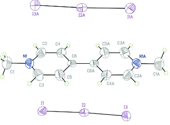
Only half of the dication of DMBP is contained in the asymmetric unit, while the other half is generated by the
inversion center at (1/2,1/2,1/2) (Fig 1.). The N,N′-dimethyl-4,4'bipyridylium(II) dication has an essentially planar
conformation, the maximum deviation of the C1 atom (the methyl group) from its mean plane being 0.010 (5) Å. The
geometry of the dication is similar to the one observed in Russell & Wallwork (1972). Meanwhile, the geometry of the
anion is comparable to that described in Marsh (2004) and Madsen et al. (1999).
Weak C3—H3···I3 interactions link two I3 anions to each dication. A weaker C2—I2···H1 interaction links each anion to
a further DMBP cation, to form sheets parallel to (121). Adjacent sheets are packed into a three-dimensional motif (Fig.
2).
S2. Experimental
C12H14N2.4Cl (0.5 mmol, 128 mg) and KI (10 mmol, 1660 mg) were added to 50 ml of CH3CN. After stirring and
refluxing for 12 h, the mixture was filtered, and the clear solution was allowed to evaporate slowly under inert
atmosphere. Prismatic crystals of the title compound were obtained after 5 days. The crystals were filtered, washed by
cool EtOH and dried in air.
S3. Refinement
All of the H atoms were positioned geometrically and refined using a riding model with C—H = 0.930 Å and 0.96 Å,
Figure 1
Molecular structure showing 50% probability displacement ellipsoids. The atoms marked with A are derived from the
reference atoms by means of the (1 - x, 1 - y, 1 - z) symmetry transformation..
Figure 2
Packing diagram viewed down the a axis. Weak C—H···I interactions are shown as dotted lines.
1,1′-Dimethyl-4,4′-bipyridinium bis(triiodide)
Crystal data
C12H14N22+·2I3− Mr = 947.65 Triclinic, P1 Hall symbol: -P 1 a = 7.5457 (4) Å b = 7.9541 (6) Å
[image:3.610.126.480.385.561.2]F(000) = 418 Dx = 2.893 Mg m−3
Mo Kα radiation, λ = 0.71073 Å Cell parameters from 4412 reflections θ = 2.6–27.6°
µ = 8.56 mm−1 T = 296 K Prism, black
0.22 × 0.16 × 0.08 mm
Data collection
Bruker SMART CCD area-detector diffractometer
Radiation source: fine-focus sealed tube Graphite monochromator
φ and ω scans
Absorption correction: multi-scan (SADABS; Sheldrick, 1996) Tmin = 0.211, Tmax = 0.504
12956 measured reflections 2683 independent reflections 1468 reflections with I > 2σ(I) Rint = 0.052
θmax = 28.3°, θmin = 3.9° h = −10→10
k = −10→10 l = −11→12
Refinement
Refinement on F2 Least-squares matrix: full R[F2 > 2σ(F2)] = 0.040 wR(F2) = 0.073 S = 1.02 2683 reflections 93 parameters 0 restraints
Primary atom site location: structure-invariant direct methods
Secondary atom site location: difference Fourier map
Hydrogen site location: inferred from neighbouring sites
H-atom parameters constrained w = 1/[σ2(F
o2) + (0.005P)2 + 2.2853P] where P = (Fo2 + 2Fc2)/3
(Δ/σ)max < 0.001 Δρmax = 0.97 e Å−3 Δρmin = −0.86 e Å−3
Extinction correction: SHELXL97 (Sheldrick, 2008), Fc*=kFc[1+0.001xFc2λ3/sin(2θ)]-1/4 Extinction coefficient: 0.0028 (3)
Special details
Geometry. All e.s.d.'s (except the e.s.d. in the dihedral angle between two l.s. planes) are estimated using the full covariance matrix. The cell e.s.d.'s are taken into account individually in the estimation of e.s.d.'s in distances, angles and torsion angles; correlations between e.s.d.'s in cell parameters are only used when they are defined by crystal symmetry. An approximate (isotropic) treatment of cell e.s.d.'s is used for estimating e.s.d.'s involving l.s. planes.
Refinement. Refinement of F2 against ALL reflections. The weighted R-factor wR and goodness of fit S are based on F2, conventional R-factors R are based on F, with F set to zero for negative F2. The threshold expression of F2 > σ(F2) is used only for calculating R-factors(gt) etc. and is not relevant to the choice of reflections for refinement. R-factors based on F2 are statistically about twice as large as those based on F, and R- factors based on ALL data will be even larger.
Fractional atomic coordinates and isotropic or equivalent isotropic displacement parameters (Å2)
x y z Uiso*/Ueq
I1 0.11371 (7) 0.64766 (7) 0.84204 (6) 0.0705 (2)
I2 0.19121 (6) 0.80427 (6) 0.56237 (6) 0.05927 (17)
I3 0.25337 (8) 0.96496 (8) 0.28546 (6) 0.0816 (2)
N1 0.3773 (9) 0.2800 (7) 0.8128 (7) 0.0588 (16)
C1 0.3276 (13) 0.1852 (11) 0.9438 (9) 0.085 (3)
H1A 0.4248 0.2166 1.0179 0.128*
H1B 0.3064 0.0638 0.9235 0.128*
H1C 0.2190 0.2131 0.9758 0.128*
C2 0.5358 (12) 0.3875 (11) 0.8116 (9) 0.074 (2)
C3 0.2652 (11) 0.2566 (10) 0.6956 (10) 0.072 (2)
H3 0.1526 0.1813 0.6969 0.086*
C4 0.5864 (9) 0.4764 (10) 0.6903 (8) 0.061 (2)
H4 0.6984 0.5532 0.6924 0.074*
C5 0.3120 (10) 0.3414 (10) 0.5722 (8) 0.066 (2)
H5 0.2309 0.3216 0.4906 0.079*
C6 0.4743 (8) 0.4540 (7) 0.5658 (7) 0.0396 (14)
Atomic displacement parameters (Å2)
U11 U22 U33 U12 U13 U23
I1 0.0689 (4) 0.0899 (4) 0.0576 (3) 0.0291 (3) 0.0010 (3) 0.0076 (3) I2 0.0503 (3) 0.0648 (3) 0.0672 (3) 0.0224 (2) 0.0040 (2) 0.0045 (2) I3 0.0838 (4) 0.0895 (4) 0.0816 (4) 0.0335 (3) 0.0267 (3) 0.0292 (3)
N1 0.066 (4) 0.051 (4) 0.063 (4) 0.016 (3) 0.016 (4) 0.010 (3)
C1 0.106 (7) 0.076 (6) 0.075 (6) 0.017 (5) 0.017 (5) 0.015 (5)
C2 0.071 (6) 0.088 (6) 0.061 (5) 0.020 (5) −0.011 (4) 0.015 (5) C3 0.062 (5) 0.066 (5) 0.078 (6) −0.010 (4) 0.011 (5) −0.003 (5) C4 0.039 (4) 0.077 (5) 0.058 (5) −0.004 (4) −0.016 (3) 0.006 (4) C5 0.052 (5) 0.080 (6) 0.054 (5) −0.007 (4) −0.003 (4) −0.001 (4) C6 0.031 (3) 0.032 (3) 0.054 (4) 0.005 (3) −0.002 (3) −0.003 (3)
Geometric parameters (Å, º)
I1—I2 2.9341 (8) C2—H2 0.9300
I2—I3 2.9061 (8) C3—C5 1.364 (10)
N1—C2 1.314 (9) C3—H3 0.9300
N1—C3 1.317 (9) C4—C6 1.371 (8)
N1—C1 1.467 (9) C4—H4 0.9300
C1—H1A 0.9600 C5—C6 1.359 (9)
C1—H1B 0.9600 C5—H5 0.9300
C1—H1C 0.9600 C6—C6i 1.464 (12)
C2—C4 1.370 (10)
I3—I2—I1 177.49 (2) N1—C3—C5 120.9 (7)
C2—N1—C3 119.7 (7) N1—C3—H3 119.5
C2—N1—C1 119.8 (7) C5—C3—H3 119.5
C3—N1—C1 120.5 (7) C2—C4—C6 121.0 (6)
N1—C1—H1A 109.5 C2—C4—H4 119.5
N1—C1—H1B 109.5 C6—C4—H4 119.5
H1A—C1—H1B 109.5 C6—C5—C3 121.6 (7)
N1—C1—H1C 109.5 C6—C5—H5 119.2
H1A—C1—H1C 109.5 C3—C5—H5 119.2
H1B—C1—H1C 109.5 C5—C6—C4 115.9 (6)
N1—C2—C4 121.0 (7) C5—C6—C6i 122.1 (7)
N1—C2—H2 119.5 C4—C6—C6i 122.1 (7)
C3—N1—C2—C4 0.3 (12) N1—C3—C5—C6 −0.7 (13)
C1—N1—C2—C4 179.4 (7) C3—C5—C6—C4 0.0 (11)
C2—N1—C3—C5 0.6 (12) C3—C5—C6—C6i −179.8 (8)
C1—N1—C3—C5 −178.5 (7) C2—C4—C6—C5 0.9 (11)
N1—C2—C4—C6 −1.0 (12) C2—C4—C6—C6i −179.3 (8)
Symmetry code: (i) −x+1, −y+1, −z+1.
Hydrogen-bond geometry (Å, º)
D—H···A D—H H···A D···A D—H···A
C3—H3···I3ii 0.93 3.05 3.951 (8) 163
C2—H2···I1iii 0.93 3.16 4.066 (8) 164
C5—H5···I2ii 0.93 3.13 3.839 (7) 135