The dehydrated copper silicate
Na
2[Cu
2Si
4O
11]: a three-dimensional
microporous framework with a linear
Si—O—Si linkage
Luı´s Cunha-Silva, Paula Branda˜o, Joa˜o Rocha and Filipe A. Almeida Paz*
Department of Chemistry, University of Aveiro, CICECO, 3810-193 Aveiro, Portugal Correspondence e-mail: [email protected]
Received 28 November 2007; accepted 15 January 2008
Key indicators: single-crystal X-ray study;T= 298 K; mean(Si–O) = 0.004 A˚;
Rfactor = 0.043;wRfactor = 0.120; data-to-parameter ratio = 11.9.
The structure of the title dehydrated copper silicate, disodium
dicopper undecaoxide tetrasilicate, Na2(Cu2O11Si4), was
determined by single-crystal X-ray diffraction from a non-merohedral twin. It exhibits an effective three-dimensional microporous framework with the major channels, in which the
Na+cations are placed, running along thea-axis direction and
smaller channels observed along the b-axis direction. The
structure is unusual in that it contains a symmetry-constrained
Si—O—Si angle of 180. The Cu centre is coordinated to five
O atoms, exhibiting a slightly distorted square-pyramidal coordination geometry. The Na cation is interacting with five neighbouring O atoms, exhibiting an uncharacteristic coordi-nation environment.
Related literature
For related literature, see: Branda˜o et al. (2005); Haile &
Wuensch (2000); Liebau (1985); Rocha & Anderson (2000);
Rocha & Lin (2005); dos Santoset al.(2005); Ananias et al.
(2001, 2006); Andersonet al.(1994); Ferreiraet al.(2003).
Experimental
Crystal data
Na2(Cu2O11Si4)
Mr= 461.44 Triclinic,P1
a= 5.190 (2) A˚
b= 6.299 (3) A˚
c= 8.196 (4) A˚
= 96.390 (7)
= 97.281 (7)
= 100.461 (7)
V= 258.9 (2) A˚3
Z= 1
MoKradiation
= 4.71 mm1
T= 298 (2) K 0.280.080.04 mm
Data collection
Bruker SMART CCD 1000 diffractometer
Absorption correction: multi-scan (TWINABS; Sheldrick, 2002)
Tmin= 0.627,Tmax= 0.834
1587 measured reflections 1043 independent reflections 782 reflections withI> 2(I)
Rint= 0.042
Refinement
R[F2> 2(F2)] = 0.042
wR(F2) = 0.119
S= 1.01 1043 reflections
88 parameters max= 1.40 e A˚
3
[image:1.610.83.261.592.700.2]min=1.58 e A˚ 3
Table 1
Selected bond lengths (A˚ ).
Cu1—O5i
1.909 (3)
Cu1—O2 1.950 (4)
Cu1—O6ii
1.970 (4)
Cu1—O6 1.974 (3)
Cu1—O2iii 2.316 (4)
Symmetry codes: (i)x;y1;z; (ii)xþ1;y;zþ2; (iii)xþ2;y;zþ2.
Data collection:SMART(Bruker, 1998); cell refinement:SMART; data reduction:SAINT-Plus(Bruker, 2003); program(s) used to solve structure:SIR92(Altomare et al., 1993); program(s) used to refine structure: SHELXTL (Sheldrick, 2008); molecular graphics:
DIAMOND(Brandenburg, 2007); software used to prepare material for publication:SHELXTL.
We are grateful to the Fundac¸a˜o para a Cieˆncia e a Tecnologia (FCT, Portugal) for their general financial support under the POCI programme (supported by FEDER) and for a Postdoctoral Fellowship (SFRH/BPD/14410/2003) to LCS.
Supplementary data and figures for this paper are available from the IUCr electronic archives (Reference: BR2065).
References
Altomare, A., Cascarano, G., Giacovazzo, C. & Guagliardi, A. (1993).J. Appl. Cryst.26, 343–350.
Ananias, D., Almeida Paz, F. A., Carlos, L. D., Geraldes, C. F. G. C. & Rocha, J. (2006).Angew. Chem. Int. Ed.45, 7938–7942.
Ananias, D., Ferreira, A., Rocha, J., Ferreira, P., Rainho, J. P., Morais, C. & Carlos, L. D. (2001).J. Am. Chem. Soc.123, 5735–5742.
Anderson, M. W., Terasaki, O., Ohsuna, T., Philippou, A., Mackay, S. P., Ferreira, A., Rocha, J. & Lidin, S. (1994).Nature (London),367, 347–351. Branda˜o, P., Almeida Paz, F. A. & Rocha, J. (2005).Chem. Commun.pp. 171–
173.
Brandenburg, K. (2007). DIAMOND. Version 3.1e. Crystal Impact GbR, Bonn, Germany.
Bruker (1998).SMART. Version 5.054. Bruker AXS Inc., Madison, Wisconsin, USA.
Bruker (2003). SAINT-Plus. Version 6.45A. Bruker AXS Inc., Madison, Wisconsin, USA.
Ferreira, A., Ananias, D., Carlos, L. D., Morais, C. & Rocha, J. (2003).J. Am. Chem. Soc.125, 14573–14579.
inorganic compounds
Acta Cryst.(2008). E64, i13–i14 doi:10.1107/S1600536808001608 #2008 International Union of Crystallography
i13
Acta Crystallographica Section E Structure Reports
Online
Liebau, F. (1985).Structural Chemistry of Silicates: Structure, Bonding, and Classification, pp. 14–29. Berlin: Springer-Verlag.
Rocha, J. & Anderson, M. W. (2000).Eur. J. Inorg. Chem.pp. 801–818. Rocha, J. & Lin, Z. (2005).Reviews in Mineralogy and Geochemistry, Vol. 57,
edited by G. Ferraris & S. Merlino, ch. 6, pp. 173–201. Washington, DC: Mineralogical Society of America, Geochemical Society.
Ferreira, L. P., Godinho, M., Volkova, O. & Vasiliev, A. (2005).Phys. Rev. B,
72, Art. No. 092403.
Sheldrick, G. M. (2002).TWINABS. Version 1.05. Bruker AXS Inc., Madison, Wisconsin, USA.
supporting information
sup-1
Acta Cryst. (2008). E64, i13–i14
supporting information
Acta Cryst. (2008). E64, i13–i14 [doi:10.1107/S1600536808001608]
The dehydrated copper silicate Na
2[Cu
2Si
4O
11]: a three-dimensional
microporous framework with a linear Si
—
O
—
Si linkage
Lu
í
s Cunha-Silva, Paula Brand
ã
o, Jo
ã
o Rocha and Filipe A. Almeida Paz
S1. Comment
Molecular sieves containing metal cations with a range of coordination geometries have been extensively studied due to
their novel topologies, interesting chemical properties and potential aplications in optoelectronics, batteries, magnetic
materials and sensors (besides the traditional applications of zeolites) (Rocha & Anderson, 2000; Rocha & Lin, 2005). In
the last decade, we have been interested in the synthesis and structural characterization of novel open-frameworks
containing Si and metal cations (such as Ti, V, Cr, Nb, Zr and Sn) in tetrahedral and (more commonly) octahedral
coordination environments, and lanthanide silicates exhibiting interesting photoluminescence properties (Anderson et al.,
1994; Ananias et al., 2001; Ferreira et al., 2003; Ananias et al., 2006). As part of this research line, we prepared and
characterized the hydrated copper silicate Na2(Cu2Si4O11).2H2O (Brandão et al., 2005). This compound was dehydrated
and the magnetic properties of both hydrated and dehydrated forms were investigated (Santos et al., 2005), however the
crystalline structure of the dehydrated compound was not reported. Here we describe the structure of the dehydrated
microporous copper silicate, Na2(Cu2Si4O11) (I).
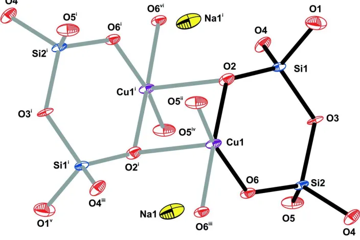
The asymmetric unit of the copper silicate (I) comprises one Cu(II) cation, two corner-shared SiO4 groups and one Na+
counter-cation (Figure 1). The crystallographic unique Cu(II) metal centre is coordinated to five O-atoms from five
distinct SiO4 tetrahedral moieties (four basal SiO4 and one apical SiO4), in a geometry resembling a distorted square
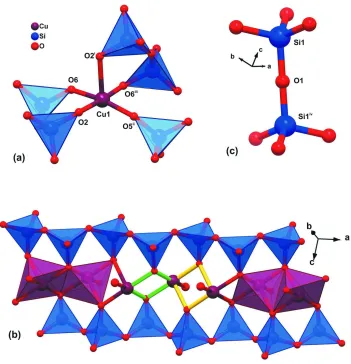
pyramid for which the apical Cu—O bond is longer than the basal ones (Figure 2a and Table 1).
Adjacent SiO4 tetrahedral moieties are linked along the a direction by corner-shared oxygen atoms (O3 and O4 are
shared alternately) leading to the formation of zigzag metallic anionic chains, [(Cu2Si4O11)∞]2-, in which the Cu···Cu
distances alternate between 2.9921 (8) Å (via bridging basal SiO4, green bonds in Fig. 2 b) and 3.1031 (10) Å (via the
apical SiO4 tetrahedron, yellow bonds in Fig. 2 b). [(Cu2Si4O11)∞]2- chains are interconnected via corner-sharing SiO4
tetrahedra through linear interactions Si1–O1–Siiv [angle is 180.0°; symmetry code: (iv) 2 - x, -y, 1 - z] to form infinite
layers (Fig. 2c). This linear Si–O–Si interaction is very rare and represents a remarkable structural feature of the copper
silicate (I) framework. We note that such occurrence was also recently reported in the lanthanide silicate K3(NdSi7O17)
(Haile & Wuensch, 2000). From the evaluation of the structures of several hundred silicates it was concluded that the
average of an unstrained Si—O—Si bond angle is ca 139° and that truly linear bonds are energetically unfavorable
(Liebau, 1985). In fact, the crystallographically determined values of 180° are more likely to represent a time average
rather than the actual value of the bond angle. The bond, at any instant in time, should have an O-atom displaced from its
average position such that the instantaneous value of Si—O—Si is less than 180° (Haile & Wuensch, 2000). This
structural feature is ultimately reflected in the anisotropic displacement parameters associated with this bridging O-atom.
Indeed, the thermal parameters associated with this atom are unusually large, with the greatest displacement occurring in
dimensional microporous framework with the major channels running along the a direction, formed by eight-membered
rings and having a cross-section of ca 7.5 × 4.3 Å (Figure 3a). Interestingly, the Na+ cations are located within the
channels but are remarkably close to the previously described layers, creating an effective porous copper framework
(Figure 3a). In addition, remarkably large channels are also observed along the b direction, which are formed by
six-membered rings and display a cros-section of ca 5.2 × 4.6 Å (Figure 3 b).
S2. Experimental
Chemicals were purchased from commercial sources and used without further purification. An alkaline solution was
prepared by mixing 13.86 g of a sodium silicate solution (Na2O 8 wt%, SiO2 27 wt%), 16.13 g H2O and 4.11 g NaOH,
and a second solution was prepared by mixing 17.87 g H2O with 7.60 g of Cu(SO4).15H2O. These two solutions were
combined, stirred thoroughly during 2 h and the resulting gel, with a molar composition of CuO: 3.1SiO2: 1.4Na2O:
94.5H2O, was autoclaved for 10 days at 503 K. A crystalline material was obtained [Na2(Cu2Si4O11).2H2O], filtered and
treated thermally at 573 K for six hours leads to the removal of the crystallization water molecules.
S3. Refinement
Even though crystals of the title compound could be indexed with the unit-cell parameters summarized in Table 1, a
visual inspection of the centered reflections using RLATT showed the presence of a rotational twin (non-merohedral). A
full sphere of reflections was collected and a partial data set was then deconvoluted using CELL_NOW (Sheldrick 2004)
into a two-component twin. Data integration was performed by assuming that the second twin domain was identical to
the first. The final structural model exhibits a large average U(i,j) tensor, most likely due to the applied twinning
[image:4.610.129.481.405.640.2]correction which ultimately seems to lead to large U3/U1 ratios.
Figure 1
Fragment of the crystal structure of the title compound with the atoms represented as thermal displacement ellipsoids
drawn at the 50% probability level [Symmetry codes: (i) 2 - x, -y, 2 - z; (ii) x, -1 + y, z; (iii) 1 - x, -y, 2 - z; (iv) 2 - x, 1 - y,
supporting information
sup-3
[image:5.610.131.482.69.433.2]Acta Cryst. (2008). E64, i13–i14
Figure 2
(a) Mixed ball-and-stick and polyhedral representation of the coordination environment of the Cu(II) cations and (b) the metallic chain [(Cu2Si4O11)n]2- running along the a direction of the unit cell. (c) Schematic representation of the linear Si
—O—Si bond connecting adjacent Si1 centres via the O1 atom. [Symmetry codes: (i) 2 - x, -y, 2 - z; (ii) x, -1 + y, z; (iii)
Perspective views of the crystal packing arrangement along the (a) [100] and (b) [010] directions of unit cell.
disodium dicopper undecaoxide tetrasilicate
Crystal data Na2(Cu2O11Si4) Mr = 461.44 Triclinic, P1 Hall symbol: -P 1 a = 5.190 (2) Å b = 6.299 (3) Å c = 8.196 (4) Å α = 96.390 (7)° β = 97.281 (7)° γ = 100.461 (7)° V = 258.9 (2) Å3
Z = 1 F(000) = 224 Dx = 2.960 Mg m−3
Mo Kα radiation, λ = 0.71073 Å Cell parameters from 758 reflections θ = 8.1–58.1°
µ = 4.71 mm−1 T = 298 K Plate, black
0.28 × 0.08 × 0.04 mm
Data collection
Bruker SMART CCD 1000 diffractometer
Radiation source: fine-focus sealed tube Graphite monochromator
ω scans
Absorption correction: multi-scan (TWINABS; Sheldrick, 2002) Tmin = 0.627, Tmax = 0.834
1587 measured reflections 1043 independent reflections 782 reflections with I > 2σ(I) Rint = 0.042
θmax = 26.4°, θmin = 3.9° h = −6→6
k = −7→7 l = 0→10
Refinement Refinement on F2 Least-squares matrix: full R[F2 > 2σ(F2)] = 0.042 wR(F2) = 0.119 S = 1.01 1043 reflections 88 parameters 0 restraints
Primary atom site location: structure-invariant direct methods
Secondary atom site location: difference Fourier map
w = 1/[σ2(F
o2) + (0.0809P)2] where P = (Fo2 + 2Fc2)/3 (Δ/σ)max < 0.001
Δρmax = 1.40 e Å−3 Δρmin = −1.58 e Å−3
Special details
Geometry. All e.s.d.'s (except the e.s.d. in the dihedral angle between two l.s. planes) are estimated using the full covariance matrix. The cell e.s.d.'s are taken into account individually in the estimation of e.s.d.'s in distances, angles and torsion angles; correlations between e.s.d.'s in cell parameters are only used when they are defined by crystal symmetry. An approximate (isotropic) treatment of cell e.s.d.'s is used for estimating e.s.d.'s involving l.s. planes.
Refinement. Refinement of F2 against ALL reflections. The weighted R-factor wR and goodness of fit S are based on F2, conventional R-factors R are based on F, with F set to zero for negative F2. The threshold expression of F2 > σ(F2) is used only for calculating R-factors(gt) etc. and is not relevant to the choice of reflections for refinement. R-factors based on F2 are statistically about twice as large as those based on F, and R- factors based on ALL data will be even larger.
Fractional atomic coordinates and isotropic or equivalent isotropic displacement parameters (Å2)
x y z Uiso*/Ueq
supporting information
sup-5
Acta Cryst. (2008). E64, i13–i14
Na1 0.8700 (4) 0.3540 (3) 1.1990 (3) 0.0235 (6) Si1 1.0175 (2) 0.1358 (2) 0.67930 (18) 0.0087 (4) Si2 0.5954 (3) 0.3456 (2) 0.80969 (18) 0.0089 (4) O1 1.0000 0.0000 0.5000 0.0217 (13) O2 1.0064 (7) −0.0190 (6) 0.8196 (5) 0.0127 (8) O3 0.7804 (6) 0.2736 (6) 0.6719 (5) 0.0126 (8) O4 0.2923 (6) 0.3209 (5) 0.7137 (5) 0.0144 (8) O5 0.7200 (7) 0.5899 (6) 0.8873 (5) 0.0170 (9) O6 0.5974 (6) 0.1760 (5) 0.9452 (5) 0.0107 (8)
Atomic displacement parameters (Å2)
U11 U22 U33 U12 U13 U23
Cu1 0.0054 (4) 0.0083 (4) 0.0159 (4) 0.0011 (2) 0.0051 (2) 0.0018 (2) Na1 0.0137 (11) 0.0161 (12) 0.0396 (16) 0.0021 (9) 0.0072 (10) −0.0030 (10) Si1 0.0048 (7) 0.0096 (7) 0.0124 (8) 0.0018 (5) 0.0034 (5) 0.0009 (5) Si2 0.0040 (6) 0.0078 (7) 0.0155 (8) 0.0013 (5) 0.0042 (5) 0.0018 (5) O1 0.020 (3) 0.024 (3) 0.021 (3) 0.006 (2) 0.007 (2) −0.006 (2) O2 0.0078 (17) 0.0137 (18) 0.018 (2) 0.0033 (13) 0.0040 (14) 0.0045 (14) O3 0.0057 (16) 0.0169 (19) 0.018 (2) 0.0068 (14) 0.0049 (14) 0.0018 (14) O4 0.0063 (16) 0.0140 (18) 0.023 (2) 0.0017 (14) 0.0014 (14) 0.0061 (15) O5 0.0145 (18) 0.0100 (18) 0.028 (2) 0.0000 (14) 0.0129 (16) 0.0016 (15) O6 0.0082 (16) 0.0111 (17) 0.016 (2) 0.0039 (13) 0.0061 (14) 0.0050 (14)
Geometric parameters (Å, º)
Cu1—O5i 1.909 (3) Si1—O4iv 1.642 (4) Cu1—O2 1.950 (4) Si2—O5 1.583 (4) Cu1—O6ii 1.970 (4) Si2—O6 1.625 (4) Cu1—O6 1.974 (3) Si2—O4 1.639 (3) Cu1—O2iii 2.316 (4) Si2—O3 1.650 (4) Cu1—Cu1ii 2.9921 (13) Si2—Cu1ii 3.1221 (19) Cu1—Cu1iii 3.1031 (15) O1—Si1v 1.5991 (14) Cu1—Si2ii 3.1221 (19) O2—Cu1iii 2.316 (4) Cu1—Si1 3.1673 (19) O4—Si1vi 1.642 (4) Si1—O2 1.588 (4) O5—Cu1vii 1.909 (3) Si1—O1 1.5991 (14) O6—Cu1ii 1.970 (4) Si1—O3 1.629 (3)
O5i—Cu1—Cu1ii 131.06 (12) O1—Si1—Cu1 115.96 (7) O2—Cu1—Cu1ii 135.03 (10) O3—Si1—Cu1 84.08 (15) O6ii—Cu1—Cu1ii 40.70 (10) O4iv—Si1—Cu1 129.55 (15) O6—Cu1—Cu1ii 40.62 (10) O5—Si2—O6 113.2 (2) O2iii—Cu1—Cu1ii 90.41 (9) O5—Si2—O4 111.75 (19) O5i—Cu1—Cu1iii 103.18 (12) O6—Si2—O4 109.3 (2) O2—Cu1—Cu1iii 48.19 (11) O5—Si2—O3 107.1 (2) O6ii—Cu1—Cu1iii 130.50 (11) O6—Si2—O3 107.00 (19) O6—Cu1—Cu1iii 91.93 (10) O4—Si2—O3 108.15 (19) O2iii—Cu1—Cu1iii 38.86 (9) O5—Si2—Cu1ii 109.62 (16) Cu1ii—Cu1—Cu1iii 116.74 (4) O4—Si2—Cu1ii 81.61 (15) O5i—Cu1—Si2ii 73.75 (12) O3—Si2—Cu1ii 134.72 (14) O2—Cu1—Si2ii 156.11 (11) Si1v—O1—Si1 180.0 O6ii—Cu1—Si2ii 26.72 (10) Si1—O2—Cu1 126.8 (2) O6—Cu1—Si2ii 103.96 (11) Si1—O2—Cu1iii 116.29 (18) O2iii—Cu1—Si2ii 78.31 (10) Cu1—O2—Cu1iii 92.95 (15) Cu1ii—Cu1—Si2ii 64.59 (4) Si1—O3—Si2 132.7 (3) Cu1iii—Cu1—Si2ii 115.04 (5) Si2—O4—Si1vi 137.0 (2) O5i—Cu1—Si1 106.73 (13) Si2—O5—Cu1vii 152.9 (2) O2—Cu1—Si1 23.68 (11) Si2—O6—Cu1ii 120.24 (18) O6ii—Cu1—Si1 153.39 (10) Si2—O6—Cu1 130.3 (2) O6—Cu1—Si1 75.74 (11) Cu1ii—O6—Cu1 98.68 (16)