catena
-Poly[[bis(pyrazine-2-carbox-amide)mercury(II)]-di-
l
-chlorido]
Alireza Azhdari Tehrani, Bahareh Mir Mohammad Sadegh and Hamid Reza Khavasi*
Department of Chemistry, Shahid Beheshti University, G.C., Evin, Tehran 1983963113, Iran
Correspondence e-mail: h-khavasi@sbu.ac.ir
Received 3 January 2010; accepted 1 February 2010
Key indicators: single-crystal X-ray study;T= 298 K; mean(C–C) = 0.011 A˚; Rfactor = 0.054;wRfactor = 0.144; data-to-parameter ratio = 19.5.
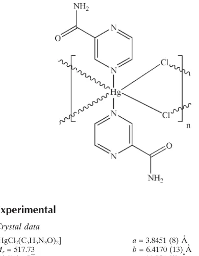
In the polymeric title compound, [HgCl2(C5H5N3O)2]n, the
HgIIatom (site symmetry 1) adopts a distortedtrans-HgN2Cl4
octahedral coordination geometry. In the crystal, adjacent mercury ions are bridged by pairs of chloride ions, generating
infinite [100] chains, and N—H O and N—H (N,N)
hydrogen bonds help to consolidate the packing.
Related literature
For related structures, see: Cati & Stoeckli-Evans (2004);
Hausmann & Brooker (2004); Mir Mohammad Sadeghet al.
(2010); Miyazakiet al.(2007).
Experimental
Crystal data
[HgCl2(C5H5N3O)2]
Mr= 517.73
Triclinic,P1
a= 3.8451 (8) A˚
b= 6.4170 (13) A˚
c= 14.854 (3) A˚
= 101.14 (3)
= 92.53 (3)
= 94.69 (3) V= 357.73 (13) A˚3
Z= 1
MoKradiation
= 11.14 mm1
T= 298 K
0.480.150.06 mm
Data collection
Stoe IPDS II diffractometer Absorption correction: numerical
[optically, byX-REDand
XSHAPE(Stoe & Cie, 2005)]
Tmin= 0.150,Tmax= 0.515
4201 measured reflections 1887 independent reflections 1880 reflections withI> 2(I)
Rint= 0.096
Refinement
R[F2> 2(F2)] = 0.054
wR(F2) = 0.144
S= 1.08 1887 reflections
97 parameters
H-atom parameters constrained max= 3.25 e A˚
3 min=3.75 e A˚ 3
Table 1
Selected geometric parameters (A˚ ,).
Hg1—N2 2.661 (7) Hg1—Cl1i
2.970 (2)
Hg1—Cl1 2.375 (2)
Hg1—Cl1—Hg1ii 91.31 (7)
[image:1.610.50.245.470.721.2]Symmetry codes: (i)x1;y;z; (ii)xþ1;y;z.
Table 2
Hydrogen-bond geometry (A˚ ,).
D—H A D—H H A D A D—H A
N3—H3A O1iii
0.86 2.01 2.864 (12) 176 N3—H3B N1 0.86 2.40 2.758 (12) 105 N3—H3B N1iv
0.86 2.54 3.198 (12) 134
Symmetry codes: (iii)xþ2;y;zþ1; (iv)xþ1;yþ1;zþ1.
Data collection:X-AREA(Stoe & Cie, 2005); cell refinement: X-AREA; data reduction:X-AREA; program(s) used to solve structure: SHELXTL (Sheldrick, 2008); program(s) used to refine structure: SHELXTL; molecular graphics:ORTEP-3(Farrugia, 1997); software used to prepare material for publication:WinGX(Farrugia, 1999).
The authors wish to acknowledge Shahid Beheshti
University, G.C., for financial support.
Supplementary data and figures for this paper are available from the IUCr electronic archives (Reference: HB5301).
References
Cati, D. S. & Stoeckli-Evans, H. (2004).Acta Cryst.E60, m177–m179. Farrugia, L. J. (1997).J. Appl. Cryst.30, 565.
Farrugia, L. J. (1999).J. Appl. Cryst.32, 837–838.
Hausmann, J. & Brooker, S. (2004).Chem. Commun.pp. 1530–1531. Mir Mohammad Sadegh, B., Azhdari Tehrani, A. & Khavasi, H. R. (2010).
Acta Cryst.E66, m158.
Miyazaki, S., Ohkubo, K., Kojima, T. & Fukuzumi, S. (2007).Angew. Chem. Int. Ed.46, 905–908.
Sheldrick, G. M. (2008).Acta Cryst.A64, 112–122.
Stoe & Cie (2005).X-AREA,X-REDandXSHAPE. Stoe & Cie, Darmstadt, Germany.
Acta Crystallographica Section E
Structure Reports Online
supporting information
Acta Cryst. (2010). E66, m261 [doi:10.1107/S1600536810003879]
catena
-Poly[[bis(pyrazine-2-carboxamide)mercury(II)]-di-
µ
-chlorido]
Alireza Azhdari Tehrani, Bahareh Mir Mohammad Sadegh and Hamid Reza Khavasi
S1. Comment
The coordination chemistry of parazineamides is rich. Examples of coordination via the pyrazine N atoms, the carbonyl O
atoms and the amide N atoms of the ligand in a non-, mono-, or bis-deprotonated form are known (Hausmann and
Brooker, 2004; Cati & Stoeckli-Evans, 2004; Miyazaki et al. 2007) and metal complexes of the ligands have been used
extensively to mimic the properties of biologically active systems. Here we synthesized the title compound, (I), and
report here its crystal structure.
The asymmetric unit of the title compound, (I), contains one half-molecule (Fig. 1). The HgII atom is six-coordinated in
a distorted octahedral configuration by two N atoms from pyrazine amides and four bridging Cl atoms. The bridging
function of chloro atoms leads to a one-dimensional chain structure. The Hg—Cl and Hg—N bond lengths and angles
(Table 1) are within normal ranges. In the crystal structure (Fig. 2), intermolecular N—H···O and N—H···N hydrogen
bonds (Table 2) result in the formation of a supramolecular structure, in which they may be effective in the stabilization
of the structure.
S2. Experimental
A solution of pyrazineamide (0.246 g, 2.0 mmol) in methanol (10 ml) was added to a solution of HgCl2 (0.272 g, 1.0
mmol) in methanol (5 ml) at room temperature. Colourless plates of (I) were obtained by slow evaporation from
methanolic solution after one week (yield; 0.359 g, 69.3%).
S3. Refinement
All of the H atoms were positioned geometrically with C—H = 0.93 and 0.86Å for aromatic ring and NH2 hydrogen
atoms respectively, and constrained to ride on their parent atoms, with Uiso(H) = 1.2Ueq(C). The largest peak and deppest
Figure 1
The molecular staucture with displacement ellipsoids drawn at 30% probability level.
Figure 2
A packing diagram of (I) in b-directrion. Hydrogen bonds are shown as dashed lines.
catena-Poly[[bis(pyrazine-2-carboxamide)mercury(II)]-di-µ-chlorido]
Crystal data
[HgCl2(C5H5N3O)2]
Mr = 517.73 Triclinic, P1 Hall symbol: -P 1
a = 3.8451 (8) Å
b = 6.4170 (13) Å
c = 14.854 (3) Å
α = 101.14 (3)°
β = 92.53 (3)°
γ = 94.69 (3)°
V = 357.73 (13) Å3
Z = 1
F(000) = 242
Dx = 2.403 Mg m−3
Mo Kα radiation, λ = 0.71073 Å Cell parameters from 976 reflections
θ = 3.3–29.1°
µ = 11.14 mm−1
[image:3.610.128.484.274.460.2]Data collection
Stoe IPDS II diffractometer
ω scans
Absorption correction: numerical
[optically, by X-RED and X-SHAPE (Stoe & Cie, 2005)]
Tmin = 0.150, Tmax = 0.515
4201 measured reflections
1887 independent reflections 1880 reflections with I > 2σ(I)
Rint = 0.096
θmax = 29.1°, θmin = 3.3°
h = −5→4
k = −8→8
l = −20→20
Refinement
Refinement on F2
Least-squares matrix: full
R[F2 > 2σ(F2)] = 0.054
wR(F2) = 0.144
S = 1.08 1887 reflections 97 parameters
0 restraints
H-atom parameters constrained
w = 1/[σ2(F
o2) + (0.110P)2 + 0.204P]
where P = (Fo2 + 2Fc2)/3
(Δ/σ)max < 0.001
Δρmax = 3.25 e Å−3
Δρmin = −3.75 e Å−3
Special details
Geometry. All e.s.d.'s (except the e.s.d. in the dihedral angle between two l.s. planes) are estimated using the full covariance matrix. The cell e.s.d.'s are taken into account individually in the estimation of e.s.d.'s in distances, angles and torsion angles; correlations between e.s.d.'s in cell parameters are only used when they are defined by crystal symmetry. An approximate (isotropic) treatment of cell e.s.d.'s is used for estimating e.s.d.'s involving l.s. planes.
Fractional atomic coordinates and isotropic or equivalent isotropic displacement parameters (Å2)
x y z Uiso*/Ueq
C1 0.397 (3) 0.5265 (12) 0.2863 (6) 0.0431 (16)
H1 0.3077 0.6583 0.3014 0.052*
C2 0.400 (3) 0.4268 (13) 0.1935 (6) 0.0431 (16)
H2 0.3177 0.4953 0.1482 0.052*
C3 0.632 (2) 0.1435 (13) 0.2363 (6) 0.0391 (14)
H3 0.7083 0.008 0.2215 0.047*
C4 0.639 (2) 0.2438 (11) 0.3279 (5) 0.0341 (12)
C5 0.793 (2) 0.1365 (12) 0.3999 (6) 0.0385 (14)
N1 0.520 (2) 0.4354 (11) 0.3536 (5) 0.0429 (14)
N2 0.519 (2) 0.2350 (11) 0.1690 (5) 0.0412 (13)
N3 0.784 (3) 0.2340 (13) 0.4863 (6) 0.0516 (19)
H3A 0.8724 0.1795 0.5296 0.062*
H3B 0.6888 0.352 0.4994 0.062*
O1 0.924 (3) −0.0327 (12) 0.3755 (5) 0.0539 (18)
Cl1 0.8689 (6) −0.2371 (3) 0.05218 (16) 0.0444 (4)
Hg1 0.5 0 0 0.03963 (18)
Atomic displacement parameters (Å2)
U11 U22 U33 U12 U13 U23
C1 0.054 (4) 0.036 (3) 0.041 (4) 0.016 (3) −0.005 (3) 0.007 (3)
C2 0.056 (4) 0.042 (3) 0.034 (4) 0.011 (3) 0.000 (3) 0.012 (3)
C4 0.038 (3) 0.036 (3) 0.029 (3) 0.009 (2) −0.001 (3) 0.006 (2)
C5 0.045 (4) 0.040 (3) 0.030 (3) 0.006 (3) −0.004 (3) 0.008 (2)
N1 0.052 (4) 0.038 (3) 0.038 (3) 0.012 (2) −0.001 (3) 0.003 (2)
N2 0.049 (4) 0.045 (3) 0.031 (3) 0.014 (2) −0.001 (3) 0.007 (2)
N3 0.077 (6) 0.045 (3) 0.035 (3) 0.032 (3) −0.003 (3) 0.003 (3)
O1 0.084 (5) 0.047 (3) 0.033 (3) 0.033 (3) 0.001 (3) 0.004 (2)
Cl1 0.0448 (9) 0.0477 (9) 0.0434 (10) 0.0146 (7) 0.0022 (8) 0.0114 (7) Hg1 0.0397 (2) 0.0505 (3) 0.0305 (2) 0.01765 (14) −0.00015 (15) 0.00733 (15)
Geometric parameters (Å, º)
C1—N1 1.340 (12) C5—N3 1.318 (11)
C1—C2 1.404 (12) N3—H3A 0.86
C1—H1 0.93 N3—H3B 0.86
C2—N2 1.338 (11) Cl1—Hg1i 2.970 (2)
C2—H2 0.93 Hg1—Cl1ii 2.375 (2)
C3—N2 1.327 (11) Hg1—N2ii 2.661 (7)
C3—C4 1.387 (10) Hg1—Cl1iii 2.970 (2)
C3—H3 0.93 Hg1—N2 2.661 (7)
C4—N1 1.338 (10) Hg1—Cl1iv 2.970 (2)
C4—C5 1.506 (11) Hg1—Cl1 2.375 (2)
C5—O1 1.232 (11)
N1—C1—C2 121.3 (7) C5—N3—H3A 120
N1—C1—H1 119.4 C5—N3—H3B 120
C2—C1—H1 119.4 H3A—N3—H3B 120
N2—C2—C1 121.3 (8) Hg1—Cl1—Hg1i 91.31 (7)
N2—C2—H2 119.4 Cl1ii—Hg1—Cl1 180.0
C1—C2—H2 119.4 Cl1ii—Hg1—N2 89.49 (17)
N2—C3—C4 122.0 (7) Cl1—Hg1—N2 90.51 (17)
N2—C3—H3 119 Cl1ii—Hg1—N2ii 90.51 (17)
C4—C3—H3 119 Cl1—Hg1—N2ii 89.49 (17)
N1—C4—C3 121.7 (8) N2—Hg1—N2ii 180.0
N1—C4—C5 119.3 (7) Cl1ii—Hg1—Cl1iii 91.31 (7)
C3—C4—C5 118.9 (7) Cl1—Hg1—Cl1iii 88.69 (7)
O1—C5—N3 124.0 (8) N2—Hg1—Cl1iii 94.05 (18)
O1—C5—C4 119.1 (7) N2ii—Hg1—Cl1iii 85.95 (18)
N3—C5—C4 116.9 (7) Cl1ii—Hg1—Cl1iv 88.69 (7)
C4—N1—C1 116.7 (7) Cl1—Hg1—Cl1iv 91.31 (7)
C3—N2—C2 117.0 (7) N2—Hg1—Cl1iv 85.95 (18)
C3—N2—Hg1 116.0 (5) N2ii—Hg1—Cl1iv 94.05 (18)
C2—N2—Hg1 126.8 (6) Cl1iii—Hg1—Cl1iv 180.0
N1—C1—C2—N2 1.5 (15) C1—C2—N2—Hg1 174.2 (7)
N2—C3—C4—N1 2.4 (13) Hg1i—Cl1—Hg1—N2 −94.04 (18)
N2—C3—C4—C5 −175.8 (8) Hg1i—Cl1—Hg1—N2ii 85.96 (18)
N1—C4—C5—O1 −174.9 (9) Hg1i—Cl1—Hg1—Cl1iii 0
N1—C4—C5—N3 4.0 (12) C3—N2—Hg1—Cl1ii 163.8 (6)
C3—C4—C5—N3 −177.8 (9) C2—N2—Hg1—Cl1ii −10.0 (8)
C3—C4—N1—C1 −0.5 (12) C3—N2—Hg1—Cl1 −16.2 (6)
C5—C4—N1—C1 177.7 (8) C2—N2—Hg1—Cl1 170.0 (8)
C2—C1—N1—C4 −1.4 (13) C3—N2—Hg1—Cl1iii −104.9 (6)
C4—C3—N2—C2 −2.3 (13) C2—N2—Hg1—Cl1iii 81.3 (8)
C4—C3—N2—Hg1 −176.7 (6) C3—N2—Hg1—Cl1iv 75.1 (6)
C1—C2—N2—C3 0.4 (13) C2—N2—Hg1—Cl1iv −98.7 (8)
Symmetry codes: (i) x+1, y, z; (ii) −x+1, −y, −z; (iii) −x+2, −y, −z; (iv) x−1, y, z.
Hydrogen-bond geometry (Å, º)
D—H···A D—H H···A D···A D—H···A
N3—H3A···O1v 0.86 2.01 2.864 (12) 176
N3—H3B···N1 0.86 2.40 2.758 (12) 105
N3—H3B···N1vi 0.86 2.54 3.198 (12) 134