ISSN Online: 2333-9721 ISSN Print: 2333-9705
Ulnar Claw-Hand Related Neglected
Post-Traumatic Anterior Shoulder Joint
Dislocation
Hermawan Nagar Rasyid
Department of Orthopaedics and Traumatology, Faculty of Medicine, Universitas Padjadjaran, Dr. Hasan Sadikin Teaching Hospital, Bandung, Indonesia
Abstract
Shoulder joint is the most frequently dislocated joint. Humeral head disloca-tion pushed the nerve toward medial side. Neglected shoulder dislocadisloca-tion is difficult to manage and requires extensive procedures to obtain good func-tional outcome. In the case of negligence, it is often found loss of the anterior capsule due to absorption of the capsule. Nerve lesions, in particular the ulnar nerve, often do not receive attention. Clinically, it often occurred from neura-praxia to severe condition like claw-hand deformity. In my experience of a neglected case, there was a 53-year-old woman who presented to the ortho-paedic clinic with a left anterior shoulder fracture dislocation following a fall onto the right shoulder and upper right arm. She had treated herself at home for around six months before visiting the clinic. She also complained of some deformities on her ring and little fingers, known as ulnar claw-hand. Numb- ness and tingling on her left hand especially on the ring and little fingers were observed. These findings were carefully documented and the patient was aware of these persistent abnormalities. She had electromyography (EMG) and nerve conduction velosity (NCV) examination evidence of persistent ul-nar nerve deficit, performed before and after surgery. We may conclude that this case highlights the need for awareness of the potential for nerve damage following shoulder dislocation. After treating the dislocation, the functional and EMG results are very satisfactory.
Subject Areas
OrthopedicsKeywords
Anterior Shoulder Dislocation, Neglected, Ulnar Claw-Hand How to cite this paper: Rasyid, H.N.
(2017) Ulnar Claw-Hand Related Neglected Post-Traumatic Anterior Shoulder Joint Dislocation. Open Access Library Journal, 4: e3454.
https://doi.org/10.4236/oalib.1103454
Received: February 13, 2017 Accepted: March 17, 2017 Published: March 20, 2017
Copyright © 2017 by author and Open Access Library Inc.
This work is licensed under the Creative Commons Attribution International License (CC BY 4.0).
1. Introduction
Ulnar claw-hand is related to neglected shoulder dislocation but is seldom dis-cussed in literature and consequently, there is no consensus regarding its treat-ment. Post-traumatic anterior shoulder dislocation is the most common type of dislocation and is caused by excessive external rotation in abduction levers, the head of the humerus out of the glenoid socket, avulsing anterior bony and soft tissue structures in the process (the Bankart lesion) [1]. In the end, posterior part of the humeral head exists the joint, and it often collides with anterior rim of the glenoid fossa, creating a bony indentation at the back of the humeral head (the Hill Sachs lesion) [2] [3]. Moreover, anterior shoulder dislocation can be fol-lowed by rotator cuff tears, especially in elderly patients, and nerve compression. Failure to reduce a dislocated shoulder successfully within 24 hours carries the risk that it will be impossible to achieve a stable closed reduction [1].
Incidence of brachial plexus injury following anterior dislocation is approx-imate 8%. In contrast, brachial plexus injury is rare and complicated, and very few cases have been reported in the literature [4]. Ulnar nerve is an extension of the medial cord of the brachial plexus. It is a mixed nerve that supplies innerva-tion to muscles in the forearm and hand and provides sensainnerva-tion over the medial half of the ring finger and entire little finger (ulnar aspect of the palm) and ulnar portion of the posterior aspect of the hand (dorsal ulnar cutaneous distribution) [5]. The ulnar nerve in his travels will control motion and sensation as well in some of the muscles of arm and hand. Pressure on or injury to the ulnar nerve may cause denervation and paralysis of the muscles supplied by this nerve. One of the most severe consequences is loss of intrinsic muscle function. They have tingling or complete loss of sensation on the fourth and little fingers. Ulnar nerves seem to be less at risk, possibly because of this unencumbered pathway through the upper arm, providing the opportunity to stretch. Claw-hand is a condition in which the fingers are noticeably curve or bent. This condition can affect one or more fingers, on one or both hands. Claw-hand can be a congenital defect, a defect present at birth, or it may be due to a certain disorders or inju-ries.
Patient evaluation consists of a focused assessment of upper extremity sense and motor function, radiological studies and most importantly, electrodiagnostic studies [4].
A case of neglected post-traumatic anterior dislocation of the shoulder joint is reported, with an associated fracture of the greater tuberosity and ulnar claw- hand, of a perfectly healthy 53-year-old woman.
[4].
2. Patient Profile
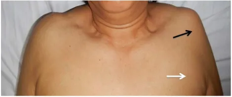
A 53-year-old woman was involved in falling on her left arm in abduction- external rotation position on January 2015, she felt discomfort and severe pain (visual analogue scale 7/10) without vascular problems and inability to use the left arm. At that time, deformity of the ring and little fingers known as claw-hand was not recognized. However two months later, she gradually started to feel bent on her ring and little fingers, since the patient had treated herself conservatively. After approximately six months following the injury, she came to our clinic at Dr. Hasan Sadikin Teaching Hospital Bandung to cure her symptoms. On the clinical examination, she demonstrated loss of normal con-tour of the left shoulder joint, prominence of acromion bone, and slight atrophy of the lateral deltoid muscle (Figure 1). There was a change of the humeral head down in front of the shoulder. There was no atrophy of the shoulder girdle mus-cles including supraspinatus, infraspinatus and teres minor. There was tender-ness over the lateral part of the shoulder.
Movement of the glenohumeral joint was recorded. It was limited in all direc-tions. Besides the limitation of the shoulder, there was an associated neurologic deficit such as numbness and tingling on her left hand especially ring and little fingers in particular on palmar and volar aspect (Figure 2).
[image:3.595.256.491.414.512.2]Conventional radiological views were taken in one plane anteroposterior (AP)
Figure 1. Clinical photograph demonstrating loss of the normal contour of the deltoid (black arrow) of the left shoulder. The humeral head itself may well be palpable anteriorly (white arrow).
Figure 2. Clinical appearance of left hand who has compression of the ulnar nerve
[image:3.595.211.539.567.667.2](a) (b)
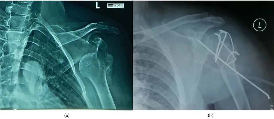
Figure 3. An AP radiograph of the left shoulder showing anteroinferior dislocation associated with fracture of greater tuberosity (a); Early postoperative AP view showing fixation using a tension band wiring (TBW) and stabilization by a Kirschner wire (b).
only. It revealed glenohumeral joint dislocation (subglenoid type), the humeral head was below the glenoid and associated with fracture of greater tuberosity (Figure 3(a)).
The EMG-NCV examinations were used to determine the severity, the pattern and the recovery of ulnar nerve lesion as a cause of this deformity. Table 2 revealed the result of EMG-NCV before and after surgery.
Surgical procedure was performed by open reduction and internal fixation to reduced humeral head to glenoid and greater tuberosity fracture was reduced to its position and TBW technique was performed to fix the fracture (Figure 3(b)). Intraoperatively findings were dislocation of the humeral head to infraglenoid, fracture of the greater tuberosity and torn of the anterior capsule due to absorption process of the middle and inferior glenohumeral ligaments (MGHL and IGHL). At surgery, the anterior capsule was obliterated and replaced by a rectangular piece of fascia (2 × 15 cm) which was harvested from a femoral bone ipsilateral, and inserted on the anterior part of the glenoid and located in two to six o’clock in zig zag position. Transfixing Kirschner wire was used to immobilized the humeral head after reduction (Figure 3(b)) to avoid stretching of the tightened and recontructed soft tissue structures. The Kirschner wire was removed at six-week postoperatively. At the latest follow-up six months postoperatively, the integrity of greater tuberosity and glenohumeral joint was maintained but the ulnar claw-hand still remained slightly in particular in the little finger. However, her numbness and tingling in the ring and little fingers disappeared completely, the bent shape of the ring finger was back to completely normal. Unfortunately, the bent shape of the little finger still remained slightly on the distal interphalangeal joint (DIP) joint, but the PIP joint diappeared completely (Table 1).
Table 1. Range of motion on the affected fingers, before and after surgery.
Affected area ROM of the affected fingers
Before surgery After surgery Ring finger
the PIP joint 45˚ - 50˚ 10˚ - 100˚
the DIP joint 45˚ - 50˚ 5˚ - 100˚
Little finger
the PIP joint 45˚ - 50˚ 0˚ - 110˚
the DIP joint 45˚ - 50˚ 5˚ - 100˚
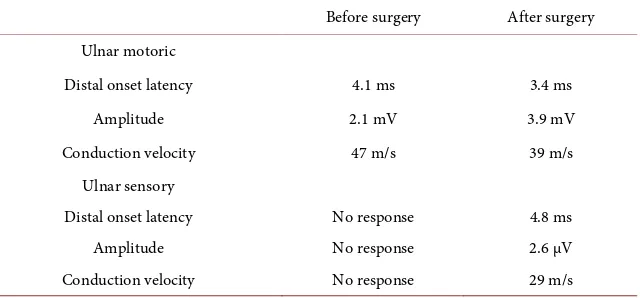
Table 2. EMG-NCV result of ulnar nerve deficit before and after surgery.
Before surgery After surgery Ulnar motoric
Distal onset latency 4.1 ms 3.4 ms
Amplitude 2.1 mV 3.9 mV
Conduction velocity 47 m/s 39 m/s Ulnar sensory
Distal onset latency No response 4.8 ms
Amplitude No response 2.6 μV
Conduction velocity No response 29 m/s
In this case, EMG evaluation demonstrated the ulnar motoric amplitude and conduction velocity still remained in the condition of before and after surgery, however the ulnar sensory in all parameters (distal onset latency, amplitude and conduction velocity), significantly improved from no response to 29 m/s for conduction velocity (Table 2).
At six-month follow-up, the patient’s condition had clinically improved in-cluded the range of motion on the left shoulder. Unfortunately, the X-ray fol-low-up demonstrated collapse of the humeral head. The upper limb was secured postoperatively according to Rowe and Zarins scoring system. This scoring sys-tem evaluates stability, motion and shoulder function [6].
3. Discussion
[image:5.595.220.540.257.406.2]pressure inside the nerve, causing delays in conducting stimuli. This discomfort is often accompanied by weakness of grip and intrinsic wasting [7] [8].
Anterior shoulder dislocations may cause several potential complications. Around 20% - 50% of patients will have some types of neurologic complication [8] [9]. Isolated fracture of the greater tuberosity accounts for approximate 20% of all proximal humeral fracture [10] [11]. They are often associated with dislo-cation of humeral head antero-inferiorly or can result from an impaction injury, against the lower surface of the acromion or superior glenoid fossa [12].
The treatment is usually open surgery to reduce humeral head to glenoid fos-sa, and capsular reconstruction, with the use of fascia which harvests from ipsi-lateral femur, performed to address a deficient anterior aspect of the capsule, by sewing it to the edge of the glenoid fossa. There are several surgical options for unstable shoulder with a deficient capsule. Warner et al. reported a good out-come after using hamstring tendons. Gallie and Le Mesurier described the use of iliotibial band with irreparable capsule, and the results were generally good, with no recurrences [13] [14]. This patient also had greater tuberosity fracture, but it was successfully reduced and fixed by TBW technique [15].
The diagnosis is based on taking a detailed history, making a detailed exami-nation and using appropriate complementary examiexami-nations. The mechanism of associated nerve damage is similar to that of the shoulder injury and results in a traction lesion of nerves stretched over the head of the humerus during the dis-location of the head [15]. In the case presented here, the ulnar claw-hand oc-curred as a result of overstretched ulnar nerve across humeral head since it was dislocated for a considerable time down anteriorly and inferiorly. Together with repeated use of the left upper extremity for around 6 months, the ulnar nerve became gradually stretched.
At six-week follow-up after removing the transfixing wire, the arm was kept anterior to the coronal plane of the body by means of an arm sling. Most reports have recommended crossed transfixing pins through the humeral head into the glenoid fossa for ten to fourteen days to prevent recurrence of dislocation fol-lowing open reduction [16] [17]. Glenohumeral fixation by Kirschner-wire was used for 4 weeks in this case and the result was evaluated according to Rowe and Zarins system [6] and it yielded good functional results. After reduction patients are generally immobilized for 4 - 6 weeks under sling. The arm support was loo-sened three times a day to allow early shoulder motion up to 90˚ of forward flexion and 10 to 20 degrees external rotation and full elbow motion. After three weeks, flexion and external rotation were gradually increased and with gradual stretching and improving subscapularis contracture, more external rotation was regained. Interestingly, the ulnar claw-hand had gradually disappeared in two weeks after the reduction of this humeral head including numbness and pain as well, meaning that compression and stretching of the ulnar nerve had been re-leased.
in comparison with healthy side. The degenerative changes occurring here in fi-nal radiographs make sense because the fracture-dislocation has occurred long enough. Even so, the patient can still perform daily activities without feeling pain.
Literatures described the association between greater tuberosity fracture, dis-location, complete axillary and brachial plexus injury in conjunction with shoulder dislocation [18] [19]. The best means to confirm a concomitant peri-phery nerve injury with shoulder dislocation includes detailed subjective and objective clinical examinations, along with EMG studies [8]. EMG is the neuro-logical examination of choice for locating the level of compression. In this nerve-conduction study, it shows reduction of the amplitude of the potential and a reduced conduction velocity. In this study, EMG shows little active denerva-tion.
In the case presented here, the patient presented improvement of her pain, tingling and paresthesia, improvement of the EMG and nerve conduction veloc-ity and correction of the ulnar claw. The latter deformveloc-ity should be treated early by hand physiotherapy until the expectations for recovery are abandoned. This is a rare event in the literature, and early surgical treatment finally, satisfactorily offers a moderate functional EMG-NCV results.
Acknowledgements
Written informed consent was obtained from the patient for publication of this paper and for the use of any accompanying images.
Conflict of Interest
The author confirms that this article content has no conflict of interest.
References
[1] Bankart, A.S.B. (1938) The Pathology and Treatment of Recurrent Dislocations of the Shoulder Joint. British Journal of Surgery, 26, 23-29.
https://doi.org/10.1002/bjs.18002610104
[2] Hovelius, L., Augustini, B.G., Fredin, H., Johansson, O., Norlin, R. and Thorling, J. (1996) Primary Anterior Dislocation of the Shoulder in Young Patients. A Ten-Year Prospective Study. The Journal of Bone and Joint Surgery, 78, 1677-1684.
https://doi.org/10.2106/00004623-199611000-00006
[3] Bost, F.C. and Inman, V.C. (1942) The Pathological Changes in Recurrent Disloca-tion of the Shoulder: A Report of Bankart’s Operative Procedure. The Journal of Bone and Joint Surgery, 23, 596-613.
[4] Mulder, J.D. and Landsmeer, J.M.F. (1968) The Mechanism of Claw Finger. The Journal of Bone and Joint Surgery, 50, 664-668.
[5] Aguiar, P.H., Bor-Seng-Shu, E., Gomes-Pinto, F., Almeida-Leme, R.J., Freitas, A.B., Martins, R.S., et al. (2001) Surgical Management of Guyon’s Canal Syndrome, an Ulnar Nerve Entrapment at the Wrist: Report of Two Cases. Arquivos de Neuro- Psiquiatria, 59, 106-111. https://doi.org/10.1590/S0004-282X2001000100022 [6] Rowe, C.R. and Zarins, B. (1982) Chronic Unreduced Dislocation of the Shoulder.
https://doi.org/10.2106/00004623-198264040-00004
[7] Ruas, E.S., Castilho, R.S., Maia, P.E.C. and Melo, G.L.R. (2010) Guyon’s Canal Syn-drome Due to a Synovial Cyst. Revista Brasileira Ortopedia, 45, 623-626.
https://doi.org/10.1590/S0102-36162010000600020
[8] Visser, C.P., Coene, L.N., Brand, R. and Tavy, H. (1999) The Incidence of Nerve Injury in Anterior Dislocation of the Shoulder and Its Influence on Functional Re-covery. A Prospective Clinical and EMG Study. The Journal of Bone and Joint Sur-gery, 81, 679-685.
[9] Blorn, S. and Dahback, L.O. (1970) Nerve Injuries in Dislocations of the Shoulder Joint and Fractures of the Neck of the Humerus. Acta Chirurgica Scandinavica, 136, 461-466.
[10] Chun, J.M., Groh, G.I. and Rockwood Jr., C. (1994) Two-Part Fractures of the Pro- ximal Humerus. Journal of Shoulder and Elbow Surgery, 3, 273-287.
https://doi.org/10.1016/S1058-2746(09)80071-2
[11] Kim, E., Shin, H.K. and Kim, C.H. (2005) Characteristics of an Isolated Greater Tuberosity Fracture of the Humerus. Journal of Orthopaedic Science, 10, 441-444. https://doi.org/10.1007/s00776-005-0924-6
[12] Court-Brown, C.M., Garg, A. and McQueen, M.M. (2001) The Epidemiology of Proximal Humeral Fractures. Acta Orthopaedica Scandinavica, 72, 365-371. https://doi.org/10.1080/000164701753542023
[13] Millet, P.J., Clavert, P. and Warner, J.P. (2005) Open Operative Treatment for An-terior Shoulder Instability: When and Why. The Journal of Bone and Joint Surgery, 87, 419-432.
[14] Gallie, W.E. and Le Mesurier, A.B. (1948) Recurring Dislocation of the Shoulder. The Journal of Bone and Joint Surgery, 30, 9-18.
[15] Platzer, P., Kutscha-Lissberg, F., Lehr, S., Vecsei, V. and Gaebler, C. (2005) The In-fluence of Displacement on Shoulder Function in Patients with Minimally Dis-placed Fractures of the Greater Tuberosity. Injury, 36, 1185-1189.
https://doi.org/10.1016/j.injury.2005.02.018
[16] Shin, A.Y., Deitch, M.A., Sachar, K. and Boyer, M.I. (2005) Ulnar-Sided Wrist Pain: Diagnosis and Treatment. Instructional Course Lectures, 54, 115-128.
[17] Milton, G.W. (1953) The Mechanism of Circumflex and Other Nerve Injuries in Dislocation of the Shoulder, and the Possible Mechanism of Nerve Injuries during Reduction of Dislocation. Australian and New Zealand Journal of Surgery, 23, 25- 30. https://doi.org/10.1111/j.1445-2197.1953.tb05013.x
[18] Wilson, J.C. and McKeever, F.N. (1949) Traumatic Posterior (Retroglenoid) Dislo-cation of the Humerus. The Journal of Bone and Joint Surgery, 31, 160-172. https://doi.org/10.2106/00004623-194931010-00018
Submit or recommend next manuscript to OALib Journal and we will pro-vide best service for you:
Publication frequency: Monthly
9 subject areas of science, technology and medicine Fair and rigorous peer-review system
Fast publication process
Article promotion in various social networking sites (LinkedIn, Facebook, Twitter,
etc.)
Maximum dissemination of your research work