Original Article
Clinical implication of UCA1 in non-small cell lung
cancer and its effect on caspase-3/7 activation
and apoptosis induction in vitro
Rui-Xue Tang*, Zhi-Min Chen*, Jing-Jing Zeng, Gang Chen, Dian-Zhong Luo, Wei-Jia Mo
Department of Pathology, First Affiliated Hospital of Guangxi Medical University, 6 Shuangyong Road, Nanning 530021, Guangxi Zhuang Autonomous Region, P. R. China. *Equal contributors.
Received February 25, 2018; Accepted March 26, 2018; Epub May 1, 2018; Published May 15, 2018
Abstract: Since urothelial cancer associated 1 (UCA1) was discovered in human bladder cancer, it has been re-ported to be dysregulated expressed in various kinds of solid tumors. But the clinical role and the function of UCA1 in non-small cell lung cancer (NSCLC) remains incompletely understood. In this study, we mined the data of UCA1 expression in NSCLC from Oncomine, Gene expression profiling interactive analysis (GEPIA) and cBioPortal to ana-lyze the contribution of UCA1 in the cancer initiation and progression of NSCLC. We also performed a series of in vitro experiments by using NSCLC cells to confirm the biological function of UCA1 in NSCLC, especially its effect on caspase-3/7 activity and apoptosis through RNA interference experiment. From Oncomine, the UCA1 levels were both up-regulated in lung adenocarcinoma (LUAD) and lung squamous carcinoma (LUSC), as compared to non-can-cerous controls. Higher levels of UCA1 pointed to a poorer overall survival in NSCLC, with the HR being 1.3. Only two genetic alterations, including amplification and deep deletion, were observed for UCA1 as provided by cBioPortal. Both MTS and Cell Titer-blue assays showed an accordant inhibitory effect of UCA1 siRNAs on the cell growth. In conclusion, lncRNA UCA1 might play a substantial role in the occurrence and development of NSCLC, especially in LUAD patients, which is partly due to its effect on caspase-3/7 activity suppression.
Keywords: UCA1, non-small cell lung cancer, caspase-3/7, apoptosis
Introduction
In the recent years, mounting evidence has showed that long non-coding RNAs (lncRNAs), which are longer than 200 nucleotides, have
become a novel star in the field of human can -cers’ occurrence and development [1-5]. Although lncRNAs are non-protein coding, they can modulate gene expression in other man-ners [6-9]. Urothelial cancer associated 1 (UCA1), the protagonist in this paper, is a
mem-ber of lncRNAs. Since UCA1 was first discov -ered in human bladder cancer, it has been found to be dysregulated expressed in various kinds of solid tumors, such as esophageal squamous cell carcinoma (ESCC), ovarian cer (OC), colorectal cancer (CRC), gastric can-cer (GC), osteosarcoma, and so on [10-12]. And UCA1 was over-expressed in the most reported malignancies mentioned above [13-15]. The up-regulation of UCA1 was even analyzed to be
associated with the clinicopathological charac-teristics of cancers, especially metastasis and survival [16-18]. Hence, UCA1 was thought to be an oncogene in certain kinds of tumors. Each year, the incidence of lung cancer (LC) is increasing with about 1.8 million new cases, and the rate of cancer-related deaths from LC is over 25% in both males and females, which makes it one of the most common cancers in the world [19-21]. Non-small cell lung cancer (NSCLC) is the main subtype of LC [22-26]. Although recent studies have reported that high expression of UCA1 could promote the pro-gression of NSCLC [27-29], the clinical function and the mechanism of UCA1 in NSCLC remains incompletely understood. Therefore, in this study, we mined the data of UCA1 expression in NSCLC from three public databases (Oncomine,
Gene expression profiling interactive analysis
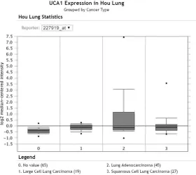
ana-Figure 1. Expression level of UCA1 in the study of Hou Lung from Oncomine. The expression of UCA1 was provided by Oncomine. The study of Hou Lung consisted of 156 samples, and was performed with Human Genome U133 Plus 2.0 Array with 19,574 measured genes. 0: non-tumorous lung tissues (N=65); 1: large cell lung carcinoma (T=19); 2: LUAD (T=45); 3: LUSC (T=27).
series experiments in vitro to
confirm the biological func -tion of UCA1 in NSCLC, espe-cially its effect on caspa- se-3/7 activity and apoptosis by making RNA interference experiment.
Materials and methods
UCA1 expression in NSCLC tissues based on Oncomine and GEPIA public databases
The Oncomine™ Platform ma- intains gene expression sig-natures, clusters and ge- ne-set modules for different diseases (https://www.onco-mine.org /resource/login. html). On Oncomine v4.5, we
first searched the UCA1
ex-pression data in NSCLC. Then, the data with comparison of UCA1 expression between lung cancers and non-cancer controls were selected. The
figures and calculated results
were obtained from the web-site. Detailed information was recorded, including the sam-ple size, subtypes of LC, de- tecting methods and mRNA numbers.
Gene expression profiling
interactive analysis (GEPIA) could help analyze the RNA sequencing data of 9,736 tumors and 8,587 normal samples from The Cancer Genome Atlas (TCGA) and the GTEx projects, with a st- andard processing pipeline (http://gepia.cancer-pku.cn) [30]. (The expression data of UCA1, as well as its compari-son in different stages and high/low survival, was obtain- ed from GEPIA).
[image:2.612.93.373.73.323.2]Genetic alterations of UCA1 in NSCLC from cBioPortal
The cBioPortal for Cancer Genomics provides visualization, analysis and download of large-scale cancer genomics data sets (http://www. cbioportal.org) [31-33]. Genetic alterations of UCA1 in NSCLC from different studies were collected from cBioPortal. Figures of OncoPrints concerning the UCA1 distribution and ratio of
UCA1 genetic alteration types
(amplifica-tion and deep dele(amplifica-tion) were obtained from the website.
Effect of UCA1 siRNAs on cell caspase activity and apoptosis of LUAD cells
The LUAD cell line A549 was purchased from the Institute of Biochemistry and Cell Bio- logy of the Chinese Academy of Sciences (Shanghai, China) and cultured as recommend-ed by the protocol.
Three UCA1 siRNAs were chemically synthe-sized by Gene Chem (Shanghai, China) and
these three siRNAs were put together to form a pool for the knock-down experiments. The sequences of these three siRNAs were as fol-lowing: UCA1-1, 5’-CCACCTGTAGAGAAGACAAA- 3’; UCA1-2, 5’-GAAGAGTAGAAG ACAGGT-3’; UC- A1-3, 5’-GCCTGGACAAGAACAGT-3’, respective-ly. The siRNA transfection was performed with
24-well plates to test the transfection
[image:4.612.91.520.73.355.2]sis, Hoechst 33342/propidium iodide (PI) dou-ble staining was performed, and the cells were
observed under a fluorescence microscope. All
the above in vitro tests were conducted as described previously [34-39].
Statistical analysis
Besides those data provided by Oncomine, GEPIA and cBioPortal, the rest of the experi-mental data were analyzed by using SPSS 22.0 (SPSS Inc., Chicago, IL, USA). Data were pre-sented as mean ± SD. The ANOVA test was selected to examine the differences of the inhibitory ratios for cell growth or inducing ratios for cell caspase activity and apoptosis among the various groups. GraphPad Prism 5.0
was applied to draw all graphs. Two-sided P<0.05 was considered to be statistically
significant.
Results
UCA1 expression in NSCLC tissues based on data from Oncomine and GEPIA
[image:5.612.93.527.71.507.2]From Oncomine, two studies (“Hou Lung” and “Okayama Lung”) provided the UCA1 expres- sion data in different subtypes of NSCLC and non-cancer controls as detected by the Hu- man Genome U133 Plus 2.0 Array. In the “Hou Lung” study, the number of cases of LUAD, LUSC, large cell lung carcinoma (LCC) and non-tumorous were 45, 27, 19 and 65, respectively
(Figure 1). In the “Okayama Lung” study, there were 226 LUAD cases and 20 non-cancerous lung cases (Figure 2). After synthetically analyz-ing the data, we found an increasanalyz-ing trend for UCA1 expression in NSCLC tissues, as com-pared to that in non-cancerous lung tissues, especially in LUAD. However, the difference was
not statistically significant. To further explore
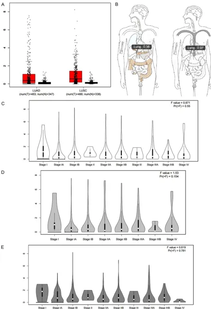
the expression level of UCA1 in NSCLC tissues, we sought the RNA-sequencing data calculat- ed by GEPIA, which had been provided by TCGA and GTEx. The database uses log2 (TPM + 1) for its log-scale. Similarly, the UCA1 levels were up-regulated both in LUAD (T=483) and LUSC (T=486), as compared to that in non-cancer controls (non-LUAD: N=347, non-LUSC: N=338, Figure 3A). When LUAD and LUSC were plac-
rank P=0.0016, Figure 4B). In LUSC, however,
no significant statistical significance was noted
between UCA1 and OS (Figure 4C). The UCA1 level was not closely related to the DFS of NSCLC, LUAD or LUSC (Figure 4D-F).
Genetic alterations of UCA1 in NSCLC from cBioPortal
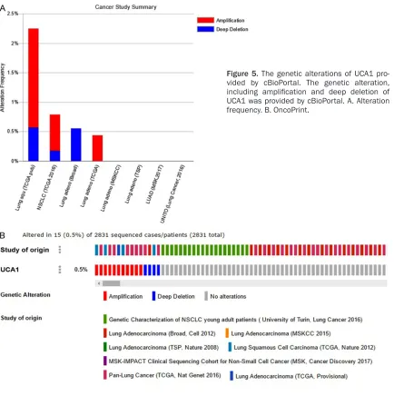
Genetic alterations of UCA1 in NSCLC were col-lected from cBioPortal, which provided various studies with the genetic alteration, including Genetic characterization of NSCLC young adult patients (University of Turin, Lung cancer 2016), Lung adenocarcinoma (Broad, Cell 2012), Lung adenocarcinoma (MSKCC 2015), Lung adeno-carcinoma (TSP, Nature 2008), MSK-IMPACT clinical sequencing cohort for non-small cell Figure 6. Effect of UCA1 siRNAs on
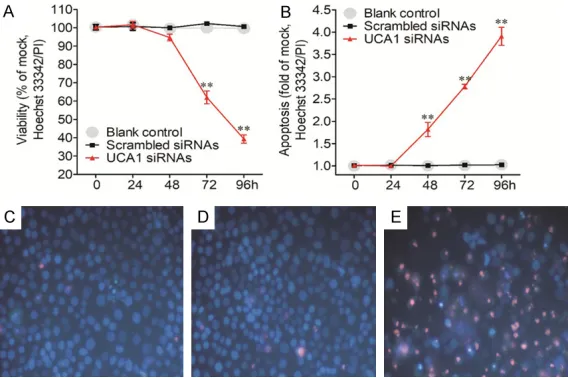
the cell growth and caspase-3/7 activity in A549 cells. A. The pro-liferation of A549 cells after trans-fection UCA1 siRNA was detected by the MTS assay. B. The viabil-ity of A549 cells after transfection UCA1 siRNA was assessed by the CellTiter-Blue assay. C. The cas-pase-3/7 activity in A549 cells after transfection UCA1 siRNA was evaluated by Homogeneous Cas-pase-3/7 Assay.
the over-expression trend of UCA1 could be found on both microarray and RNA-seque- ncing data. Then, we tried to analyze the potential function of UCA1 in the progression of NSCLC but found no obvi-ous relationship between the UCA1 level and the clinical stages in NSCLC (Figure 3C), LUAD (Figure 3D) or LUSC (Figure 3E). Next, the survival analysis of UCA1 was calcu-lated using the GEPIA data-base in NSCLC, LUAD and LUSC. The survival indicators were overall survival (OS) and disease-free survival (DFS). The cut-off of the high/low expression group was the median transcripts per million reads (TPM) value. In NSCLC, a higher level of UCA1 pointed to a poorer overall survival, with the HR being 1.3 (logrank P=0.0039, Figure 4A). In LUAD, the poorer prognostic role of UCA1 expression was more apparent considering the OS. The HR was 1.6, which
further confirmed that UCA1
cancer (MSK, Cancer Discovery 2017), Pan-lung cancer (TCGA, Nat Genet 2016), etc. Two
genetic alterations, amplification and deep
deletion, were observed in UCA1 as provided by cBioPortal. Figure 5A displays the two altera-tions’ frequencies in each study; Figure 5B shows the total frequency (0.5%) of alteration in all studies.
Effect of UCA1 siRNAs on cell caspase activity and apoptosis of LUAD cells
As the prognostic effect of UCA1 expression was more potent in LUAD than in LUSC, we then validated the function of UCA1 siRNAs on the biological reaction of LUAD cells in vitro. The A549 cell line was taken as an example for LUAD and after UCA1 siRNAs were transfected, different experiments were carried out to detect the variation of cell growth, caspase-3/7 activity and apoptosis. Both MTS and Cell Titer-blue assays showed the accordant inhibitory effect of UCA1 siRNAs on cell growth. From 48 h post transfection of UCA1 siRNAs, the growth speed was decreased for the A549 cells. At 96 h post transfection, the cell growth inhibitory rate reached 30% (Figure 6A, 6B), which was accompanied by the increase of caspase-3/7 activity (Figure 6C). The caspase-3/7 activity was more than 2.5 times the blank and scram-bled siRNA controls. To further verify the above
[6]. In some studies, UCA1 has been report- ed to be more over-expressed in NSCLC tissu- es than in normal lung tissues [27-29]. However, the clinical function and the mechanism of UCA1 in NSCLC remains incompletely un-
derstood. In this study, we verified that UCA1 was indeed significantly up-regulated in
NS-CLC after composite analysis the UCA1 ex- pression in Oncomine, GEPIA and cBioPortal. Calculated by GEPIA, higher level of UCA1 in LUAD could lead to lower patient survival ra-
tes. The amplification and deep deletion were
the main types of genetic alteration of UCA1 in NSCLC, which was provided by cBioPortal. We also performed in vitro experiments to explore the function of UCA1 in tumor proli- feration and apoptosis. After transfection of UCA1 siRNA in the A549 cell line, the experi-ment’s results showed that UCA1 could induce caspase-3/7 activity and apoptosis in A549 cells.
UCA1 was over-expressed in some lung cancer cell lines, including A549, H517, H446, H4006, H460, H1299 and H1650, as compared to cul-tured human lung epithelial cells (BEAS-2B), normal embryonic lung WI-38, and HEL-1 cells [27, 29] which was consistent with our previous detection (data not shown). As for the expres-sion level of UCA1 in NSCLC tissues, two researches published the concordant
up-regu-finding, the cell growth, cell necrosis and apoptosis were tested by another indepen-dent approach, which focus- ed on the morphological change of cells and could be observed under microscope. This Hoechst 33342/PI
dou-ble staining confirmed the
aforementioned data, which suggested that UCA1 may play a substantial part in the cell growth and cell apoptosis in LUAD cells (Figure 7). Discussion
Urothelial cancer associated 1 (UCA1), a member of lnc-
RNAs, was first discovered in
[image:7.612.91.375.72.261.2]human bladder cancer. It is found on human chromosome 19p13.12 and consists of three exons and two introns Figure 7. Validation of the influence of UCA1 siRNAs on the cell growth and
also be observed in the data of some other detection methods: microarray and RNA-sequ-
encing. The findings in our paper, together with
those from earlier studies [27, 28] and our in-house RT-qPCR discovery (data not shown),
verified the oncogenic role of UCA1 in the occur
-rence of NSCLC. Significantly, UCA1 also has
the potential to be used as a non-invasive bio-marker for the early screening of NSCLC, as it was reported that the circulating UCA1 level was also markedly up-regulated in NSCLC patients, with the AUC being 0.886 [28]. However, this diagnostic role was based on a single cohort with a small size of patients, so it remains to be validated with a larger sample size.
In addition to the role of UCA1 in the tumorigen-esis of NSCLC, UCA1 has also been reported to play an essential part in the development of NSCLC. For instance, over-expression of UCA1 was closely related to histological grade and the status of lymph node metastasis [28]. UCA1 over-expression could cause an appar-ently poorer outcome of NSCLC patients, and multivariate analysis found that UCA1 could act as an independent risk factor of the progno-sis of NSCLC [27]. A meta-analyprogno-sis summariz- ed all available studies concerning the prognos-tic role of UCA1 in NSCLC with three studies being involved [27, 28, 40]. Indeed, the result
confirmed the risky role of UCA1 in the progno -sis prediction ability of NSCLC, with the HR of 1.49 (95% CI: 1.16-1.90) [41]. This is in line
with the current finding based on
RNA-sequencing data with a large sample size of 955 patients, which led to the HR of 1.3. More importantly, the HR became more apparent in the subtype of LUAD, which reached 1.6. The
current finding, with previous reports [41], fur -ther unveil the vital role of UCA1 in the progres-sion and survival of LUAD, but not LUSC. The etiology of this difference still needs further investigation.
Due to the potent influence of UCA1 on the
survival of LUAD, we then performed in vitro experiments to investigate the relevant bio- logical function of UCA1 on LUAD A549 cells.
ingly increase the caspase-3/7 activity, which are executioner proteins of apoptosis in A549 cells. This effect on caspase-3 could also be found in other cancer types, such as cholangio-carcinoma [42], cervical cancer [14], prostate cancer [43] and gastric cancer [44]. Hence, UCA1 might play similar roles in different can-cers to restrain the caspase-3/7 and enhance cell growth.
A relationship between UCA1 and EGFR muta-tion has also been reported. The UCA1 level was notably up-regulated in lung cancer cells and patients with acquired resistance to
EGFR-TKIs. UCA1 silencing rescued gefitinib
sen-sitivity in those acquired resistant cells which had non-T790M mutations. UCA1 may prompt non-T790M acquired resistance to EGFR-TKIs [40]. The molecular mechanism of UCA1 in NSCLC could also rely on the ceRNA axis. Two ceRNA axes of UCA1 in lung cancer have
been clarified. One is UCA1/miR-144/Pre-B cell
leukemia homeobox 3 (PBX3) [29], the other one is UCA1/miR-193-3p/ERBB4 [27]. Other axes still need to be explored for UCA1 in NSCLC.
To sum up, lncRNA UCA1 might play a substan-tial role in the occurrence and development of NSCLC, especially in LUAD patients, which is partly due to its effect on caspase-3/7 activity suppression. Additional potential molecular mechanisms of UCA1 in NSCLC require further assessment.
Acknowledgements
This work was supported by Natural Science Foundation of China (NSFC 81560448) and Innovation Project of Guangxi Graduate Education (YCBZ2017040).
Disclosure of conflict of interest
None.
Autonomous Region, P. R. China. E-mail: [email protected] (WJM); [email protected] (DZL)
References
[1] Simion V, Haemmig S and Feinberg MW. Ln-cRNAs in vascular biology and disease. Vascul Pharmacol 2018; [Epub ahead of print]. [2] Sui J, Li YH, Zhang YQ, Li CY, Shen X, Yao WZ,
Peng H, Hong WW, Yin LH, Pu YP and Liang GY. Integrated analysis of long non-coding RNAas-sociated ceRNA network reveals potential ln-cRNA biomarkers in human lung adenocarci-noma. Int J Oncol 2016; 49: 2023-2036. [3] Yao J, Huang JX, Lin M, Wu ZD, Yu H, Wang PC,
Ye J, Chen P, Wu J and Zhao GJ. Microarray ex-pression profile analysis of aberrant long non-coding RNAs in esophageal squamous cell car-cinoma. Int J Oncol 2016; 48: 2543-2557. [4] Xu CZ, Jiang C, Wu Q, Liu L, Yan X and Shi R. A
feed-forward regulatory loop between hur and the long noncoding RNA HOTAIR promotes head and neck squamous cell carcinoma pro-gression and metastasis. Cell Physiol Biochem 2016; 40: 1039-1051.
[5] Zhang J, Yin M, Peng G and Zhao Y. CRNDE: an important oncogenic long non-coding RNA in human cancers. Cell Prolif 2018; [Epub ahead of print].
[6] Xue M, Chen W and Li X. Urothelial cancer as-sociated 1: a long noncoding RNA with a cru-cial role in cancer. J Cancer Res Clin Oncol 2016; 142: 1407-1419.
[7] Li Z, Yu X and Shen J. Long non-coding RNAs: emerging players in osteosarcoma. Tumour Biol 2016; 37: 2811-2816.
[8] Li Y and Wang X. Role of long noncoding RNAs in malignant disease (Review). Mol Med Rep 2016; 13: 1463-1469.
[9] Zhang F, Zhang L and Zhang C. Long noncod-ing RNAs and tumorigenesis: genetic associa-tions, molecular mechanisms, and therapeutic strategies. Tumour Biol 2016; 37: 163-175. [10] Chen S, Zhu J, Wang F, Guan Z, Ge Y, Yang X
and Cai J. LncRNAs and their role in cancer stem cells. Oncotarget 2017; 8: 110685-110692.
[11] Wang H, Guan Z, He K, Qian J, Cao J and Teng L. LncRNA UCA1 in anti-cancer drug resis-tance. Oncotarget 2017; 8: 64638-64650. [12] Li Z, Dou P, Liu T and He S. Application of long
noncoding RNAs in osteosarcoma: biomarkers and therapeutic targets. Cell Physiol Biochem 2017; 42: 1407-1419.
[13] Wang Y, Gao W, Xu J, Zhu Y and Liu L. The long noncoding RNA urothelial carcinoma-associat-ed 1 overexpression as a poor prognostic bio-marker in clear cell renal cell carcinoma. Tu-mour Biol 2017; 39: 1010428317698377.
[14] Wang B, Huang Z, Gao R, Zeng Z, Yang W, Sun Y, Wei W, Wu Z, Yu L, Li Q, Zhang S, Li F, Liu G, Liu B, Leng L, Zhan W, Yu Y, Yang G and Zhou S. Expression of long noncoding RNa urothelial cancer associated 1 promotes cisplatin resis-tance in cervical cancer. Cancer Biother Radio-pharm 2017; 32: 101-110.
[15] Qian Y, Liu D, Cao S, Tao Y, Wei D, Li W, Li G, Pan X and Lei D. Upregulation of the long non-coding RNA UCA1 affects the proliferation, in-vasion, and survival of hypopharyngeal carci-noma. Mol Cancer 2017; 16: 68.
[16] Groenink L, Van der Gugten J, Mos J, Maes RA and Olivier B. The corticosterone-enhancing ef-fects of the 5-HT1A receptor antagonist, (S)-UH301, are not mediated by the 5-HT1A recep-tor. Eur J Pharmacol 1995; 272: 177-183. [17] Wang YH, Wang F, Zhang L and Lu JC. Long
non-coding RNA UCA1 can predict tumor lymph node metastasis. Tumour Biol 2017; 39: 1010428317706208.
[18] He A, Hu R, Chen Z, Liao X, Li J, Wang D, Lv Z, Liu Y, Wang F and Mei H. Role of long noncod-ing RNA UCA1 as a common molecular marker for lymph node metastasis and prognosis in various cancers: a meta-analysis. Oncotarget 2017; 8: 1937-1943.
[19] Li Q, Hou J, Hu Z, Gu B and Shi Y. Multiple mu-tations of lung squamous cell carcinoma shared common mechanisms. Oncotarget 2016; 7: 79629-79636.
[20] Siegel RL, Miller KD and Jemal A. Cancer sta-tistics, 2018. CA Cancer J Clin 2018; 68: 7-30. [21] Gurel D, Ulukus C, Karacam V, Ellidokuz H,
Umay C, Oztop I and Sarioglu S. The prognostic value of morphologic findings for lung squa-mous cell carcinoma patients. Pathol Res Pract 2016; 212: 1-9.
[22] Wang LY, Cui JJ, Guo AX and Yin JY. Clinical ef-ficacy and safety of afatinib in the treatment of non-small-cell lung cancer in Chinese patients. Onco Targets Ther 2018; 11: 529-538. [23] Chen Y, Peng X, Zhou Y, Xia K and Zhuang W.
Comparing the benefits of chemoradiotherapy and chemotherapy for resectable stage III A/ N2 non-small cell lung cancer: a meta-analy-sis. World J Surg Oncol 2018; 16: 8.
[24] Wang L, Chen Z, An L, Wang Y, Zhang Z, Guo Y and Liu C. Analysis of long non-coding RNA ex-pression profiles in non-small cell lung cancer. Cell Physiol Biochem 2016; 38: 2389-2400. [25] Roviello G, Zanotti L, Cappelletti MR, Gobbi A,
Dester M, Paganini G, Pacifico C, Generali D and Roudi R. Are EGFR tyrosine kinase inhibi-tors effective in elderly patients with EGFR-mutated non-small cell lung cancer? Clin Exp Med 2018; 18: 15-20.
by targeting miR-193a-3p. Cancer Lett 2016; 371: 99-106.
[28] Wang HM, Lu JH, Chen WY and Gu AQ. Upregu-lated lncRNA-UCA1 contributes to progression of lung cancer and is closely related to clinical diagnosis as a predictive biomarker in plasma. Int J Clin Exp Med 2015; 8: 11824-11830. [29] Li D, Li H, Yang Y and Kang L. Long noncoding
RNA urothelial carcinoma associated 1 pro-motes the proliferation and metastasis of hu-man lung tumor cells by regulating microR-NA-144. Oncol Res 2017; [Epub ahead of print].
[30] Tang Z, Li C, Kang B, Gao G, Li C and Zhang Z. GEPIA: a web server for cancer and normal gene expression profiling and interactive anal-yses. Nucleic Acids Res 2017; 45: W98-W102. [31] Gao J, Aksoy BA, Dogrusoz U, Dresdner G,
Gross B, Sumer SO, Sun Y, Jacobsen A, Sinha R, Larsson E, Cerami E, Sander C and Schultz N. Integrative analysis of complex cancer ge-nomics and clinical profiles using the cBioPor-tal. Sci Signal 2013; 6: pl1.
[32] Cerami E, Gao J, Dogrusoz U, Gross BE, Sumer SO, Aksoy BA, Jacobsen A, Byrne CJ, Heuer ML, Larsson E, Antipin Y, Reva B, Goldberg AP, Sander C and Schultz N. The cBio cancer ge-nomics portal: an open platform for exploring multidimensional cancer genomics data. Can-cer Discov 2012; 2: 401-404.
[33] Huang R, Liao X and Li Q. Identification of key pathways and genes in TP53 mutation acute myeloid leukemia: evidence from bioinformat-ics analysis. Onco Targets Ther 2018; 11: 163-173.
[34] Chen G, Umelo IA, Lv S, Teugels E, Fostier K, Kronenberger P, Dewaele A, Sadones J, Geers C and De Greve J. miR-146a inhibits cell growth, cell migration and induces apoptosis in non-small cell lung cancer cells. PLoS One 2013; 8: e60317.
[35] He R, Yang L, Lin X, Chen X, Lin X, Wei F, Liang X, Luo Y, Wu Y, Gan T, Dang Y and Chen G. MiR-30a-5p suppresses cell growth and enhances apoptosis of hepatocellular carcinoma cells via targeting AEG-1. Int J Clin Exp Pathol 2015; 8: 15632-15641.
[37] Li JJ, Luo J, Lu JN, Liang XN, Luo YH, Liu YR, Yang J, Ding H, Qin GH, Yang LH, Dang YW, Yang H and Chen G. Relationship between TRAF6 and deterioration of HCC: an immuno-histochemical and in vitro study. Cancer Cell Int 2016; 16: 76.
[38] Zhong DN, Luo YH, Mo WJ, Zhang X, Tan Z, Zhao N, Pang SM, Chen G, Rong MH and Tang W. High expression of long noncoding HOTAIR correlated with hepatocarcinogenesis and me-tastasis. Mol Med Rep 2018; 17: 1148-1156. [39] Chen G, Noor A, Kronenberger P, Teugels E,
Umelo IA and De Greve J. Synergistic effect of afatinib with su11274 in non-small cell lung cancer cells resistant to gefitinib or erlotinib. PLoS One 2013; 8: e59708.
[40] Cheng N, Cai W, Ren S, Li X, Wang Q, Pan H, Zhao M, Li J, Zhang Y, Zhao C, Chen X, Fei K, Zhou C and Hirsch FR. Long non-coding RNA UCA1 induces non-T790M acquired resistance to EGFR-TKIs by activating the AKT/mTOR pathway in EGFR-mutant non-small cell lung cancer. Oncotarget 2015; 6: 23582-23593. [41] Wang X, Peng F, Cheng L, Yang G, Zhang D, Liu
J, Chen X and Zhao S. Prognostic and clinico-pathological role of long non-coding RNA UCA1 in various carcinomas. Oncotarget 2017; 8: 28373-28384.
[42] Xu Y, Yao Y, Leng K, Li Z, Qin W, Zhong X, Kang P, Wan M, Jiang X and Cui Y. Long non-coding RNA UCA1 indicates an unfavorable prognosis and promotes tumorigenesis via regulating AKT/GSK-3beta signaling pathway in cholan-giocarcinoma. Oncotarget 2017; 8: 96203-96214.
[43] Wang X, Yang B and Ma B. The UCA1/miR-204/Sirt1 axis modulates docetaxel sensitivity of prostate cancer cells. Cancer Chemother Pharmacol 2016; 78: 1025-1031.