Original Article
ASK1 induces retinal microvascular endothelial cell
apoptosis through ER stress-associated pathway
Wenjun Zou1,2, Chen Zou3, Minjie Zhao4, Zhi Zheng2
1Department of Ophthalmology, Nanjing Medical University Affiliated Wuxi Second Hospital, Wuxi, China; 2 Depart-ment of Ophthalmology, Shanghai First People’s Hospital of Nanjing Medical University, Shanghai, China; 3 Depart-ment of Ophthalmology, Shanghai First People’s Hospital, Shanghai Jiao Tong University School of Medicine, Shanghai, China; 4Department of Ophthalmology, Yixing People’s Hospital, Yixing, China
Received December 26, 2018; Accepted January 21, 2019; Epub April 1, 2019; Published April 15, 2019
Abstract: Diabetic retinopathy (DR) is a major microvascular complication in patients with diabetes mellitus; it can cause a variety of eye problems in a high percentage of diabetic patients. The purpose of this study was to deter-mine the role of apoptosis signal-regulating kinase 1 (ASK1) in the regulation of the endoplasmic reticulum (ER) stress-associated apoptosis pathway in microvascular endothelial cells. For in vivo studies, a streptozotocin (STZ)-induced diabetes model was used to assess apoptosis in retinal tissues. Apoptotic cell death was determined by TU-NEL assay. For in vitro studies, a high glucose (HG)-induced retinal microvascular endothelial cell injury model was
generated to evaluate apoptosis. Apoptotic rates were measured by flow cytometry and apoptosis-related proteins
were detected by western blotting. We found that retinal microvascular endothelial cell apoptosis was increased in both animal and cell models. induced apoptosis primarily occurred in an ER stress-dependent manner.
HG-induced apoptosis was alleviated by inhibiting ASK1 with shRNA or a specific inhibitor, NQDI-1. TUNEL and western
blot assays showed that ASK1 promoted the expression of ER stress-related proteins that are the master regulators of DR. Our study suggests that ASK1 functions as a promoter of DR through the ER stress-induced apoptosis path-way, and it may be a therapeutic target for DR.
Keywords: ASK1, diabetic retinopathy, ER stress, invasion, apoptosis
Introduction
Diabetic retinopathy (DR) is a chronically pro-gressive, potentially vision-threatening disease of the retinal microvasculature; it is a major cause of blindness among working-age individ-uals [1]. Chronic hyperglycemia-associated low-grade aseptic inflammation is thought to play a key role in the initiation and progression of DR [2-4]. Despite a vast amount of investigative effort, the pathogenesis of DR remains unclear. It has been reported that the endoplasmic reticulum (ER) stress-associated apoptosis pathway is involved in DR progression [5-7]. However, the regulatory role of ASK1 on DR occurrence has not been reported.
Apoptosis signal-regulating kinase 1 (ASK1) is a member of the mitogen-activated protein kinase (MAPK) kinase family that activates the p38 MAPK and c-Jun NH2-terminal kinase sig-naling pathways in response to various stimuli,
including oxidative stress, endoplasmic reticu-lum stress, infection and calcium influx [8, 9]. Under these stress conditions, ASK1 plays piv-otal roles in cellular signaling pathways and bio-logical functions. Hyperglycemia increases oxi-dative stress in various tissues, and it has been reported that high glucose-induced activation of ASK1 contributes to endothelial cell senes-cence, leading to diabetes-related vascular aging mediated by oxidative stress [10]. Previous studies have shown that ASK1 plays a regulatory role in diabetes complications, including diabetic cardiomyopathy, diabetic kid-ney disease, and diabetic embryopathy [11-13]. Nevertheless, the effect of ASK1 on DR has not yet been elucidated. Therefore, we investigated whether ASK1 contributed to high glucose-induced apoptosis of retinal microvascular endothelial cells.
trans-port and protein synthesis, and it acts as a res-ervoir for Ca2+ [14]. There is evidence suggest-ing that ER stress can lead to pancreatic β-cell apoptosis, inhibition of insulin receptor expres-sion, and insulin resistance, and ultimately the development of type 2 diabetes [2, 15]. A study reported that ER stress in cultured human reti-nal pericytes was increased after hypoglycemia and when glucose concentrations were reduced from high to low levels. Apoptotic pathways are ultimately induced if ER stress cannot be relieved by protein folding and degradation [16]. While it has been suggested that early pathological changes associated with DR may involve apoptosis, it is not clear whether ASK1 is involved in ER stress-induced apoptosis. In the present study, we first found that ASK1 promoted ER stress-induced apoptosis in DR. We therefore sought to clarify the contribution of the ER stress-mediated apoptosis signaling pathways to the high glucose-induced inflam -matory response and apoptosis using human retinal microvascular endothelial cells (HR- MECs) and to assess the protective effect of NQDI-1 on hyperglycemia-induced injury.
Materials and methods
Animal model
All experimental procedures were conducted in accordance with institutional guidelines for the care and use of laboratory animals, and proto-cols were approved by the Institutional Ani- mal Care and Use Committees of Shanghai Laboratory Animal Center of the Chinese Academy of Sciences. Six-week-old male inbred Sprague Dawley (SD) rats weighing 100-120 g (Shanghai Laboratory Animal Center of the Chinese Academy of Sciences) were housed 5 per cage in an animal colony facility for 2 weeks. The animals were maintained in a room with a constant temperature (22 ± 2°C). All animals were born and raised in a 12-h-light/12-h-dark environment with an average illumination of 80 lx. Tap water and food pellets were provided. SD rats were randomly divided into the DM group and the non-diabetes group. The DM group was intraperitoneally injected with 60 mg/kg STZ. Tail vein blood glucose levels were measured at 48 h after the injection, and those with blood glucose ≥ 16.7 mmol/L were consid -ered DM rats. Diabetes was induced in rats by STZ treatment for 8 weeks. The experiments were conducted between 10:00 and 14:00.
STZ (Sigma) was dissolved in cold 50 mM citric acid buffer (pH 4.5).
Cell lines and cell culture
HRMECs were purchased from Cell Systems (Kirkland, WA, USA), and cells from passages 3-7 were used in the experiments. Cells were grown in M199 medium with 45 ng/ml bFGF, heparin, 20% fetal bovine serum, and 1% peni-cillin-streptomycin solution (10,000 units of penicillin and 10 mg of streptomycin in 0.9% NaCl) in a humidified atmosphere of 5% CO2 and 95% air at 35°C. The passage number range for this cell line was maintained between 10 and 15. The cells were cultured in a 10 cm 2 cell culture dish; 90% confluent cells were switched to serum-free medium for 24 h before the experiment was conducted. The cells were incubated with normal glucose (5 mmol/L) plus 100 nM ASK1 inhibitor, NQDI-1 (100 nmol/L, Selleck, China) or with high glucose (30 mmol/L) plus 100 nM NQDI-1 for 48 h. NLRP3 was inhib -ited with 50 nmol/L MCC950 (Selleck, China).
TUNEL assay
The TUNEL method was employed to detect apoptotic cells according to manufacturer’s instructions. Briefly, cultured HRMECs were infected with lentivirus vectors expressing shScramble or shASK1 (or pretreated with 100 nM ASK1 inhibitor NQDI-1) and then incubated with low glucose (5 mM) or high glucose (30 mM) medium for 48 h. The percentage of apop-totic cells was assessed by terminal deoxyribo-nucleotidyl transferase-mediated dUTP-biotin nick end labeling (TUNEL) method using an apoptosis in situ detection kit (DeadEnd™ Colorimetric TUNEL System, Promega, USA). After TUNEL labeling, the nuclei were labeled with 4’,6-diamidino-2-phenylindole (DAPI; Mo- lecular Probes, Invitrogen, Carlsbad, CA, USA). The FITC-labeled, TUNEL-positive cells were imaged under a fluorescence microscope with 488 nm excitation and 530 nm emission. After four cortical fields were randomly selected from each section, 100 cells were successively counted for each field by a blinded observer. The ratio of TUNEL-positive cells to the total cell number is shown.
MTT assay
assay was optimized for the cell lines used in these experiments. Briefly, for the purpose of these experiments, at the end of the incubation time, the cells were incubated for 4 h with 0.8 mg/ml of MTT dissolved in serum free medium. After washing with PBS (1 ml), DMSO (1 ml) was added, followed by gentle shaking for 10 min so that complete dissolution was achieved. Aliquots (200 µl) of the resulting solutions were transferred into 96-well plates, and the absor-bance was recorded at 560 nm using a micro-plate spectrophotometer system (Spectra max190-Molecular Devices). The results were analyzed and presented as a percentage of the control values.
Western blot analysis and antibodies
Total cell extracts were obtained by lysing the cells in RIPA buffer and boiling for 5 min. Protein concentrations were measured by the Bradford assay (Bio-Rad, Hercules, CA, USA). Cellular proteins were extracted and separated on 4-10% Tris glycine/SDS-polyacrylamide gels and electrotransferred to ECL nitrocellulose membranes (#IPFL00010, Millipore). The mem-branes were blocked with 5% nonfat milk and incubated with specific antibodies. β-actin pro -tein was used as the endogenous control. Antibodies against the following proteins were purchased from Cell Signaling Technology: Caspase 4 and cytochrome c. The following antibodies were purchased from Santa Cruz Biotechnology: IRE1, CHOP, ASK1 and β-actin. Immunocomplexes were visualized by ECL (Pharmacia-Amersham, Freiburg, Germany).
Lentivirus-mediated gene silencing
The following short hairpin RNA (shRNA) was cloned into a pGIPZ vector and used for targeting human ASK1 (Asiavector Biology, China): TGCTGTTGACAGTGAGCGATTGGTCGAA- TCTACAAAGATATAGTGAAGCCACAGATG TATATC- TTTGTAGATTCGACCAACTGCCTACTGCCTCGGA. Lentivirus was generated by transfection of HEK293T cells with pLKO.1 or ASK1 shRNA plasmids and packaging plasmids (PSPA and PMD2G, obtained from Asiavector Biology, China) using Lipofectamine 2000 (Invitrogen, Carlsbad, CA, USA). After 48 h, the virus-con-taining cell culture supernatants were collect-ed, filtered and concentrated. The infection of HRMECs with the ASK1-lentivirus or scramble-lentivirus (MOI = 10) was performed in the presence of 5 µg/ml polybrene (Sigma-Aldrich,
USA). After 48 h, the efficiency of gene silencing was determined by semi-quantitative RT-PCR and western blot.
Flow cytometry analysis
Cells were washed twice with PBS before the experiments. The cells were collected via cen-trifugation at 1000 rpm for 5 min and then stained with Annexin V/propidium iodide (PI), Annexin V-APC/7-AAD and binding buffer (KeyGEN Biotech) at room temperature for 15 min. After mixing, the samples were analyzed using a flow cytometer. The data were analyzed on BD Accuri C6.
Statistical analysis
Each experiment was performed at least 3 times, on independent passages, usually in triplicate. Data were analyzed by the Newman-Keuls test using Statistica software as indicat-ed and were presentindicat-ed as the mean ± SEM. P < 0.05 was considered statistically significant. The results of time-lapse microscopy experi-ments were analyzed with the Wilcoxon test in R software.
Results
Retinal microvascular endothelial cell apopto
-sis is frequently increased in DR tissues and
cells
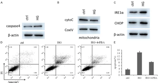
We first investigated apoptosis of retinal tis -sues in DR rat models. The TUNEL assay was used to determine apoptosis in control and DR samples. The apoptotic rates of DR tissues were higher than those of the control group (Figure 1A). We then examined whether HG pro-moted apoptosis of HRMECs. As expected, HG increased the expression level of cleaved cas-pase-3 (CLCasp-3) after HG incubation for 48 h (P < 0.05) in HRMECs, as determined by flow cytometry and western blotting (Figure 1B, 1C). We observed that HG induced a time-depen-dent decrease in cell viability (P < 0.01) (Figure 1D). Taken together, these data suggest that caspase-3 is upregulated in DR, possibly con-tributing to DR pathogenesis.
High glucose-induced apoptosis primarily oc
-curred in an ER stress-dependent manner
The expression levels of caspase-4, cyto C, IRE1α and CHOP in HRMECs were examined by western blotting (Figure 2A-C). Notably, levels of caspase-4, IRE1α and CHOP, hallmarks of endoplasmic stress, were dramatically elevat-ed, whereas levels of cyto C, a mitochondrial apoptosis marker, were not significantly altered in HG groups. We then used flow cytometry to measure apoptosis rates among ctrl, HG-in- duced and HG+4-PBA groups (Figure 2D). We found that ER stress-related apoptosis was the primary pathway in the high-glucose-induced HRMECs model.
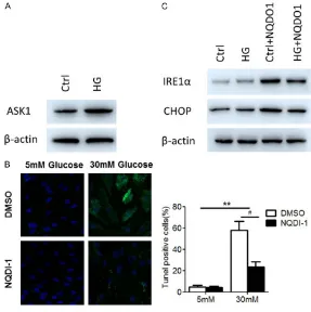
ASK1 induced apoptosis via the ER stress pathway
To further evaluate how ASK1 regulates the apoptosis pathway in DR, we first examined ASK1 expression levels in NG and HG-induced HRMECs cells by WB. The results from the WB indicated that HG significantly increased ASK1 expression levels (Figure 3A). Then, HRMECs were incubated with LG plus NQDI-1 or HG plus NQDI-1 for 48 h. We found that NQDI-1 treat
-rates of DR tissues were higher than those of the control group, and 4-PBA rescued HG-induced apoptosis (Figure 4A). The tissues were then subjected to WB assays to identify whether 4-PBA rescued the HG-induced in- creases of mitochondrial apoptosis. We found that treatment with 4-PBA inhibited HG-prom- oted apoptosis (Figure 4B, 4C).
Discussion
It is well-known that ER stress plays important roles in the microenvironment of diabetes mel-litus (DM), including DR [17]. Here, we found that the apoptosis rate was increased in DR in vivo and in vitro, with respect to rates in the control group. Moreover, we demonstrated that restoration of ASK1 suppressed HG-promoted apoptosis, suggesting a fundamental role of ASK1 as an ER stress-specific marker in DR.
[image:6.612.88.375.72.360.2]Numerous studies have shown that ASK1 was preferentially activated in response to various types of stress, including ROS, TNF-α, lipopoly -saccharides and ER stress, and has potential Figure 3. ASK1 promoted apoptosis through the ER stress pathway. A.
West-ern blotting analysis of ASK1 in normal HRMECs cells and HG-induced HR-MECs cells, β-actin serving as the loading control. B. Apoptosis rates were determined by TUNEL staining. C. Western blotting analysis of IRE1α and CHOP in HRMECs cells, in response to HG, NQDP-1 or HG+NQDP-1. β-actin was used as loading control.
ment attenuated HG-mediat- ed apoptosis and increased viability (Figure 3B). These results suggest that ASK1 alleviates, at least in part, the level of apoptosis induced by HG in vitro. Furthermore, WB was carried out to investigate whether ASK1 was involved in apoptotic ER stress in various experimental groups. As ex- pected, levels of CHOPα and IRE1, key mediators of apop-totic ER stress, were signifi -cantly higher after treatment with NQDI-1. HG rescued ASK-1-induced apoptosis through the ER stress pathway. These results confirmed that ASK1 positively regulated IRE1α and CHOP expression (Figure 3C).
ER stress promoted DR by upregulating ASK1
roles in a wide variety of cellular responses, including apoptosis, inflammation and differen -tiation [18-20]. Nevertheless, the expression and distribution of ASK1 in HRMECs has not yet been elucidated. We found that HG contributed to ASK1 activation in HRMECs. Then, we com-pared the injury responses and biochemical reactions in normal HRMECs and ASK1 inhibi-tor-treated HRMECs cultured in HG medium. We demonstrated that activation of ASK1 by HG upregulated caspase-3 and induced endo-plasmic stress protein activation, promoting apoptosis in HRMECs, whereas the inhibition of ASK1 by shRNA decreased HRMEC sensitivity to HG stress.
Several studies suggested that ER stress-medi-ated cell death is associstress-medi-ated with the death of
[image:7.612.91.522.71.441.2]in HG-induced cells alleviated ER stress-induced apoptosis.
Given that ASK1 is a key element in ER stress-induced cell apoptosis, levels of ER stress markers were measured in HG cells treated with an inhibitor of endoplasmic stress. 4-PBA treatment significantly decreased expression levels of IRE1α and CHOP. These results sug -gest that 4-PBA treatment may reverse the DR process to inhibit cell apoptosis. To address the molecular mechanisms involved in ASK1-mediated promotion of apoptosis, caspase-4, a molecule predicted to be downstream of ER stress, was selected for further study. To inves-tigate the therapeutic potential of inhibiting ASK1 to protect HRMECs exposed to HG, we incubated HRMECs with the recently-identified ASK1 inhibitor NQDI-1 in HG, inhibiting cellu-lar apoptosis in HRMECs. NQDI-1 significantly inhibited activation of the endoplasmic stress protein, and attenuated HG-induced apoptosis. Taken together with the previous findings, these data suggest that ASK1 serves as a pivotal mediator in regulation of DR progression. On the basis of these studies, our data sug-gested that restoring ASK1 expression attenu-ated protein levels of IRE1α and CHOP by regu -lating ER stress-related apoptosis. Targeting the ASK1/CHOP interaction or rescuing ASK1 expression may be a new therapeutic applica-tion to treat DR patients in the future.
Acknowledgements
This work was supported by grants from the National Natural Science Foundation of China (81700852), the Young Talent’s Subsidy Pro- ject in Science and Education of the Depart- ment of Public Health of Jiangsu Province (QNRC2016140) and Funded Project of the Wuxi Municipal Health and Family Planning Commission (Q201623).
Disclosure of conflict of interest
None.
Address correspondence to: Wenjun Zou, Depart- ment of Ophthalmology, Nanjing Medical University
Affiliated Wuxi Second Hospital, Zhongshan Road
68, Wuxi 214002, China. Tel: 86-510-68562222-3091; Fax: 86-510-68562222-86-510-68562222-3091; E-mail: wen-dyzwj0805@163.com; Zhi Zheng, Department of
Ophthalmology, Shanghai First People’s Hospital of Nanjing Medical University, Haining Road 100, Shanghai 200080, China. Tel: 86-21-63240090-6823; Fax: 86-21-63240090-86-21-63240090-6823; E-mail: zzh- eng88@sjtu.edu.cn
References
[1] Noonan JE, Jenkins AJ, Ma JX, Keech AC, Wang JJ and Lamoureux EL. An update on the
mo-lecular actions of fenofibrate and its clinical
effects on diabetic retinopathy and other mi-crovascular end points in patients with diabe-tes. Diabetes 2013; 62: 3968-3975.
[2] Biden TJ, Boslem E, Chu KY and Sue N. Lipo-toxic endoplasmic reticulum stress, beta cell failure, and type 2 diabetes mellitus. Trends Endocrinol Metab 2014; 25: 389-398. [3] Capitao M and Soares R. Angiogenesis and
in-flammation crosstalk in diabetic retinopathy. J
Cell Biochem 2016; 117: 2443-2453.
[4] Rubsam A, Parikh S and Fort PE. Role of
in-flammation in diabetic retinopathy. Int J Mol
Sci 2018; 19.
[5] Chung YR, Choi JA, Koh JY and Yoon YH. Urso-deoxycholic acid attenuates endoplasmic re-ticulum stress-related retinal pericyte loss in streptozotocin-induced diabetic mice. J Diabe-tes Res 2017; 2017: 1763292.
[6] Elmasry K, Ibrahim AS, Saleh H, Elsherbiny N, Elshafey S, Hussein KA and Al-Shabrawey M. Role of endoplasmic reticulum stress in 12/15-lipoxygenase-induced retinal microvas-cular dysfunction in a mouse model of diabetic retinopathy. Diabetologia 2018; 61: 1220-1232.
[7] Roy S, Trudeau K, Roy S, Tien T and Barrette KF. Mitochondrial dysfunction and endoplas-mic reticulum stress in diabetic retinopathy: mechanistic insights into high glucose-induced retinal cell death. Curr Clin Pharmacol 2013; 8: 278-284.
[8] Sekine Y, Takeda K and Ichijo H. The ASK1-MAP kinase signaling in ER stress and neuro-degenerative diseases. Curr Mol Med 2006; 6: 87-97.
[9] Shiizaki S, Naguro I and Ichijo H. Activation mechanisms of ASK1 in response to various
stresses and its significance in intracellular
signaling. Adv Biol Regul 2013; 53: 135-144. [10] Yokoi T, Fukuo K, Yasuda O, Hotta M, Miyazaki
J, Takemura Y, Kawamoto H, Ichijo H and Ogi-hara T. Apoptosis signal-regulating kinase 1 mediates cellular senescence induced by high glucose in endothelial cells. Diabetes 2006; 55: 1660-1665.
in-hibitor, GS-4997, in patients with diabetic kid-ney disease. Nephron 2015; 129: 29-33. [12] Thandavarayan RA, Watanabe K, Ma M,
Veer-aveedu PT, Gurusamy N, Palaniyandi SS, Zhang S, Muslin AJ, Kodama M and Aizawa Y. 14-3-3 protein regulates Ask1 signaling and protects against diabetic cardiomyopathy. Bio-chem Pharmacol 2008; 75: 1797-1806. [13] Wang F, Reece EA and Yang P. Advances in
re-vealing the molecular targets downstream of oxidative stress-induced proapoptotic kinase signaling in diabetic embryopathy. Am J Obstet Gynecol 2015; 213: 125-134.
[14] Hotamisligil GS. Endoplasmic reticulum stress
and the inflammatory basis of metabolic dis -ease. Cell 2010; 140: 900-917.
[15] Marroqui L, Masini M, Merino B, Grieco FA,
Mil-lard I, Dubois C, Quesada I, Marchetti P, Cnop
M and Eizirik DL. Pancreatic alpha cells are resistant to metabolic stress-induced apopto-sis in type 2 diabetes. EBioMedicine 2015; 2: 378-385.
[16] Sano R and Reed JC. ER stress-induced cell death mechanisms. Biochim Biophys Acta 2013; 1833: 3460-3470.
[17] Evans-Molina C, Hatanaka M and Mirmira RG. Lost in translation: endoplasmic reticulum stress and the decline of beta-cell health in diabetes mellitus. Diabetes Obes Metab 2013; 15 Suppl 3: 159-169.
[18] Fujisawa T and Ichijo H. ASK1-MAP kinase sig-naling pathway as a therapeutic target for hu-man diseases. Nihon Rinsho 2014; 72: 957-965.
[19] Ma J, Zhao D, Lu H, Huang W and Yu D. Apop-tosis signal-regulating kinase 1 (ASK1) activa-tion is involved in silver nanoparticles induced apoptosis of A549 lung cancer cell Line. J Biomed Nanotechnol 2017; 13: 349-354. [20] Tartey S, Gurung P, Dasari TK, Burton A and
Kanneganti TD. ASK1/2 signaling promotes
inflammation in a mouse model of neutrophilic
dermatosis. J Clin Invest 2018; 128: 2042-2047.
[21] Lipson KL, Fonseca SG and Urano F. Endoplas-mic reticulum stress-induced apoptosis and auto-immunity in diabetes. Curr Mol Med 2006; 6: 71-77.
[22] Fu D, Yu JY, Yang S, Wu M, Hammad SM, Con-nell AR, Du M, Chen J and Lyons TJ. Survival or death: a dual role for autophagy in stress-in-duced pericyte loss in diabetic retinopathy. Di-abetologia 2016; 59: 2251-2261.