Review Article
Expression of VEGF, COX-2 and MMP-9 in breast cancer
and their relationship with ultrasound findings
Zhangshen Ran1*, Lixia Hou4*, Hongxia Guo1, Keqiang Wang2, Xiangqi Li3
Departments of 1Physical Examination Center, 2Clinical Laboratory, 3Breast Surgery, The Affiliated Hospital of Tais-han Medical University, Tai’an, STais-handong Province, China; 4Taishan Medical University Radiation Institute, Tai’an, Shandong Province, China. *Equal contributors.
Received June 6, 2018; Accepted August 14, 2018; Epub September 1, 2018; Published September 15, 2018
Abstract: Objective: We wished to ascertain the relationship between expression of vascular endothelial growth fac-tor (VEGF), cyclo-oxygenase (COX)-2, and matrix metallopeptidase (MMP)-9 and various features of ultrasound
im-ages in breast cancer (BC) patients. Methods: Eighty-nine breast lesions were confirmed to have BC by ultrasound, surgery and pathology. According to the Breast Imaging Reporting and Data System classification method published by the American College of Radiology, six groups were created: spiculation and Burr sign; calcification and non-calcification; vascular anomaly syndrome and non-vascular anomaly syndrome; lymph node metastasis (LNM) and
non-LNM. In each case, the expression of VEGF, COX-2 and MMP-9 was detected by immunohistochemistry. Results: Expression of VEGF, COX-2 and MMP-9 was higher in BC patients with the Burr sign than in those without it (P<0.05).
There was no significant difference in expression of VEGF, COX-2, and MMP-9 between the calcification group and non-calcification group of BC patients (P>0.05). Expression of VEGF, COX-2, and MMP-9 was higher in those with
vascular abnormalities than in those without them (P<0.05), as well as in the LNM group compared with the
non-LNM group (P<0.05). Conclusions: The high expression of VEGF, COX-2, and MMP-9 could have an influence on the
Burr sign, abnormal blood vessels, and LNM in BC patients using ultrasound.
Keywords: Breast cancer, ultrasound features, VEGF, COX-2, MMP-9
Introduction
Cancer statistics released in 2012 revealed that the worldwide prevalence of female bre- ast cancer (BC) had reached 29% [1]. In 2012, approximately 1.7 million people world-wide were diagnosed with breast cancer and 500,000 people died of breast cancer, which is one of the leading causes of death in develop-ing countries [2, 3]. The prognosis for early BC is quite good, but that of advanced BC is poor. Hence, the early diagnosis of BC is extremely important. Ultrasound of the breast is a simple, reliable and non-invasive method for BC diag-nosis. The occurrence and development of BC is a multi-factor, multi-stage, multi-step pro-cess. Studies [4] have suggested that angio-genesis and lymphangioangio-genesis have impor-tant roles in the growth of BC and other tumors. Some researchers [5-7] have pointed out that vascular endothelial growth factor (VEGF)
pro-motes angiogenesis during BC progression. Studies have demonstrated that cyclo-oxygen-ase (COX)-2, prostaglandin E2 (PGE2) and thromboxane A2 inhibit the apoptosis of endo-thelial cells and promote tumor angiogenesis [8, 9]. Matrix metallopeptidase (MMP)-9 leads to membrane damage and creates the condi-tions for tumor-cell invasion into surrounding tissue [10]. We wished to study the relationship between the expression of VEGF, COX-2 and MMP-9 to more accurately assess the progno-sis of BC and to provide a theoretical baprogno-sis to formulate a rational treatment plan.
Materials and methods
Patients
A total of 89 women with invasive ductal carci-noma of the breast underwent ultrasound examination and resection from March 2014 to
cording to the Breast Imaging Reporting and
Data System classification method published
by the American College of Radiology, six gr- oups were created: spiculation and non-Burr
sign; calcification and non-calcification; vascu -lar anomaly syndrome and non-vascu-lar anom-aly syndrome; lymph node metastasis (LNM) and non-LNM.
Statistical analyses
[image:2.612.91.376.73.277.2]Statistical analyses were undertaken using SPSS v13 (IBM, Armonk, NY, USA). The chi-Figure 1. Ultrasound of the breast.
Figure 2. VEGF showed strong expression in BC patients with the Burr sign
(×400 magnification).
Ultrasound of the breast
Ultrasound of the breast was undertaken by two very experi-enced physicians (deputy ch- ief physician and attending physician).
Immunohistochemical (IHC)
analyses
Immunohistochemistry was us- ed to detect the expression of VEGF, COX-2 and MMP-9 pro-teins in 89 specimens using the streptavidin peroxidase conjugated method.
The results of IHC staining we- re evaluated using phosphate-buffered saline as a blank con-trol. Under microscope obser-vation, the positive signals of VEGF, COX-2 and MMP-9 in normal cells were located in the cytoplasm and expressed in brown granules. No staining denoted a negative result. The proportion of positive cells in cancer cells was counted ac- cording to the method of Mi- yake et al., the proportion of
positive cells were ≤30% indi -cated negative outcome, and
>30% indicated positive.
Imaging
The two ultrasonographers we-
re blinded to the final patho
-logic findings of breast lesions
and expression of IHC mark-ers. A double-blind imaging diagnosis was carried out. Ac-
Table 1. Expression of VEGF, COX-2 and MMP-9 in BC patients with and without the Burr sign
N VEGF COX-2 MMP-9
Burr 60 90.00% 93.33% 100%
Non-Burr sign 29 72.41% 65.52% 58.62%
P 0.033 0.001 <0.001
χ2 4.561 11.411 28.697
[image:2.612.90.376.320.534.2] [image:2.612.91.289.622.690.2]square test was used to ascertain if there was
a significant relationship between two categori
-oup with calcification and in those without cal
-cification (Table 2).
Expression of VEGF, COX-2 and MMP-9 and its
correlation with vascular abnormalities
Another important ultrasound feature of BC is vascular abnormalities (Figures 5, 6). Ex- pression of VEGF, COX-2 and MMP-9 in BC patients with a vascular anomaly group was
significantly higher than that in BC patients
[image:3.612.90.376.72.272.2] [image:3.612.92.372.315.528.2]without a vascular abnormality (Table 3). Figure 3. Ultrasound of a BC patient with multiple calcifications.
Figure 4. A BC patient with calcification showed high expression of COX-2 (×400 magnification).
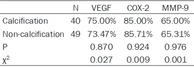
Table 2. Expression of VEGF, COX-2 and
MMP-9 in BC patients with and without calcification
N VEGF COX-2 MMP-9
Calcification 40 75.00% 85.00% 65.00%
Non-calcification 49 73.47% 85.71% 65.31%
P 0.870 0.924 0.976
χ2 0.027 0.009 0.001
cal variables. P<0.05 was
con-sidered significant. Results
Expression of VEGF, COX-2
and MMP-9 in BC and its rela -tionship with the Burr sign
The Burr sign is an important feature in an ultrasound diag-nosis of BC, and is used to dif-ferentiate benign and malig-nant breast tumors (Figures 1,
2). Expression of VEGF, COX-2 and MMP-9 was higher in BC patients with the Burr sign compared with BC patients without the Burr sign, and the
difference was significant (Ta-
ble 1).
Relationship between
expres-sion of VEGF, COX-2, MMP-9 and calcification
Calcification of breast tissue
is important in BC diagnosis.
Calcification of breast tissue
was made by pathology in 60-83 of patients, and by ultrasound in 40-60 of pa-
tients. Calcification within a
tumor can be used as an important index to judge be- nign and malignant tumors [6]. Expression of VEGF, COX-2, and MMP-9 showed no obvious correlation with
calci-fication (Figures 3, 4). There
was no significant difference
[image:3.612.90.287.604.672.2]Figure 5. Ultrasound of a BC patient with vascular abnormalities.
Figure 6. High expression of COX-2 in a BC patient with a vascular anomaly
(×400 magnification).
Table 3. Expression of VEGF, COX-2 and MMP-9 in BC patients with and without a vascular anomaly
Group N VEGF COX-2 MMP-9
Vascular anomaly 31 100% 100% 90.32%
Non-vascular anomaly 58 65.52% 55.17% 67.24%
P <0.001 <0/001 0.021
X2 13.788 14.349 5.301
Expression of VEGF, COX-2
and MMP-9 and its relation -ship with LNM
Accurate preoperative staging of BC is the key to treatment success. Checking for LNM from BC is an important com-ponent of preoperative stag-ing (Figures 7, 8). Expression of VEGF, COX-2, and MMP-9 in
the LNM group was signifi -cantly higher than that in the non-LNM group (Table 4).
Discussion
Breast tumors can secrete many vasoactive substances. VEGF can enhance angiogen-esis and vascular and lym-phatic permeability, and aids the proliferation and metasta-sis of tumor cells [5-7]. COX-2 is an inducible enzyme which, under normal physiologic con-ditions, is not detected in most tissues. However, if cells receive the corresponding sti- mulus after PG synthesis, COX-2 expression can inc- rease and promote the forma-tion of tumor vessels [8, 9]. MMP-9 can dissolve in type-IV collagen, leading to damage to basement membranes, and has a very important role in the metastasis of cancer cells [10]. Neovascularization of malignant tumors is affected by interactions between stim-ulatory and inhibitory factors. VEGF, COX-2, and MMP-9 have important roles in the migra-tion and proliferamigra-tion of endo-thelial cells, degradation of basement membranes, angio-genesis, induction of tumor metabolism and increase in the blood supply of tumors [11].
[image:4.612.90.374.653.720.2]and MMP-9 reflected hyperplasia, proliferation
[image:5.612.90.377.73.286.2]of BC tissue, and vascular abnormalities. Such provide a theoretical basis for the evaluation, treatment and prognosis of BC. Figure 7. Ultrasound of a BC patient with lymph node metastasis.
Figure 8. MMP-9 expression in a BC patient with lymph node metastasis
(×200 magnification).
Table 4. Expression of VEGF, COX-2 and MMP-9 in BC patients with and without lymph node metastasis
N VEGF COX-2 MMP-9
Lymph node metastasis 39 87.18% 82.05% 100%
Non-lymph node metastasis 50 64.00% 62.00% 76.00%
P 0.013 0.049 0.001
χ2 6.143 4.260 11.510
high expression may be corre-lated with a poor prognosis in BC patients. Due to increased expression of VEGF and COX-2 and accelerated microvessel formation in tumors, the risk of tumor proliferation is in- creased.
Calcification is another fea -ture of BC and is different in different parts of the breast. The central area of tumor
cal-cification may be due to tumor
metabolism and blood supply, which can lead to necrosis
and calcinosis. Calcification in
tissues adjacent to the tumor may be due to secretion of substances from cells. VEGF
expression in the calcification
group was higher than that in
the non-calcification group,
but the difference was not
sig-nificant. This finding may have been because of slight calcifi -cation in tumor tissue. Se- cretion of vasoactive
sub-stances without calcification
foci is weak, so the ability to induce the formation of new blood vessels will be relatively poor [12]. Whether there is a correlation between tumor
calcification and expression
of COX-2 and MMP-9 merits further study.
Calcification, the Burr sign,
[image:5.612.91.376.326.536.2] [image:5.612.88.376.612.680.2]Acknowledgements
The research was supported by the National Natural Science Foundation of China (No 81473687), Shandong Provincial Natural Sci- ence Foundation of China (No ZR2013HM038, ZR2014HL086), Shandong Provincial Medical &Health Science and Technology Development Plan (No 2011HWS084).
Disclosure of conflict of interest
None.
Address correspondence to: Dr. Xiangqi Li, Depart- ment of Breast Surgery and Clinical Laboratory, The
Affiliated Hospital of Taishan Medical University, Tai’an 271000, Shandong Province, China. Tel: +86-538-6230822; Fax: +86-538-8420042; E-mail:
drlixqi@hotmail.com; Dr. Keqiang Wang, Department
of Clinical Laboratory, The Affiliated Hospital of Taishan Medical University, Tai’an, Shandong Pro-vince, China. Tel: 538-6236422; Fax:
+86-538-8420042; E-mail: wkqsd@163.com
References
[1] Siegel R, Naishadham D, Jemal A. Cancer sta-tistics, 2012. CA Cancer J Clin 2012; 62: 10-29.
[2] Nadia H, Michael G. Breast cancer. Lancet 2016; 389: 1134-50.
[3] Torre LA, Bray F, Siegel RL, Ferlay J, Lortet-Tieu-lent J, Jemal A. Global cancer statistics 2012. CA Cancer J Clin 2015; 65: 87-108.
[4] Kurmyshkina OV, Belova LL, Kovchur PI, Volko-va TO. Remodeling of angiogenesis and lym-phangiogenesis in cervical cancer develop-ment. Biomed Khim 2015; 61: 579-597.
[5] Zhang L, Wang H, Li C, Zhao Y, Wu L, Du X, Han Z. VEGF-A/Neuropilin 1 pathway confers
can-cer stemness via activating Wnt/β- catenin
axis in breast cancer cells. Cell Physiol Bio-chem 2017; 44: 1251-1262.
[6] Chen Y, Liu Y, Wang Y, Li W, Wang X, Liu X, Chen
Y, Ouyang C, Wang J. Quantification of STAT3
and VEGF expression for molecular diagnosis of lymph node metastasis in breast cancer. Medicine (Baltimore) 2017; 96: e8488. [7] Soukup V, Ĉapoun O, Peŝl M, Sobotka R,
Vávřová L, Hanuš T, Zima T, Kalousová M. Pla -cental growth factor in bladder cancer com-pared to the diagnostic accuracy and prognos-tic performance of vascular endothelial growth factor A. Anticancer Res 2018; 38: 239-246. [8] Tian J, Hachim MY, Hachim IY, Dai M, Lo C,
Raffa FA, Ali S, Lebrun JJ. Cyclooxygenase-2 regulates TGFB-induced cancer stemness in triple-negative breast cancer. Sci Rep 2017; 7: 40258.
[9] Tury S, Becette V, Assayag F, Vacher S, Benoist C, Kamal M, Marangoni E, Bièche I, Lerebours F, Callens C. Combination of COX-2 expression and PIK3CA mutation as prognostic and pre-dictive markers for celecoxib treatment in breast cancer. Oncotarget 2016; 7: 85124-41. [10] Puzovic VJ, Brcic I, Ranogajec I, Jakic-Razu-movic J. Prognostic values of ETS-1 MMP-2 and MMP-9 expression and CO expression in breast cancer patients. Neoplasma 2014; 61: 439-446.
[11] Hall FM, Storella JM, Silverstone DZ, et al. Non-palpable breast lesion: recommend at sonog-raphy 2000; 167: 356-359.
[12] Fondrinier E, Lrimier G, Guerin-Boblet V. Breast