Original Article
miR-124 mediated neuron apoptosis in spinal cord
injury by targeting the 3’ UTR end of calpain 1
Xianshu Yi, Dianming Jiang
Department of Orthopedic Surgery, First Affiliated Hospital of Chongqing Medical University, Chongqing, China
Received January 7, 2016; Accepted March 20, 2016; Epub June 1, 2016; Published June 15, 2016
Abstract: Spinal cord injury (SCI) is a devastating and common neurologic disorder. Different sets of minas are usu-ally deregulated in different cancers. Here, we determined the function of miR-124 in SCI and study showed the expression level of miR-124 in spinal cord tissues of rats treated with miR-124 agomir before the construction of SCI models was significantly higher than rats treated with agomir-NC and saline. Overexpression of miR-124 induced by miR-124 agomir resulted in the activation extent of caspase 3 was inhibited, the decrease of calpain level and the number of apoptotic neurons. MiR-124 targeted the 3’ UTR end of calpain 1, which were found to be important for the regulation of cell growth and differentiation. Luciferase report assay showed the normalized luciferase activity of calpin 1-UTR in neurons transfected with miR-124 mimic was markedly decreased compared with the negative control group. Our study also showed the percentage of apoptotic neurons cells decreased after transfection of miR-124 mimic. Moreover, the protein levels of neurons cells isolated from spinal cord tissues after transfection with miR-124 mimic were decreased observably. Therefore, we concluded miR-124 could regulate neuron apoptosis in spinal cord injury by targeting the 3’ UTR end of calpain 1.
Keywords: Spinal cord injury, MiR-124, Calpain 1, 3’ UTR, Caspase
Introduction
Spinal cord injury (SCI) is a devastating and common neurologic disorder that has profound influences on modern society from physical, psychosocial, and socioeconomic perspectives [1]. It results in loss of oligodendrocytes demy-elination of surviving axons and severe func-tional impairment [2]. Many individuals with SCI do not regain their ability to walk, even though it is a primary goal of rehabilitation [3]. SCI is characterized by an immediate, irreversible loss of tissue at the lesion site, as well as a sec-ondary expansion of tissue damage over time [4]. It has been recognized as a complication of cervical transforaminal injections, and at pres-ent, there is no universally accepted treatment and the mechanism of injury is uncertain [5]. The recent discovery of microRNAs (miRNAs) suggests a novel regulatory control over gene expression during plant and animal develop-ment. MiRNAs play important roles during development and also in adult organisms by
site of 124. Therefore, we predicted miR-124 may express its role in regulating neuronal apoptosis by targeting calpain 1.
In this study, we constructed SCI mice models and determined the mRNA levels of miR-124, calpain 1, 68-kD NFP and cleaved caspase 3 to explore the influence of miR-124 on SCI. Materials and methods
Rat models of spinal cord injury
Adult male SD rats (230-270 g; Animal Center of Shandong University) with damage of T10 segment were used for all experiments. The rats were maintained in a climate-controlled vivarium with a 12 h light-dark cycle with free access to food. Before SCI models were con-structed by improved Allen for 1 day, 20 nmol miR-124 agomir, 20 nmol agomir-NC and saline were injected into the T10 segment of spinal cord tissues. Then, SCI models were construct-ed by Allen method: SD rats were injectconstruct-ed intra-peritoneally with 100 mg/mL chloral hydrate (300 mg/kg) in the left 1 cm of abdomen mid-line. Rats were prostrate lied, fixed on the oper-ating table and back wool was cut. Then, the back of rats was sterilized with iodophor and cut along spine spines. Vertebral plate and spine on T10 segment were removed. The T10 segment of spinal cord was struck by NYU Blow device (Curtiss-Wright, Grimethorpe) with 60 gcm (10 g × 6 cm) to result in the injury of spinal cord in T10 segment. Therefore, SD models were divided into 3 groups: miR-124 agomir group, agomir group and sham group.
The original generation of neurons HEK 293T were isolated from spinal cord of newborn rats. Newborn rats were soaked in 75% alcohol for 3~5 min. The head of rats were cut and rats were dissected along median line to expose spinal column. Rats were put into D-hanks and vascular tunic and meninge were stripped by separating spinal cord. Spinal cord tissues were cut into 0.5~1 mm3 and 2 mg/ml papain was added to culture in 37°C incubator for 20~30 min. Isopycnic fetal calf serum of 20% was used to end digestion. After blowing and beating with tubularis for some times, the solu-tion was centrifuged at 1000 rpm for 5 min. The supernatant was cultured in 37°C incuba-tor for 30 min and the cells at exponential phase were used for current experiments. All
the samples were stored at -80°C for further use.
RNA extraction and qRT-PCR assay
QRT-PCR was used to determine the relative level of miR-124 in spinal cord in sham, agomir-NC and miR-124 agomir group. The relative lev-els of miR-124 and calpain1 in spinal cord before SCI and after SCI 1, 3 and 7 days were also determined.
TRIzol solubilization and extraction is a relative-ly recentrelative-ly developed general method for de- proteinizing RNA [10]. In this study, the total RNA was extracted and isolated from BMSCs using the Trizol reagent (Invitrogen, Carlsbad, CA). RNA quality was assessed with the Ther- moScientific NanoDrop1000 (Thermo Fisher Scientific, Inc., Waltham, MA, USA). RT-PCR was performed using QuantiTect Primer Assay (Qiagen GmbH, Hilden, Germany) and Quanti- Tect SYBR Green RT-PCR Kit (Qiagen GmbH, Hilden, Germany) on a LightCycler 480 Instr- ument (Roche Diagnostics, Mannheim, Ger- many). The detection and quantification con-tained the following steps: first, reverse tran-scription was performed at 55°C for 30 min, initial activation for 15 min at 95°C, next 40 cycles of denaturation were conducted at 94°C for 15 s, then annealing for 30 s at 55°C, exten-sion for 30 s at 72°C. Fluorescence data was collected at the extension step. The relative expression of the target gene was determined using the 2-ΔCt method.
Western blot analysis
The protein levels of activated caspase 3, cal-pain 1 and its specific substrate 68-kD NFP in spinal cord tissues in sham, agomir-NC and miR-124 agomir group were determined by western blot assay. The protein levels of acti-vated caspase 3, calpain 1 and 68-kD NFP in neurons cells HEK 293T in DMSO, negative control and miR-124 mimic group were also determined.
trans-ferred to nitrocellulose filter membranes (Hybond, Escondido, CA, USA). The membrane was blocked with Tris-buffered saline Tween-20 (TBST) containing 5% skimmed milk powder for 1 h at room temperature, followed by incuba-tion in TBST containing 5% BSA (Sigma, St. Louis, MO, USA) and primary antibodies over-night at 4°C. Primary antibodies were detected using a peroxidase-coupled goat anti-rabbit secondary antibody (1:8000, ZSBio, Beijing, China) and EZ-ECL chemiluminescence Dete- ction kit for HRP (Biological Industries, Beit-Haemek, Israel). The following primary antibod-ies were used: rabbit mAb miR-381 (1:1000, Cell Signaling Technology, Danvers, MA, USA) and rabbit pAb β-actin (1:1000, Santa Cruz Biotechnology, Inc., Santa Cruz, CA, USA).
Luciferase reporter assay
A fragment of the 3’ UTR segment of calpain 1 containing the predicted miR-124 binding site was amplified by RT-PCR. The primers used were 5’-CATACTAGT GTCTTCTGGACAGGCTCTG-3’ (sense) and 5’-CTTAAGCTTAGAATCTCTCA CATA- CACAC-3’ (antisense). Restriction sites are bolded and underlined. Site-directed mutagen-esis of the miR-124 target site was carried out using Stratagene Quik-Change site-directed mutagenesis kit (Stratagene, Heidelberg, Germany). The construct was sequenced and named calpain 1-UTR-Mut. The pMIR-report luciferase vector was used for the construction of calpain 1-UTR or calpain 1-UTR-Mut plas-mids (Ambion, USA). HEK293T cells were cul-tured in 24-well plates. In each well, 10 ng of phRL-TK renilla luciferase vector (Promega, USA) was co-transfected to normalize transfec-tion efficiency. A concentratransfec-tion of 500 ng of cal-pain 1-UTR or calcal-pain 1-UTR-Mut plasmids together with 10 nM miR-1124 mimics or nega-tive control was also co-transfected. Trans- fection was done using Lipofectamine 2000 and Opti-MEM I-reduced serum medium (Life Technologies, California, USA). Briefly lucifer-ase activity was measured using the Dual lucif-erase assay kit (Promega). Normalized relative luciferase activity (RLA) was calculated as the following formula: RLA = [firefly luciferase]/ [renilla luciferase].
Cell transfection
The pre-miR-124 precursor molecule (miR-124 mimics), siRNA targeting calpain 1, activated
caspase 3 and negative control RNA-oligo-nucleotides were gained from Ambion corpora-tion (Ambion, Austin, USA). The day before transfection, HEK 293T cells were seeded in antibiotic-free medium. Transfection of miRNAs was carried out using Lipofectamine 2000 in accordance with the manufacturer’s procedure (Invitrogen, California, USA). The level of miR-124 mimics expression in the HEK 293T cells was assayed by real-time PCR.
Basso-Beaie-Bresnehan scale (BBB) score
A large dataset was constructed by pooling BBB data from 236 subjects scored at Texas A&M University and 407 subjects scored at Ohio State University. The subjects scored at Texas A&M were male Sprague Dawley rats (Harlan, Houston, TX) with moderate (12.5 mm, MASCIS impactor) spinal contusion injuries. All contusion surgeries were performed by the same individual. BBB scores were assigned by three raters that had high inter-rater reliabilities (all r 0.9). Subjects were scored daily for the first 14 days after surgery, and then every other day until day 26. After day 26, subjects were scored every 4th day until day 42. The BBB data collected at Ohio State were generated by scoring 407 male and female Long Evans hood-ed rats (Simonsen Laboratories, Gilroy, CA). All subjects were contused using a MASCIS impac-tor with weight-drop heights of 12.5 (n = 223), 25 (n = 130), or 50 mm (n = 54). Data was col-lected by raters that had high inter-rater reli-ability (r > 0.88).
Flow cytometry
per-formed after cultivation 10 min. Sample was obtained after discarding supernatant and 10 μL propidium iodide (PI) was added. Afterwards, the sample was stilled in dark for 30 min. Finally, the apoptosis was detected using flow cytometry (FCM) on the Moflo (Dako Cytomation, Glostrup, Denmark).
Statistical analysis
Statistical analysis was performed using SPSS 18.0. Data are expressed as the mean ± stan-dard error of the mean (SEM) of three indepen-dent experiments. Pearson’s product-moment correlation coefficient was used to assess the correlation between different groups. A paired Student’s t-test was used to evaluate differ-ences between two groups. Multiple group comparisons were analyzed using ANOVA with a post hoc test. P < 0.05 was considered to indicate a statistically significant result.
Results
MiR-124 promoted the function of spinal cord
MiR-124 was found to inhibit the activation of microglial cells. To determine whether miR-124 play an essential role in the cholinergic anti-inflammatory action, BALB/c mice were inject-ed intravenously with the miR-124 mimic
score in agomir-NC group was evidently higher than that in sham group (P ﹤ 0.01), while BBB score in miR-124 agomir group was significantly higher than that in agomir-NC group (P ﹤ 0.01). It indicated miR-124 agomir could effectively protect mice from septic shock.
MiR-124 inhibited apoptosis by targeting cal-pain
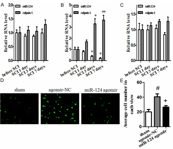
To directly assess the effect of miR-124 on the production of pro-inflammatory cytokines, miR-124 agomir was transfected into SD rats. The mRNA levels of miR-124 and calpain 1 in sham, agomir-NC and miR-124 agomir group before and after the SCI models were con-structed 1, 3 and 7 days. As shown in Figure 2A, the expression values of the miR-124 and calpain 1 in sham group before and after SCI 1, 3 and 7 days had no obvious changes. When the spinal cord of rats was injured by bitting, the level of miR-124 decreased with the time of SCI increased. The miR-124 levels in agomir-NC group after SCI 3 and 7 days were evidently downregulated compared with the value before SCI and after SCI 1 day (Figure 2B), while the level of calpain increased distinctly. However, if mice were treated with miR-124 agomir before SCI models were constructed, the mRNA levels of miR-124 and calpain 1 in sham, agomir-NC and miR-124 agomir group had no obvious dif-Figure 1. The level of miR-124 was decreased in spinal cord injury (SCI).
A: The relative level of miR-124 in sham, agomir-NC and miR-124- agomir group. B: BBB score in sham, agomir-NC and miR-124- agomir group after SCI construction for 28 days. **P < 0.01, compared with sham group, the
value in agomir-NC or miR-124- agomir group had significant difference; #P
< 0.05 or ##P < 0.01, compared with agomir-NC group, the value in miR-124-
agomir group had significant difference.
agomir. Therefore, we treated SD mice with 20 nmol miR-124 agomir or agomir-NC before bitting the spinal cord of rats 1 day. The mRNA level of miR-124 in T10 segment of spinal cord tissues in sham, agomir-NC and miR-124 ago- mir group was determined by qRT-PCR and results were shown in Figure 1A. As shown, the level of 124 in miR-124 agomir group was signi- ficantly higher than that in sham and agomir-NC group (p
[image:4.612.92.375.73.252.2]ference (Figure 2C). Therefore, we speculated the downregu-lation of miR-124 may upregu-late the level of calpain 1. Calpains are a family of calci-um-dependant cysteine-pro-teases involved in cytoskele-ton remodelling and muscle differentiation. Previous study reported calpain-1 activation contribute to cardiomyocyte apoptosis [13]. In this study, we used flow cytometry to observe the cell apoptosis situation in T10 segment of spinal cord tissues. As Figure 2D showed SCI promoted cell apoptosis in spinal cord tis-sues and the increase of miR-124 inhibited the cell apopto-Figure 2. A-C: Relative mRNA level of miR-124 and calpain 1 in sham,
agomir-NC and miR-124- agomir group before and after SCI for 1, 3 and 7 days, re-spectively. *P < 0.05 and **P < 0.01, compared with sham group, the relative
mRNA level of miR-124 or calpain 1 in agomir-NC group after SCI for 3 or 7 days had significant difference. D, E: The cell apoptosis situation in T10 seg-ment of spinal cord tissues in sham, agomir-NC and miR-124- agomir group after SCI for 7 days. #P < 0.05, compared with sham group, average
num-ber in each view in agomir-NC group had significant difference. +P < 0.05,
[image:5.612.90.378.69.316.2]compared with agomir-NC group, average number in each view in miR-124- agomir group had significant difference.
Figure 3. A: The protein levels of activated caspase 3, calpain 1 and its specific substrate 68 kD-NFP in T10 segment of spinal cord tissues in sham, agomir-NC and miR-124 agomir group after SCI for 7 days by western blotting. B: Density analysis results in sham, agomir-NC and miR-124- agomir group after SCI for 7 days by Western blotting. *P ﹤ 0.05 and **P ﹤ 0.01,
compared with sham group, rela-tive protein levels of activated caspase 3, calpain 1 and 68 kD-NFP in agomir-NC group had significant difference. #P ﹤ 0.05
and ##P ﹤ 0.01, compared with
agomir-NC group, relative protein levels of activated caspase 3, cal-pain 1 and 68 kD-NFP in miR-124 agomir group had significant dif-ference.
sis. To further verify the effect, we observed the number of apoptotic cells in each view. As shown in Figure 2E, the average cell number in each view in agomir-NC group was much higher than that in sham group, while the value in miR-124 agomir group was evidently decreased compared with agomir-NC group and the num-ber was almost the same with that in sham group.
MiR-124 inhibited the activation of caspase 3
[image:5.612.91.287.442.651.2]showed, the protein expression levels of cal-pain 1 and activated caspase 3 were increased with the downregulation of miR-124 in agomir-NC group, and the values were decreased with the increase of miR-124 in miR-124 agomir. The level of 68-KD NFP was decreased with the decrease of miR-124 in agomir-NC group and increase with the upregulation of miR-124 in miR-124 agomir group.
MiR-124 downregulated calpain 1 by targeting the 3’ UTR end
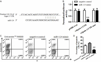
To study the molecular mechanism about the influence of miR-124 on SCI, we predicted the targets and result was shown in Figure 4A. Neuronal cell HEK 293T was isolated from the spinal cord of newborn rats and luciferase reporter assay was used to determine the lev-els of pMIR-Ctrl, calpain 1-UTR and calpain 1-UTR-mut in negative control and miR-124 mimic group. As Figure 4B showed the normal-ized luciferase activity of calpain 1-UTR was decreased significantly in miR-124 mimic group
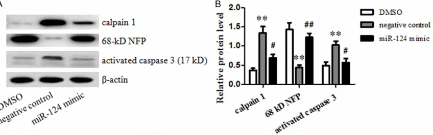
compared with that in the negative control group. The normalized luciferase activity of pMIR-Ctrl and calpain 1-UTR-mut in negative control group or miR-124 mimic group had no obvious difference. Then after the original gen-eration of neurons of spinal cord was transfect-ed with miR-124 mimic for 24 h, 1 mM L-sodium glutamate was used to treat neurons for 24 h to induce SCI and flow cytometry was used to analyze the proportion of apoptotic neurons. Neurons after treating with DMSO and negative control were taken as comparison. As Figure 4C and 4D showed, the apoptosis rate of neu-rons was significantly decreased in miR-124 mimic group compared with that in the negative control group and EntransterTM-R4000 group. Then, the protein expression levels of calpain 1, 68-kD NFP and activated caspase 3 group in DMSO, negative control and miR-124 mimic group were determined by Western blotting. As Figure 5A and 5B showed, the level of calpain 1 and activated caspase 3 is significantly reduced in miR-124 mimic group compared with nega-Figure 4. A: The prediction on the targeting of miR-124 by online software www.targetscan.org. B: The normalized luciferase activity of pMIR-Ctrl, calpain 1-UTR and calpain 1-UTR-mut in negative control and miR-124 mimic group.
**P ﹤ 0.01, compared with negative control, normalized luciferase activity of calpain 1-UTR in miR-124 mimic group
had significant difference. C: The cell growth situation of neuron cells HEK 293T, isolated from the spinal cord of newborn rats, in Entranster-R4000, negative control and miR-124 mimic group. D: The percentage of apoptotic cells HEK 293T in Entranster-R4000, negative control and miR-124 mimic group. ##P ﹤ 0.01, compared with negative
[image:6.612.93.523.74.338.2]tive control group. It indicated miR-124 may induced the degradation of calpain 1 mRNA and downregulate the protein expression of cal-pain 1 by targeting the 3’ UTR end of calcal-pain 1. Discussion
Spinal cord injury (SCI) is an insult to the spinal cord resulting in a change, either temporary or permanent, in its normal motor, sensory, or autonomic function. It is devastating and has profound influences on modern society from physical, psychosocial, and socioeconomic per-spectives [1]. It can result in loss of oligoden-drocytes demyelination of surviving axons and severe functional impairment [14]. SCI patients experience varying degrees of wrist and shoul-der pain. MicroRNAs (miRNAs) play important roles during development and also in adult organisms by regulating the expression of mul-tiple target genes. Yu et al reported miR-133b is essential for functional recovery after SCI in adult zebra fish [15]. MiR-124 is expressed spe-cifically in muscle and the central nervous sys-tem, and is considered to play an important role in SCI [9]. Several studies demonstrated that the overexpression of miR-124 in neural stem cells (NSCs) could lead the NSCs to dif-ferentiate into neurons and astrocytes, which may be important for functional recovery in SCI [16]. MiR-124 had a modest role in neuronal differentiation and cell cycle [10]. Therefore, in this study we aimed to study the role of miR-124 in SCI.
SD rats were selected for research and divided into 3 groups: 124 agomir: 20 nmol
[image:7.612.97.521.75.208.2]miR-124 agomir was injected into the injured spinal cord of rats before SCI models were construct-ed; Agomir-NC group: Rats were injected 20 nmol normal agomir at the injured spinal cord before SCI models were constructed; Sham group: the spinal cords of rats were not injured and the SCI models were not constructed suc-cessfully. The levels of miR-124, calpain 1, 68-kD NFP and caspase 3 were determined, motor function was evaluated by BBB score method and cell apoptosis situation in spinal cord tissues were determined. Results showed the level of miR-124 in injured spinal cord in agomir-NC group decreased with the increase of time, while the level of calpain 1 increased gradually. MiR-124 had evident inhibiting effect on cell apoptosis in spinal cord tissues. Moreover, when the overexpression of miR-124 was induced by miR-124 agomir, SCI could not result in the prominent decrease of miR-124 which might result in the efficient inhibition of the increase of calpain 1 level. The overexpres-sion of miR-124 resulted in the evident inhibi-tion on the level of activated caspase 3 and decreased the level of 68-kD NFP. Calpains are ubiquitous calcium-regulated cysteine proteas-es that have been implicated in cytoskeletal organization, cell proliferation, apoptosis, cell motility, and hemostasis [17]. Calpain 1 was one of them and was recently shown to bind tightly to the proximal end of the I-band titan segment in a calcium-dependent manner [18]. It was activated in neuronal apoptosis [19]. SCI evoked an increase in intracellular free Ca2+ level resulting in activation of calpain 1 [20]. The 68-kD protein forms the backbone of the Figure 5. A, B: The electrophoretogram and relative protein levels of activated caspase 3, calpain 1 and 68 kD-NFP in negative control and miR-124 mimic group. *P < 0.05 and **P < 0.01, compared with DMSO group, the relative
protein level of activated caspase 3, calpain 1 and 68 kD-NFP in negative control group had significant difference.
#P ﹤ 0.05 and##P ﹤ 0.01, compared with negative control group, the percentage of apoptotic cells in miR-124 mimic
neurofilaments and the 200- and 150-kD pro-teins appear to be more peripherally located [21]. The loss of 68-kD NFP is found to be high-est at the site of lesion, and decreases in neigh-boring regions in inverse relation to the dis-tance from the lesion epicenter [22]. 68-KD NFP was often used to indicate the activity of calpain. Indicated prevention of NFP (68 kD) by estrogen treatment of SCI for 48 h in lesion and penumbra could inhibit the increased calpain activity and activation, a common finding after SCI [23]. Caspase-3 is a newly characterized mammalian cysteine protease that promotes cell death during brain development [24]. It has been identified as a key protease in the execu-tion of apoptosis, whereas calpains have main-ly been implicated in excitotoxic neuronal injury [25]. Caspase-1 and caspase-3 were often acti-vated following SCI [26].
To explore the molecular mechanism, online software www.targetscan.org, showed 3’ UTR end of calpain 1 was the target site of miR-124. Therefore, we speculated miR-124 may express its regulating role in neuronal apoptosis by directly targeting calpain 1. Luciferase reporter assay was used to determine the expression of pMR-Ctd, calpain 1-UTR and calpain 1-UTR-mut in negative control and miR-124 mimic groups. The firefly luciferase reporter activity is signifi-cantly reduced in calpain 1-UTR vector com-pared with calpain 1-UTR-mut. Flow cytometry assay showed the number of apoptotic cells decreased significantly in neuronal cells HEK 293T infected with miR-124 mimic. It indicated miR-124 could inhibit the apoptosis of spinal cord cells. miR-124 could promote neuronal dif-ferentiation of neural progenitor cells [27]. The down-regulation of miR-124 was observed in injured hypoglossal motor neurons after nerve injury [28]. Moreover, western blotting showed miR-124 mimic could decreased the levels of calpain 1 and activated caspase 3, which fur-ther proved miR-124 could induce the degrada-tion of calpain 1 and downregulate the expres-sion of calpain 1 by targeting the 3’ UTR end of calpain.
Disclosure of conflict of interest
None.
Address correspondence to: Dr. Dianming Jiang, De- partment of Orthopedics, First Affiliated Hospital of Chongqing Medical University, Chongqing, China. E-mail: [email protected]
References
[1] Dumont RJ, Okonkwo DO, Verma S, Hurlbert RJ, Boulos PT, Ellegala DB, Dumont AS. Acute spinal cord injury, part I: pathophysiologic mechanisms. Clin Neuropharmacol 2001; 24: 254-264.
[2] Karimi-Abdolrezaee S, Eftekharpour E, Wang J, Morshead CM, Fehlings MG. Delayed trans-plantation of adult neural precursor cells pro-motes remyelination and functional neurologi-cal recovery after spinal cord injury. J Neurosci 2006; 26: 3377-3389.
[3] Behrman AL, Harkema SJ. Locomotor training after human spinal cord injury: a series of case studies. Phys Ther 2000; 80: 688-700. [4] Spinal cord injury facts and figures at a glance.
J Spinal Cord Med 2014; 37: 659-60.
[5] Baker R, Dreyfuss P, Mercer S, Bogduk N. Cervical transforaminal injection of corticoste-roids into a radicular artery: a possible mecha-nism for spinal cord injury. Pain 2003; 103: 211-215.
[6] Furuta M, Kozaki KI, Tanaka S, Arii S, Imoto I, Inazawa J. miR-124 and miR-203 are epige-netically silenced tumor-suppressive microR-NAs in hepatocellular carcinoma. Carcino- genesis 2009; 31: 766-76.
[7] Yu YM, Gibbs KM, Davila J, Campbell N, Sung S, Todorova TI, Otsuka S, Sabaawy HE, Hart RP, Schachner M. MicroRNA miR-133b is essential for functional recovery after spinal cord injury in adult zebrafish. Eur J Neurosci 2011; 33: 1587-1597.
[8] Izumi B, Nakasa T, Tanaka N, Nakanishi K, Kamei N, Yamamoto R, Nakamae T, Ohta R, Fujioka Y, Yamasaki K, Ochi M. MicroRNA-223 expression in neutrophils in the early phase of secondary damage after spinal cord injury. Neurosci Lett 2011; 492: 114-118.
[9] Nakanishi K, Nakasa T, Tanaka N, Ishikawa M, Yamada K, Yamasaki K, Kamei N, Izumi B, Adachi N, Miyaki S, Asahara H, Ochi M. Responses of microRNAs 124a and 223 fol-lowing spinal cord injury in mice. Spinal cord 2010; 48: 192-196.
[10] Cheng LC, Pastrana E, Tavazoie M, Doetsch F. miR-124 regulates adult neurogenesis in the subventricular zone stem cell niche. Nat Neurosci 2009; 12: 399-408.
[11] Yu JY, Chung KH, Deo M, Thompson RC, Turner DL. MicroRNA miR-124 regulates neurite out-growth during neuronal differentiation. Exp Cell Res 2008; 314: 2618-2633.
[13] Li Y, Ma J, Zhu H, Singh M, Hill D, Greer PA, Arnold JM, Abel ED, Peng T. Targeted inhibition of calpain reduces myocardial hypertrophy and fibrosis in mouse models of type 1 diabetes. Diabetes 2011; 60: 2985-2994.
[14] Siddall PJ, Loeser JD. Pain following spinal cord injury. Spinal cord 2001; 39: 63-73.
[15] Liu NK, Wang XF, Lu QB, Xu XM. Altered mi-croRNA expression following traumatic spinal cord injury. Exp Neurol 2009; 219: 424-429. [16] Xu W, Li P, Qin K, Wang X, Jiang X. miR-124
regulates neural stem cells in the treatment of spinal cord injury. Neurosci Lett 2012; 529: 12-17.
[17] Kuchay SM, Kim N, Grunz EA, Fay WP, Chishti AH. Double knockouts reveal that protein tyro-sine phosphatase 1B is a physiological target of calpain-1 in platelets. Mol Cell Biol 2007; 27: 6038-6052.
[18] Coulis G, Becila S, Herrera-Mendez CH, Sen- tandreu MA, Raynaud F, Richard I, Benyamin Y, Ouali A. Calpain 1 Binding Capacities of the N1-Line Region of Titin Are Significantly Enhanced by Physiological Concentrations of Calcium. Biochemistry 2008; 47: 9174-9183. [19] Huang Y, Wang KK. The calpain family and
hu-man disease. Trends Mol Med 2001; 7: 355-362.
[20] Ray SK, Hogan EL, Banik NL. Calpain in the pathophysiology of spinal cord injury: neuro-protection with calpain inhibitors. Brain Res Rev 2003; 42: 169-185.
[21] Rinderer TE, Hellmich RL 2nd, Danka RG, Collins AM. Male reproductive parasitism: a factor in the Africanization of European honey-bee populations. Science 1985; 228: 1119-1121.
[22] Ray SK, Wilford GG, Matzelle DC, Hogan EL, Banik NL. Calpeptin and methylprednisolone inhibit apoptosis in rat spinal cord injury. Ann N Y Acad Sci 1999; 890: 261-269.
[23] SHIN SJ, TREABA DO and DELELLIS RA. Imm- unohistology of endocrine tumors. Diagnostic Immunohistochemistry 2013; 322.
[24] Chen J, Nagayama T, Jin K, Stetler RA, Zhu RL, Graham SH, Simon RP. Induction of caspase-3-like protease may mediate delayed neuronal death in the hippocampus after transient cere-bral ischemia. J Neurosci 1998; 18: 4914-4928.
[25] Blomgren K, Zhu C, Wang X, Karlsson JO, Leverin AL, Bahr BA, Mallard C, Hagberg H. Synergistic activation of caspase-3 by m-cal-pain after neonatal hypoxia-ischemia: a mech-anism of “pathological apoptosis”? J Biol Chem 2001; 276: 10191-10198.
[26] Li M, Ona VO, Chen M, Kaul M, Tenneti L, Zhang X, Stieg PE, Lipton SA, Friedlander RM. Functional role and therapeutic implications of neuronal caspase-1 and-3 in a mouse model of traumatic spinal cord injury. Neuroscience 2000; 99: 333-342.
[27] Bhalala OG, Srikanth M, Kessler JA. The emerg-ing roles of microRNAs in CNS injuries. Nat Rev Neurol 2013; 9: 328-339.