ORIGINAL RESEARCH
Side Matters: Diffusion Tensor Imaging
Tractography in Left and Right Temporal Lobe
Epilepsy
M.E. Ahmadi D.J. Hagler, Jr C.R. McDonald E.S. Tecoma V.J. Iragui A.M. Dale E. Halgren
BACKGROUND AND PURPOSE: Noninvasive imaging plays a pivotal role in lateralization of the seizure focus in presurgical patients with temporal lobe epilepsy (TLE). Our goal was to evaluate the utility of diffusion tensor imaging (DTI) tractography in TLE.
MATERIALS AND METHODS: Twenty-one patients with TLE (11 right, 10 left TLE) and 21 controls were enrolled. A 1.5T MR imaging scanner was used to obtain 51 diffusion-gradient-direction images per subject. Eight pairs of white matter fiber tracts were traced, and fiber tract fractional anisotropy (FA) was calculated and compared with controls. Fiber tract FA asymmetry and discriminant function analysis were evaluated in all subjects and fiber tracts respectively.
RESULTS: Compared with controls, patients with TLE demonstrated decreased FA in 5 ipsilateral fiber tracts. Patients with left TLE had 6 ipsilateral and 4 contralateral fiber tracts with decreased FA. Patients with right TLE had 4 ipsilateral but no contralateral tracts with decreased FA compared with controls. Right-sided FA asymmetry was demonstrated in patients with right TLE for 5 fiber tracts, and left-sided asymmetry, for patients with left TLE for 1 fiber tract. Discriminant function analysis correctly categorized patients into left-versus-right TLE in 90% of all cases (100% correct in all patients without hippocampal sclerosis) by using uncinate fasciculus and parahippocampal fiber tracts.
CONCLUSIONS:We found widespread reductions in fiber tract FA in patients with TLE, which were most pronounced ipsilateral to the seizure focus. Patients with left TLE had greater, more diffuse changes, whereas patients with right TLE showed changes that were primarily ipsilateral. Disease was lateralized to a high degree independent of identifiable hippocampal pathology noted on conventional MR imaging.
E
pilepsy affects approximately 2.5 million people in the United States, making it the fourth most common neuro-logic condition in all ages.1Temporal lobe epilepsy (TLE) is the most common form and is the most frequent type of par-tial epilepsy refractory to medical therapy in adults.2,3For many of these patients, surgical resection of the epileptic focus offers a viable treatment option to eliminate seizures, and noninvasive imaging plays a pivotal role in correctly lateraliz-ing the epileptogenic zone.4-8Diffusion tensor imaging (DTI) is a relatively new nonin-vasive technique, which allows the detection and examination of the composition, integrity, and orientation of discrete white matter fiber bundles not optimally evaluated with conven-tional MR imaging.9-12It does so by quantifying the random motion of water molecules driven by Brownian motion and correlating the degree of diffusion to various neural compart-ments.13Numerous diffusion-based indices have been pro-posed, with fractional anisotropy (FA) as one of the most
widely used.14FA in white matter is high because diffusion of water parallel to fiber tracts is less restricted than diffusion perpendicular to the fiber tracts.10Brain fiber tractography by using FA and other diffusion data allows depiction of white matter tracts, and comparison between normal and diseased fiber tracts enables quantification of white matter changes due to damage.15-18However, changes in FA and other DTI indices not only reflect damage to white matter and loss of myelin but other entities such as encephalopathy, various psychiatric dis-orders, cytotoxic or vasogenic edema, postictal state, gliosis, and inflammation.14,16,19-21
Recently, DTI and tractography have been applied to the study of epilepsy and have demonstrated diffusion changes in gray and white matter tissue.12,16,18,22-31There is general in-creased mean diffusivity and dein-creased FA in subcortical structures such as the amygdala,23,31hippocampus,23,27-31and thalamus29ipsilateral to the seizure focus. Recent work evalu-ating focal white matter regions12,18,25and fiber tracts16,24,32 has generally shown reduced FA in multiple fiber tracts in-cluding the ipsilateral uncinate fasciculus (UF), fornix (FORX), and cingulum.
Because there is widespread propagation of synchronized neuronal firing in seizure disorders via neuronal networks, cortical and subcortical regions in the brain can be affected despite a single seizure focus.23,29,31Therefore, evaluation of white matter tracts connecting these various regions may pro-vide useful information as to the diffuse changes in the brain that accompany TLE. In this study, we used DTI tractography to investigate the total disease burden in patients with TLE in both temporal and extratemporal lobe fiber tracts. We hy-pothesized that patients with TLE would show diffuse white
Received January 1, 2009; accepted after revision April 2.
From the Multimodal Imaging Laboratory (M.E.A., D.J.H., C.R.M., A.M.D., E.H.) and Departments of Radiology (M.E.A., D.J.H., A.M.D., E.H.), Psychiatry (C.R.M.), and Neuro-sciences (E.S.T., V.J.I., A.M.D.), University of California, San Diego, Calif.
Supported by grant R01NS018741 from the National Institute of Neurological Disorders and Stroke (NINDS). We also greatly acknowledge support from GE Healthcare.
The content is solely the responsibility of the authors and does not necessarily represent the official views of the NINDS or the National Institutes of Health.
Please address correspondence to Mazyar E. Ahmadi, MD, Radiology Department, UCSD Medical Center, 200 West Arbor Dr, San Diego, CA 92103-8756; e-mail: mazyar.ahmadi@ gmail.com or [email protected]
Indicates open access to non-subscribers at www.ajnr.org
matter changes affecting multiple fiber tracts both ipsilateral and contralateral to the seizure focus and that ipsilateral tracts would be more affected in patients with TLE, providing useful information for lateralization of the seizure focus.
Materials and Methods
Human Subjects
The study was approved the institutional review board of our univer-sity and was performed in compliance with the Health Insurance Pri-vacy and Portability Act. All participants provided written consent before enrollment in the study. Twenty-one healthy subjects along with 21 age- and sex-matched patients with TLE (11 right, 10 left TLE) were enrolled in the study (Table). Handedness in all participants was assessed with the Edinburgh Handedness Inventory.33Two control subjects and 2 patients with left TLE were left-handed. All patients were recruited from the Epilepsy Center of our institution and clini-cally diagnosed by board-certified neurologists with expertise in epi-leptology. In all 21 patients, the diagnosis of left-versus-right TLE was based on the presence of unilateral ictal and interictal temporal lobe epileptiform activity as evidenced by video-electroencephalography (video-EEG) telemetry by using scalp and foramen ovale electrodes. Patients with bilateral seizure onset on video-EEG were excluded from our study. In 16 of the patients, seizure lateralization was sup-ported by the presence of unilateral mesial temporal sclerosis (MTS) as read by a board-certified neuroradiologist with expertise in epi-lepsy. In no case was there evidence of dual pathology on MR imaging.
Image Acquisition and Processing
MR imaging was performed with a 1.5T Signa HDx system (GE Healthcare, Waukesha, Wis) by using an 8-channel phased array head coil. A 1.5T scanner was used to decrease the possibility of suscepti-bility artifacts in the anteroinferior temporal and frontal lobes as seen in some subjects. Diffusion data were acquired by using single-shot echo-planar imaging with isotropic 2.5-mm voxels (matrix size⫽ 96⫻96, FOV⫽24 cm, 47 axial sections, section thickness⫽2.5 mm) covering the entire cerebrum and brain stem without gaps. Fifty-one diffusion-gradient directions by using a b-value of 1000 mm2/s (TE/ TR, 75.6/12,300 ms) with an additionalb⫽0 volume (approximately 11 minutes) were acquired. For use in nonlinear B0 distortion correc-tion, 2 additional volume series were acquired with 1b⫽0 volume and a single diffusion direction, with either forward or reverse phase-encode polarity (approximately 1 minute each). The imaging proto-col was identical for all participants, and all patients were seizure-free for a minimum of 24 hours before the MR imaging to avoid the possible effects of acute postictal changes on multiple diffusion pa-rameters.12,21,34There is, however, recent work that demonstrates
lack of significant change in anisotropy between the post- and inter-ictal states after 24 hours; therefore, that duration was selected as a minimum of seizure-free time required.21Patient and control scans were obtained in random order so that any drift with time in the scanner diffusion gradients would not systematically bias the group data.
Image files in DICOM format were transferred to a Linux work-station for processing with a customized automated processing stream written in Matlab (MathWorks, Natick, Mass) and C⫹⫹. Im-age distortion in the diffusion-weighted volumes caused by eddy cur-rents was minimized by nonlinear optimization with 4 free parame-ters for each diffusion-weighted volume (translation along the phase-encode direction and scaling along the phase-phase-encode direction as a linear function of x, y, or z). Image distortion caused by magnetic susceptibility artifacts was minimized with a nonlinear B0-unwarping method by using paired images with opposite phase-encode polari-ties.35,36This method corrects for nearly all image distortion caused by magnetic susceptibility artifacts. Images were resampled by using linear interpolation to have 1.875-mm isotropic voxels (47 axial sec-tions). Even though images were acquired with an in-plane resolution of 2.5 mm, they were automatically zero-padded ink-space from 96⫻ 96 to 128⫻128, and reconstructed with 1.875⫻1.875⫻1.875 mm3 voxels. Fiber tract FAs were calculated by using the algorithm in DTI-Studio (Johns Hopkins University, Baltimore, Md),37which essen-tially performs a weighted average of all voxels within the fiber tract of interest.
Semiautomated Fiber Tracking by Using DTIStudio One rater (M.E.A.), who performed tracing of entire fiber tracts, was blinded to all clinical data, including group membership of subjects in control or patient groups and the side of the seizure focus. The following fiber tracts were traced (Fig 1): cingulum fibers within cingulate gyrus (CG), parahippocampal fibers within parahippocampal gyrus (PH), superior longitudinal fasciculus (SLF), inferior longitudinal fasciculus (ILF), UF, FORX, anterior thalamic radiations (ATR), and inferior fronto-occipital fasciculus (IFOF). The algorithms for obtaining most of the fiber tracts are described by Wakana et al.38 This multiple region of interest method uses “OR,” “AND,” and “NOT” operations to show all fiber tracts within a region of interest, find shared tracts within 2 regions of interest, and remove unnecessary fibers, respectively. This method has shown high reproducibility and was used to ob-tain the CG, PH, SLF, ILF, UF, ATR, and IFOF. However, the SLF and CG were slightly modified as follows: All SLF fiber tracts within the external capsule were removed by multiple “NOT” op-erations. For CG, “OR” regions of interest were drawn in the coro-nal plane in the region of CG at the level just posterior to the genu of the corpus callosum (CC) and anterior to the splenium of CC, with “AND” argument placed at the midpoint of CC (Fig 2A).
The FORX was isolated by selecting the most posterior coronal section where the corticospinal tract (CST) can be seen contiguously from the motor cortex to the brain stem. An “OR” region of interest was drawn encompassing a focal high-intensity region in the FA map (corresponding to the crus of FORX) just lateral to the CST and me-dial and inferior to the temporal stem. At the same level, an “AND” region of interest was drawn at the level of the body of the FORX (Fig 2B). Non-FORX fibers extending anterosuperiorly to the frontal lobe, posteroinferiorly to the third ventricle, and anteriorly in the temporal lobe beyond amygdala were removed.
Demographic characteristics and epilepsy features of the TLE and control groups*
Characteristics
TLE (n⫽21)
Controls (n⫽21) Age (yr) 37.3 (10.0) 33.0 (10.2) Education 13.2** (2.2) 16.5 (2.3) Sex (females-males) 11:10 11:10 Age of seizure onset (yr) 14.3 (11.5) – Duration of illness (yr) 23.0 (14.6) – Seizure frequency (per month) 6.7 (7.4) –
Note:— – indicates not applicable; TLE, temporal lobe epilepsy. * SDs are in parentheses.
** Group mean is statistically different from that of controls atP⬍.05.
BRAIN
ORIGINAL
Statistical Analysis
Independentttests were used to test for group differences in age and education level. Due to the non-normal distribution of the seizure-related variables, nonparametric tests (Mann-WhitneyUtests) were used to evaluate group differences between patients with right-ver-sus-left TLE in illness duration, number of anticonvulsant medica-tions, and seizure frequency. Second, ipsilateral and contralateral fi-ber tract FA values in the patients with TLE were transformed intoz scores on the basis of the mean of the controls. Differences in
ipsilat-eral and contralatipsilat-eral tract FA values between the patients with TLE and controls were tested by using multivariate analysis of variances (MANOVAs). In addition, follow-up MANOVAs were performed between controls and patients with right and left TLE to determine whether there were differences in ipsilateral-versus-contralateral fiber tract FAs when patients with right and left TLE were considered sep-arately. Univariate analyses were only performed when the omnibus multivariate analysis was significant. Pairedttests were performed between the left and right fiber tracts within each group to investigate
[image:3.594.51.536.41.588.2]whether there were significant asymmetries in tract FA values. To control for type I error rates, we corrected all post hoc comparisons by using Tukey Honestly Significant Difference (HSD) tests with P⬍.01.
Individual subject analyses were performed in the patient popu-lation by using linear stepwise discriminant function analysis to de-termine whether a combination of tracts could correctly categorize patients as having left or right TLE. A “leave-one-out” procedure was used to cross-validate the model. In this procedure, each case in the analysis is classified by functions derived from all cases other than that case, making it optimal for applying the function to a new sample of cases and reducing the likelihood that any 1 case will significantly bias the model.
Results
There were no group differences between the patients and controls in age (TLE mean⫽37.3 years; control mean⫽33 years) or sex distribution (10 men, 11 women for both groups). The control population, however, had a slightly higher level of education (TLE mean⫽13.2 years; control mean⫽16 years,P⬍.05). Mann-WhitneyUtests revealed no group differences between patients with right and left TLE in illness duration, number of anticonvulsant medications, sei-zure frequency, or age of seisei-zure onset (Table).
Combined TLE Group versus Controls
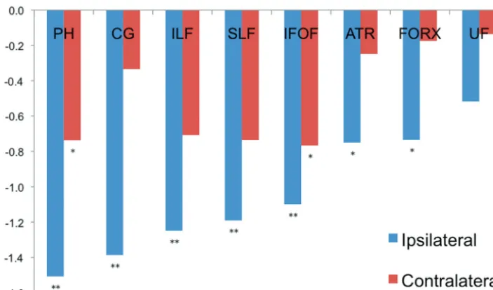
Results from the MANOVA with ipsilateral fiber tracts re-vealed a main effect for group [Wilks’F(8,29)⫽3.4,P⬍ .01], demonstrating that patients with TLE had lower overall FA values for the ipsilateral tracts relative to the control group (Fig 3). Univariate analyses corrected for multiple compari-sons revealed that patients with TLE showed decreased FA in the ipsilateral CG (P⬍.001), PH (P⬍.001), ILF (P⬍.005), IFOF (P⬍.01), and SLF (P⬍.005) relative to controls, with ATR and FORX showing a strong trend (P⬍.05).
The MANOVA with contralateral fiber tracts was not sig-nificant [F(8,29)⫽0.7,P⬎.01], suggesting no overall differ-ences in contralateral tract FA values between patients and controls. However, inspection of Fig 3 demonstrates a pattern toward reduced contralateral FA values in patients with TLE relative to controls across fiber tracts, with PH and IFOF reaching only a trend (P⬍.05).
TLE Subgroups versus Controls
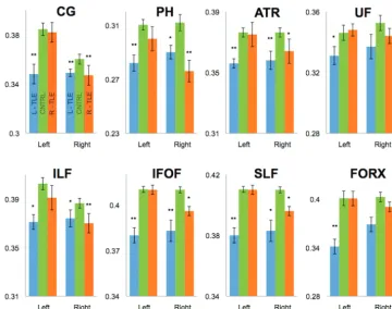
To test whether there were differences among controls and patients with right and left TLE, ipsilateral and contralateral MANOVAs were performed with patient subgroups (Fig 4). A MANOVA with ipsilateral fiber tracts was significant [F (16,58)⫽2.8,P⬍.01]. Univariate ANOVAs revealed group Fig 2.A, For CG, “OR” regions of interest were drawn in the coronal plane in the region of CG at the level just posterior to the genu of the CC and anterior to the splenium of CC, with “AND” argument placed at the midpoint of CC.B, Order of “OR” region-of-interest selection in a coronal section to obtain right FORX fiber.
[image:4.594.136.453.43.171.2] [image:4.594.117.469.209.416.2]differences in the FA of the ipsilateral CG (P⬍.001), PH (P⬍ .001), ILF (P⬍.01), IFOF (P⬍.01), SLF (P⬍.001), and FORX (P⬍.005). Post hoc tests adjusted for multiple com-parisons revealed that controls had higher FA values than pa-tients with right and left TLE in the ipsilateral CG and PH (all Pvalues ⬍.005). Conversely, controls differed from those with left TLE in the ipsilateral IFOF, SLF, FORX, and ATR) (all Pvalues⬍.005), with only a trend in the ILF and UF (allP values⬍.05). Controls differed from those with right TLE in the ipsilateral ILF (P⬍.01) but also with a trend in the ATR, SLF, and IFOF (Pvalues⬍.05). The MANOVA with con-tralateral tracts was also significant [F(16,58)⫽2.8,P⬍.05)]. Univariate tests revealed significant group differences in FA of the contralateral CG (P⬍.005), ATR (P⬍.005), and IFOF (P⬍.001). Post hoc tests revealed that controls had higher FA values than patients with left TLE in the contralateral CG, ATR, and IFOF (Pvalues⬍.01), with a trend in the PH, ILF, and SLF (Pvalues⬍.05). In no case did the controls differ from patients with right TLE in the contralateral tract FA val-ues. These data suggest that group differences in contralateral FA values were due to reductions in those with left TLE only (Fig 4).
Fiber Tract Asymmetry
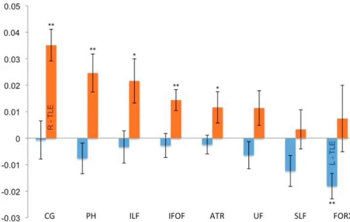
On the basis of evidence that fiber asymmetries may provide an important index of seizure lateralization, fiber FA asymme-tries were also analyzed.24Because individual tracts were of most interest, pairedttests were performed within each group (Fig 5). In controls, left-greater-than-right FA was observed in the CG (P⬍.0001) and ILF (P⬍.0001), with a trend in the UF (P⬍.05). Patients with right TLE demonstrated significant fiber tract asymmetries with right-less-than-left FA in the
IFOF (P⬍.005), PH (P⬍.007), and CG (P⬍.0001), with a trend in the ILF and ATR (Pvalues⬍.05). Patients with left TLE demonstrated left-less-than-right FA in the FORX only (P⬍.004).
Discriminant Function Analysis
With all 8 fiber tract pairs as predictors in the model, the best linear classifier included the right UF, left UF, right PH, and left PH (2⫽18.2,P⬍.003). This combination of tracts by using cross-validation of the results correctly classified 90.0% of the patients (100% of those with right TLE and 80%, with left TLE). Furthermore, all 5 patients without MTS were cor-rectly classified by using this method (3 right TLE, 2 left TLE).
Discussion
To date, several studies have shown bilateral temporal and extratemporal abnormalities of gray and white matter in pa-tients with TLE relative to controls.18,29,39-45In addition, re-cent DTI studies have demonstrated pathology within a small number of white matter tracts.16,18,25,46,47In the current study, we extend the literature by demonstrating the diffuse nature of white matter pathology in patients with TLE by evaluating FA values within 8 white matter tracts. We examined the different disease burdens in the 2 TLE subgroups and their differences compared with age-matched healthy controls. We also exam-ined fiber FA asymmetries in the controls and patients with TLE. Last, we evaluated whether the information obtained from DTI measurements could assist in lateralization of the seizure focus in individual patients.
though many of the contralateral tracts also showed a similar but nonsignificant trend. The widespread nature of fiber dam-age in TLE has been demonstrated previously in a few fiber tracts, including the UF, PH, and FORX.16,47Our results ex-tend these findings by revealing compromise to additional fi-bers, including the CG, IFOF, SLF, and ATR that was primarily accounted for by reductions in patients with left TLE.
Ipsilateral and contralateral fiber tracts were further ana-lyzed in patients with left or right TLE versus controls, reveal-ing several interestreveal-ing results. Six fiber tracts ipsilateral to the seizure focus in patients with left TLE showed decreased FA, with the remaining 2 showing a strong trend. Many ipsilateral fiber tracts in those with right TLE demonstrated decreased FA, with 3 reaching significance and another 3, only a trend. Three of 8 contralateral fiber tracts in the patients with left TLE were also affected (with an additional 3 demonstrating a strong trend), whereas none of the contralateral fiber tracts in the patients with right TLE showed significant compromise relative to controls. This finding suggests bilateral widespread white matter changes in patients with left TLE, and mostly unilateral white matter changes in patients with right TLE compared with the controls.
There is prior work demonstrating bihemispheric (though predominantly ipsilateral) gray and white matter changes by histology, volumetry, and DTI in patients with epilep-sy.16,18,25,40,48,49Voxel-based morphometry has demonstrated more widespread and extensive gray matter volume changes in patients with left TLE as opposed to right TLE regardless of the presence or absence of MTS.49,50Voxel-based DTI in patients with TLE with MTS has also demonstrated more extensive changes in patients with left TLE versus right.51Furthermore, it has been shown by DTI that white matter connectivity ap-pears more extensive if the focus is in the speech-dominant hemisphere.52Although there is no definitive reason why pa-tients with left TLE show more widespread cerebral changes than those with right TLE, our results are in agreement with
other prior work obtained by various imaging modalities and hint at the possibility that left and right TLE represent distinct patient groups. Perhaps neuronal connections in the left hemisphere are more likely to support seizure propagation to the contralateral hemisphere. It is also possible that left and right TLE are etiologically distinct and pathologically different syndromes from the outset. Regardless, the finding of diffuse changes in patients with left TLE and unilateral changes in those with right TLE is relatively new and unexplored, and further work is necessary to evaluate this trend.
We found left-sided fiber tract FA asymmetry in the CG38,53and extended the results in the literature by demon-strating left-sided asymmetry in the ILF. We only found a trend in right-sided asymmetry in the UF as elucidated by prior work.24,54,55However, there is literature that demon-strates conflicting results with left-greater-than-right fiber FA in the UF in controls,54,56which is likely related to how the fiber was traced and which subsegment of the UF was evaluated.46
[image:6.594.115.473.42.269.2]trend in controls showed a right-sided trend in patients with left TLE, including the IFOF, SLF, ATR, and FORX. However, only the FORX reached statistical significance in this patient group.
This asymmetric decrease of fiber FA in the ipsilateral hemisphere in patients with TLE can be a useful clue in the lateralization of seizure focus. In fact, there has been prior work in functional MR imaging showing loss of leftward func-tional asymmetry for language lateralization in patients with left TLE and increased asymmetry in patients with right TLE.52,57 Furthermore, bilateral pathology in patients with left-relative-to-right TLE has been demonstrated previously in regions of the temporal neocortex.49We provide preliminary evidence that the lateralization of individual patients can be increased by using tract FA values. Although presence of MTS or hippocampal volume loss may alone suffice in the lateral-ization of many patients,58our discriminant function analysis successfully lateralized all of the patients without MTS and all patients with right TLE and most of patients with left TLE with or without MTS. Therefore, it appears that in patients with normal-appearing hippocampi, microstructural change in fi-ber tracts that project from the medial temporal lobe (ie, the PH and UF) can assist in seizure lateralization.
We suspect the misclassification of 2 of 10 patients with left TLE was due to decreased fiber asymmetry due to bilateral white matter change as has been reported in previously men-tioned studies. These results provide additional evidence for anatomic asymmetries that may underlie functional reorgani-zation observed in patients with TLE. Furthermore, our data suggest that bilateral reductions in fiber integrity may charac-terize many patients with unilateral left TLE but do not neces-sarily imply a bilateral seizure focus, whereas patients with right TLE are more likely to show asymmetric fiber tract integrity.
We acknowledge multiple limitations to our current study. We used DTI methodology due to its increasing availability and use in mainstream clinical settings. However, other more sophisticated methods such as diffusion spectrum imaging and Q-ball imaging are now available, which can overcome current limitations of DTI, including the resolution of cross-ing fibers.59In addition, although we scanned only patients who had been seizure-free for a minimum of 24 hours, it is possible that postictal changes in some patients persisted for longer periods of time and that these changes are reflected in the data. These changes likely reflect cellular swelling in the area of seizure onset and possibly areas of seizure spread, though it appears that FA is fairly insensitive to this transient change.21Nevertheless, postictal diffusivity changes are com-plex and dynamic, and timing after the seizure may be critical. Moreover, additional research is needed to evaluate fully how long postictal changes persist and which DTI measurements they affect.
The power of our data can be further improved by increas-ing the number of controls and patients in our study. Because the fibers were manually drawn, there is the possibility of in-traoperator error. Furthermore, although every attempt was made, it is possible that the left and right regions of interest were not completely symmetric because freehand region of interest selection was used. We used the methodology put forth by Dr. Mori and Wakana,38which has shown excellent
inter- and intraoperator reproducibility. We chose to follow their methodology so as to stay as consistent as possible to prior work published in the literature and to minimize possi-ble rater error.
Conclusions
Intractable TLE is marked by widespread involvement of fiber tracts and asymmetries of white matter fibers, with lower fiber FA ipsilateral versus contralateral to the seizure focus. Some-what different patterns of tracts affected were observed in pa-tients with right TLE (solely ipsilateral) and left TLE (many bilateral), and combined evaluation of these patterns im-proved the ability to lateralize the seizure focus regardless of the presence or absence of MTS. This noninvasive lateraliza-tion of the seizure focus can perhaps be used in conjunclateraliza-tion with other established methods of diagnosis with the hope of decreasing the need for invasive presurgical diagnostic proce-dures and increasing the rate of postsurgical success in TLE.
References
1. Hirtz D, Thurman DJ, Gwinn-Hardy K, et al.How common are the “common” neurologic disorders?Neurology2007;68:326 –37
2. Hauser WA, Annegers JF, Kurland LT.Prevalence of epilepsy in Rochester, Minnesota: 1940 – 80.Epilepsia1991;32:429 – 45
3. Engel J Jr.Mesial temporal lobe epilepsy: what have we learned?Neuroscientist
2001;7:340 –52
4. McIntosh AM, Kalnins RM, Mitchell LA, et al.Temporal lobectomy: long-term seizure outcome, late recurrence and risks for seizure recurrence.Brain
2004;127:2018 –30
5. Juhasz C, Chugani HT.Imaging the epileptic brain with positron emission tomography.Neuroimaging Clin N Am2003;13:705–16, viii
6. Imbesi SG.Proton magnetic resonance spectroscopy of mesial temporal sclerosis: analysis of voxel shape and position to improve diagnostic accuracy.
J Comput Assist Tomogr2006;30:287–94
7. Lowe AJ, David E, Kilpatrick CJ, et al.Epilepsy surgery for pathologically proven hippocampal sclerosis provides long-term seizure control and im-proved quality of life.Epilepsia2004;45:237– 42
8. Antel SB, Li LM, Cendes F, et al.Predicting surgical outcome in temporal lobe epilepsy patients using MRI and MRSI.Neurology2002;58:1505–12 9. Le Bihan D, Breton E, Lallemand D, et al.MR imaging of intravoxel incoherent
motions: application to diffusion and perfusion in neurologic disorders. Ra-diology1986;161:401– 07
10. Pierpaoli C, Jezzard P, Basser PJ, et al.Diffusion tensor MR imaging of the human brain.Radiology1996;201:637– 48
11. Basser PJ, Mattiello J, LeBihan D.Estimation of the effective self-diffusion tensor from the NMR spin echo.J Magn Reson B1994;103:247–54
12. Rugg-Gunn FJ, Eriksson SH, Symms MR, et al.Diffusion tensor imaging of cryptogenic and acquired partial epilepsies.Brain2001;124:627–36 13. Beaulieu C.The basis of anisotropic water diffusion in the nervous system: a
technical review.NMR Biomed2002;15:435–55
14. Assaf Y, Pasternak O.Diffusion tensor imaging (DTI)-based white matter mapping in brain research: a review.J Mol Neurosci2008;34:51– 61 15. Arfanakis K, Gui M, Lazar M.Optimization of white matter tractography for
pre-surgical planning and image-guided surgery.Oncol Rep2006;15(Spec no):1061– 64
16. Concha L, Beaulieu C, Gross DW.Bilateral limbic diffusion abnormalities in unilateral temporal lobe epilepsy.Ann Neurol2005;57:188 –96
17. Mori S, van Zijl PC.Fiber tracking: principles and strategies—a technical review.NMR Biomed2002;15:468 – 80
18. Gross DW, Concha L, Beaulieu C.Extratemporal white matter abnormalities in mesial temporal lobe epilepsy demonstrated with diffusion tensor imaging.
Epilepsia2006;47:1360 – 63
19. Oster J, Doherty C, Grant PE, et al.Diffusion-weighted imaging abnormalities in the splenium after seizures.Epilepsia2003;44:852–54
20. Vermathen P, Laxer KD, Schuff N, et al.Evidence of neuronal injury outside the medial temporal lobe in temporal lobe epilepsy: N-acetylaspartate con-centration reductions detected with multisection proton MR spectroscopic imaging—initial experience.Radiology2003;226:195–202
21. Diehl B, Symms MR, Boulby PA, et al.Postictal diffusion tensor imaging.
Epilepsy Res2005;65:137– 46
temporal lobe epilepsy with hippocampal sclerosis. Neuroimage
2005;28:682–90
24. Rodrigo S, Oppenheim C, Chassoux F, et al.Uncinate fasciculus fiber tracking in mesial temporal lobe epilepsy: initial findings.Eur Radiol2007;17:1663– 68 25. Arfanakis K, Hermann BP, Rogers BP, et al.Diffusion tensor MRI in temporal
lobe epilepsy.Magn Reson Imaging2002;20:511–19
26. Assaf BA, Mohamed FB, Abou-Khaled KJ, et al.Diffusion tensor imaging of the hippocampal formation in temporal lobe epilepsy.AJNR Am J Neuroradiol
2003;24:1857– 62
27. Salmenpera TM, Simister RJ, Bartlett P, et al.High-resolution diffusion tensor imaging of the hippocampus in temporal lobe epilepsy. Epilepsy Res
2006;71:102– 06
28. Yu AH, Li KC, Yu CS, et al.Diffusion tensor imaging in medial temporal lobe epilepsy.Chin Med J (Engl) 2006;119:1237– 41
29. Kimiwada T, Juhasz C, Makki M, et al.Hippocampal and thalamic diffusion abnormalities in children with temporal lobe epilepsy. Epilepsia
2006;47:167–75
30. Londono A, Castillo M, Lee YZ, et al.Apparent diffusion coefficient measure-ments in the hippocampi in patients with temporal lobe seizures.AJNR Am J Neuroradiol2003;24:1582– 86
31. Hakyemez B, Erdogan C, Yildiz H, et al.Apparent diffusion coefficient mea-surements in the hippocampus and amygdala of patients with temporal lobe seizures and in healthy volunteers.Epilepsy Behav2005;6:250 –56
32. Concha L, Beaulieu C, Wheatley BM, et al.Bilateral white matter diffusion changes persist after epilepsy surgery.Epilepsia2007;48:931– 40
33. Oldfield RC.The assessment and analysis of handedness: the Edinburgh in-ventory.Neuropsychologia1971;9:97–113
34. Yogarajah M, Duncan JS.Diffusion-based magnetic resonance imaging and tractography in epilepsy.Epilepsia2008;49:189 –200
35. Morgan PS, Bowtell RW, McIntyre DJ, et al.Correction of spatial distortion in EPI due to inhomogeneous static magnetic fields using the reversed gradient method.J Magn Reson Imaging2004;19:499 –507
36. Reinsberg SA, Doran SJ, Charles-Edwards EM, et al.A complete distortion correction for MR images. II. Rectification of static-field inhomogeneities by similarity-based profile mapping.Phys Med Biol2005;50:2651– 61 37. Jiang H, van Zijl PC, Kim J, et al.DtiStudio: resource program for diffusion
tensor computation and fiber bundle tracking.Comput Methods Programs Biomed2006;81:106 –16. Epub 2006 Jan 18
38. Wakana S, Caprihan A, Panzenboeck MM, et al.Reproducibility of quantita-tive tractography methods applied to cerebral white matter.Neuroimage
2007;36:630 – 44
39. Margerison JH, Corsellis JA.Epilepsy and the temporal lobes: a clinical, elec-troencephalographic and neuropathological study of the brain in epilepsy, with particular reference to the temporal lobes.Brain1966;89:499 –530 40. Babb TL.Bilateral pathological damage in temporal lobe epilepsy.Can J
Neu-rol Sci1991;18:645– 48
41. Marsh L, Morrell MJ, Shear PK, et al.Cortical and hippocampal volume defi-cits in temporal lobe epilepsy.Epilepsia1997;38:576 – 87
42. Townsend TN, Bernasconi N, Pike GB, et al.Quantitative analysis of temporal lobe white matter T2 relaxation time in temporal lobe epilepsy.Neuroimage
2004;23:318 –24
43. Lin JJ, Salamon N, Lee AD, et al.Reduced neocortical thickness and complexity mapped in mesial temporal lobe epilepsy with hippocampal sclerosis.Cereb Cortex2007;17:2007–18. Epub 2006 Nov 6
44. Seidenberg M, Kelly KG, Parrish J, et al.Ipsilateral and contralateral MRI volu-metric abnormalities in chronic unilateral temporal lobe epilepsy and their clinical correlates.Epilepsia2005;46:420 –30
45. Araujo D, Santos AC, Velasco TR, et al.Volumetric evidence of bilateral dam-age in unilateral mesial temporal lobe epilepsy.Epilepsia2006;47:1354 –59 46. Rodrigo S, Naggara O, Oppenheim C, et al.Human subinsular asymmetry
studied by diffusion tensor imaging and fiber tracking.AJNR Am J Neurora-diol2007;28:1526 –31
47. Diehl B, Busch RM, Duncan JS, et al.Abnormalities in diffusion tensor imag-ing of the uncinate fasciculus relate to reduced memory in temporal lobe ep-ilepsy.Epilepsia2008;49:1409 –18. Epub 2008 Apr 4
48. Corsellis JA.The neuropathology of temporal lobe epilepsy.Mod Trends Neu-rol1970;5:254 –70
49. Keller SS, Mackay CE, Barrick TR, et al.Voxel-based morphometric compari-son of hippocampal and extrahippocampal abnormalities in patients with left and right hippocampal atrophy.Neuroimage2002;16:23–31
50. Riederer F, Lanzenberger R, Kaya M, et al.Network atrophy in temporal lobe epilepsy: a voxel-based morphometry study.Neurology2008;71:419 –25 51. Focke NK, Yogarajah M, Bonelli SB, et al.Voxel-based diffusion tensor
imag-ing in patients with mesial temporal lobe epilepsy and hippocampal sclerosis.
Neuroimage2008;40:728 –37
52. Powell HW, Parker GJ, Alexander DC, et al.Abnormalities of language net-works in temporal lobe epilepsy.Neuroimage2007;36:209 –21
53. Kubicki M, Westin CF, Nestor PG, et al.Cingulate fasciculus integrity disrup-tion in schizophrenia: a magnetic resonance diffusion tensor imaging study.
Biol Psychiatry2003;54:1171– 80
54. Buchel C, Raedler T, Sommer M, et al.White matter asymmetry in the human brain: a diffusion tensor MRI study.Cereb Cortex2004;14:945–51
55. Park HJ, Westin CF, Kubicki M, et al.White matter hemisphere asymmetries in healthy subjects and in schizophrenia: a diffusion tensor MRI study. Neu-roimage2004;23:213–23
56. Kubicki M, Westin CF, Maier SE, et al.Uncinate fasciculus findings in schizophrenia: a magnetic resonance diffusion tensor imaging study.Am J Psychiatry2002;159:813–20
57. Thivard L, Hombrouck J, du Montcel ST, et al.Productive and perceptive language reorganization in temporal lobe epilepsy. Neuroimage
2005;24:841–51
58. Liu Z, Mikati M, Holmes GL.Mesial temporal sclerosis: pathogenesis and significance.Pediatr Neurol1995;12:5–16