REVIEW ARTICLE
A Decade of DTI in Traumatic Brain Injury:
10 Years and 100 Articles Later
M.B. Hulkower, D.B. Poliak, S.B. Rosenbaum, M.E. Zimmerman, and M.L. Lipton
ABSTRACT
SUMMARY: The past decade has seen an increase in the number of articles reporting the use of DTI to detect brain abnormalities in patients with traumatic brain injury. DTI is well-suited to the interrogation of white matter microstructure, the most important location of pathology in TBI. Additionally, studies in animal models have demonstrated the correlation of DTI findings and TBI pathology. One hundred articles met the inclusion criteria for this quantitative literature review. Despite significant variability in sample characteristics, technical aspects of imaging, and analysis approaches, the consensus is that DTI effectively differentiates patients with TBI and controls, regardless of the severity and timeframe following injury. Furthermore, many have established a relationship between DTI measures and TBI outcomes. However, the heterogeneity of specific outcome measures used limits interpretation of the literature. Similarly, few longitu-dinal studies have been performed, limiting inferences regarding the long-term predictive utility of DTI. Larger longitulongitu-dinal studies, using standardized imaging, analysis approaches, and outcome measures will help realize the promise of DTI as a prognostic tool in the care of patients with TBI.
ABBREVIATIONS:FA⫽fractional anisotropy; GCS⫽Glasgow Coma Scale; MD⫽mean diffusivity; TAI⫽traumatic axonal injury; TBI⫽traumatic brain injury; TBSS⫽tract-based spatial statistics
T
he clinical pathology underlying TBI-related impairment is traumatic axonal injury.1TAI, referred to as diffuse axonalinjury when damage is extensive, is a microscopic injury that oc-curs even in the absence of frank tissue disruption. Therefore, patients may experience significant impairment despite the ab-sence of abnormal findings on conventional CT and MR imaging. Moreover, focal imaging abnormalities that can be detected by using CT and MR imaging are poor predictors of outcome.1
Di-agnostic tests that can discriminate significant TAI are needed to effectively allocate patients to follow-up and treatment, to accu-rately assess injury severity and safety in sports and military set-tings, and to guide clinical trials of novel therapeutic agents. DTI is a relatively new MR imaging technique that measures the direc-tional coherence of water diffusion in vivo. Because of the highly uniform collinear structure of normal white matter, DTI is uniquely able to probe its microscopic structure and is, therefore,
particularly well-suited for the assessment of TAI. Although gross abnormalities can be identified in some cases of TAI by using CT and conventional MR imaging, DTI can both qualitatively and quantitatively (Fig 1) demonstrate pathology not detected by other modalities and is, therefore, an important tool not only in the research setting but in the clinical setting as well.
Most studies of TBI report fractional anisotropy, a summary measure derived from DTI, which describes the directional coher-ence (anisotropy) of water diffusion within tissue. However, mean diffusivity, axial diffusivity, and radial diffusivity may more specifically describe the direction and magnitude of tissue water diffusion. Animal studies have shown a direct correspondence between even very subtle TAI pathology and decreases in white matter anisotropy that can be imaged in vivo by using DTI (eg, Mac Donald et al2). Numerous clinical studies have assessed TBI
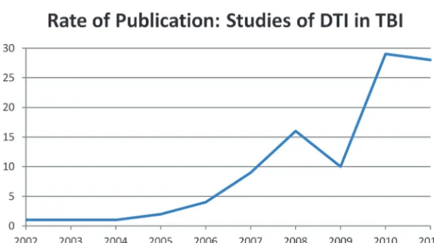
by using DTI. Since the earliest research article reporting DTI applied to TBI was published in 2002,3there has been an overall
exponential increase in the number of articles published on this topic (Fig 2).
The purpose of this review was to systematically summarize and detail the landscape of DTI applied to the study of TBI and to highlight both the salient conclusions to be drawn from this large literature and its limitations, which can serve as impor-tant considerations for future research. We summarize a num-ber of different aspects of the articles, including the demo-From the Gruss Magnetic Resonance Research Center (M.L.L.) and Saul R. Korey
Department of Neurology (M.E.Z.), Albert Einstein College of Medicine (M.B.H., D.B.P., S.B.R.), Bronx, New York.
Please address correspondence to Michael Lipton, MD, PhD, The Gruss Magnetic Resonance Research Center, Albert Einstein College of Medicine, 1300 Morris Park Ave, Bronx, NY 10461; e-mail: [email protected]
Indicates open access to non-subscribers at www.ajnr.org
Indicates article with supplemental on-line tables
graphics of TBI subjects and controls, the timing and severity of TBI, technical factors related to image acquisition and anal-ysis, the nature and location of abnormalities, and findings relating DTI to outcome measures. We also note that a valid meta-analysis of this literature is not feasible due to the great diversity in study design and measurement approaches used across the articles.
A structured search was performed by using PubMed to in-clude all relevant articles through 2011. The search used the fol-lowing key word combinations: “diffusion tensor imaging and traumatic brain injury,” “DTI and TBI,” and “DTI and concus-sion.” The total results included 391 articles with 293 unique ar-ticles. We further examined the references cited by these articles to identify additional relevant articles. After we eliminated articles on the basis of our exclusion criteria (below), 100 articles3-102
remained and were systematically analyzed and included in this review. Exclusion criteria included the following: language other than English (n⫽7); animal or in vitro studies (n⫽30); studies
of diseases other than TBI (eg, spinal cord injury, brain tumors) (n⫽57); case re-ports (n⫽37), reviews (n⫽48), editori-als (n⫽3), posters (n⫽1), or abstracts (n⫽ 1); and use of diffusion-weighted imaging or other MR imaging measures, but not DTI (n⫽8).
SUBJECTS WITH TBI
The population studied or substrate of in-jury is perhaps as important as the TBI itself in determining the nature and extent of consequent pathology.103 An
impor-tant consideration in the study of TBI is thus the choice of the study sample and the feasibility of attaining a homogeneous cohort. A total of 2337 subjects was stud-ied across all 100 articles. The average number of patients per study was 23 (range, 5– 83 subjects). Our review identified several articles that described patient samples with extremely similar or identical demographic characteristics (eg, McCauley et al55and Wilde et al,87,88,90) but reported either
different abnormal brain regions or different analyses of DTI in relation to outcome. Thus, some of the subjects may have been reported in multiple studies published by the same group of researchers. Our best estimate is that the number of subjects reported in multiple studies may be up to 140 individuals. All except 8 articles reported sex breakdown; 65% of reported sub-jects were male.6,30,41,46,52,77,81,83
Most commonly, abnormalities on DTI are defined on the basis of comparison with a control group because universal thresholds for abnormality have not yet been established. All studies, except 5, compared subjects with TBI with a control group.17,18,24,83,85Three of these exceptions used a longitudinal
within-subjects design.24,83,85In all except 7 studies, control
sub-jects were healthy individuals.28,43,49,53,55,81,90In 3 of the studies
FIG 1.FA image (A) reveals no abnormality in a patient with TBI. Tractography (B) can be used to delineate a region of interest for analysis. In this case, the forceps major (red) appears normal, but quantitative analysis of FA within this tract showed lower FA in the TBI group compared with controls. Whole-brain voxelwise analysis (C) reveals areas of low (blue) and high (red) FA. Low FA, consistent with TAI, is present within the forceps major at the splenium of the corpus callosum, as well as elsewhere.
[image:2.594.55.535.48.227.2] [image:2.594.54.370.293.470.2]that did not use healthy individuals as control subjects, 2 groups of subjects with TBI were compared (eg, with and without major depressive disorders53). In 3 pediatric studies, the control groups
were children who were hospitalized for orthopedic injury but had no evidence of head injury.43,55,90The average number of
control subjects per study was 18 (range, 6 – 47). Several articles, in addition to comparing patients with TBI with controls by using DTI, looked at subgroups of patients with TBI, including patients with TBI with major depressive disorders,53,99with raised
intra-cranial pressures,81with spinal cord injuries,86or veterans with
blast injuries.41,49,53,79,99An important consideration in the
selec-tion of control subjects, particularly when studying symptom en-dorsement, is that comparison of subjects with TBI with healthy controls fails to eliminate the potential confound of morbidity due to the experience of trauma itself, rather than adverse out-comes specifically due to physical injury to the brain. At the same time, it is unclear whether a purportedly non-head-injured pa-tient sustained neurologic trauma during the course of an injury, even if not reported by the patient or witnesses of the event or detected by clinical assessment of the patient.
The ages of subjects studied across all articles ranged from 2 through 70 years, with each individual article reporting on a more limited age range. Children were studied exclusively in 29 of 72 articles.4,5,11-14,17,19-22,25,27,42,43,55,66,75,81,87-94,96,98The
total number of children studied across all 29 articles was 564. In addition, 20 studies included adults and children (younger than 19 years).8-11,26,31,49,50,56,59,60,62,64,65,67,70,83,84,97,99However, varied
age thresholds were used to define the pediatric population. Most studies defined the pediatric population as individuals younger than 17 years of age, but other studies included children up to 18 years,4,5,9219 years,7520 years,12-14or 22.5 years of age.81White
matter changes associated with normal development might con-found detection of white matter injury; moreover, because develop-mental changes can occur at different rates even in children of the
same chronologic age, use of DTI in the pe-diatric population is a challenging undertaking.
Many studies reported the mechanism of injury of patients with TBI such as mo-tor vehicle collisions, falls, and assaults. However, because patients with different mechanisms were almost always consoli-dated into a single patient group, it is im-possible to draw conclusions about imag-ing findimag-ings as they relate to different mechanisms of injury.
Because age, sex, anthropometrics, and injury mechanism can greatly influ-ence outcomes, it is important that these issues be considered in study design and interpretation of results. Two major is-sues therefore emerge in consideration of the demographics of a TBI population. First, it is important to frame the compar-ison of results from multiple studies in the context of demographic differences be-tween the studied samples. Second, within a single study, a group analysis involving a demographically diverse sample might mask important findings unique to a particular demographic subset or lead to spurious group differences.
SEVERITY, CHRONICITY, AND STUDY DESIGN
Traumatic injury to the brain can result in a spectrum of injuries. There is a lack of consensus regarding whether they represent subsets of a single-entity or distinct pathologic processes. We found a wide variation in the injury severity studied, ranging from mild, in which there is a complete absence of abnormalities on conventional imaging, to severe, in which subjects remain in a vegetative state. While some studies were restricted to patients of a specific injury severity, many studies included patients of vary-ing severities. For studies reportvary-ing the GCS, we defined severity as mild (GCS, 13–15), moderate (GCS, 9 –12), or severe (GCS, 3– 8). For articles that did not report the GCS but characterized severity as mild, moderate, or severe, we accepted the authors’ report of severity. Four studies did not report injury sever-ity.26,33,53,60Several articles distinguished between
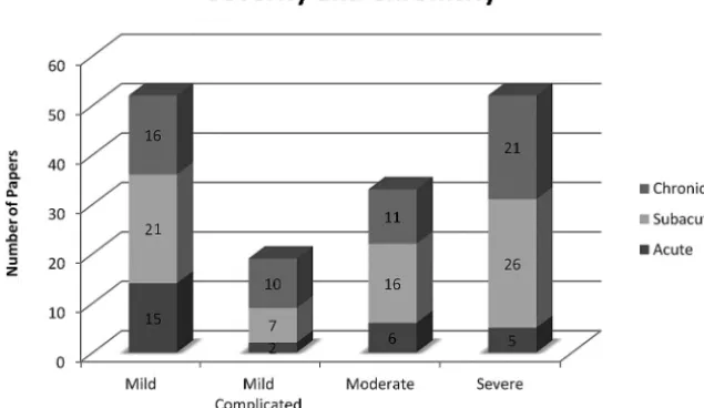
mild-compli-cated and mild-uncomplimild-compli-cated TBI. While both groups of pa-tients exhibited GCS in the mild range (⬎13), patients with mild-complicated TBI had findings of TBI on conventional imaging (CT and structural MR imaging), whereas patients with “true” mild or mild-uncomplicated TBI did not. Some articles specifi-cally reported a distinction between patients with mild-compli-cated and mild-uncomplimild-compli-cated TBI. Many articles, though not making the mild-complicated versus mild-uncomplicated dis-tinction, specifically noted that subjects with mild TBI lacked sig-nificant findings on conventional imaging. However, other arti-cles simply reported “mild” severity without mentioning the results of conventional brain imaging. There has been an increas-ing trend toward differentiatincreas-ing complicated versus mild-uncomplicated injury beginning in 2009. The earlier literature did not make the distinction, referring to the injury as “mild” without FIG 3. The number of articles that studied patients at each timeframe and level of injury
[image:3.594.53.371.59.243.2]qualification. This likely represents an increasing awareness of the distinction between mild-complicated and mild-uncomplicated, because studies have demonstrated that the clinical features of mild-complicated injuries often bear closer resemblance to those of moderate TBI than they do to mild-uncomplicated TBI.104
Finally, some articles did not differentiate moderate and severe TBI but, rather, grouped all such patients into 1 category.
Similar to severity, chronicity (Fig 3) is an important factor in the development of the study design. Primary injury and secondary injury play different roles in the evolution of pathol-ogy as a function of time postinjury. Similarly, microstructural pathology, as detected with DTI, may change with time. We found a wide variation among studies as to the timing of DTI relative to TBI, ranging from days to years. To systematically assess the timing of DTI after injury, which we termed “injury chronicity,” we divided subjects into 3 generally accepted cat-egories. Acute injury included patients imaged within 2 weeks of TBI, while chronic injury included patients imaged at least 1 year following injury. The subacute period included imaging per-formed between 2 weeks and 1 year after TBI. All articles reported the timing of DTI after TBI.
Figure 3 details the total number of articles reporting DTI for the study of TBI within a given timeframe and of a particular injury severity and includes articles that report mixed severities, mixed chronicities, or both. Most subjects were studied in the
subacute setting (Fig 3), most commonly having severe TBI followed by mild TBI. This trend might be attributable to the greater ease of subject enrollment during the subacute period, rather than to the clinical importance of this timeframe for the study. Patients are more easily en-rolled from rehabilitation centers, where they were actively seeking treatment for their injuries. In the acute setting, patients with mild TBI might not even seek medi-cal treatment and patients with severe TBI were too involved in urgent treatment to participate in a research study. Unless identified retrospectively, patients are easily lost to follow-up at chronic time points. When categorized according to severity, in-dependent of chronicity, the total number of patients with moderate TBI represented approximately half the total number of pa-tients with mild TBI and half the total num-ber of patients with severe TBI (Fig 3). Although the minority of patients stud-ied across all severities was examined in the acute setting, use of DTI as a prog-nostic tool is dependent on the identifi-cation of early biomarkers; therefore, the acute setting is an important area for future research, as are longitudinal studies examining patients at both the acute and chronic time points.
Most articles used cross-sectional designs. Eighteen articles reported multiple groups of sub-jects who were imaged at various times postin-jury,3,5,8,24,25,31,32,34,39,54,56,65,68,72,76,85,93,102whereas 79
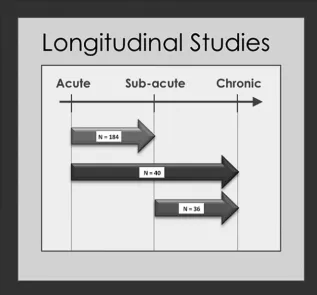
arti-cles reported a single homogeneous group of patients all within a 1-injury chronicity timeframe. Only 13 articles, reporting a total of 283 patients, used longitudinal designs and imaged the same group of patients at 2 different time points (Fig 4).3,24,29,39,47,49,54,56,67,83,85,93,102One additional article reported
imaging of the same group of patients acutely and again at least 1 year following injury but used a cross-sectional analysis at each of these time points and did not report change with time within subjects.85
A prospective study of patients with TBI is difficult; attrition during follow-up is a significant challenge. Of those studies that used a longitudinal design, only 8 reported attrition. The average attrition rate across these studies was 0.32 (range, 0.11– 0.60).3,24,29,49,54,56,83,85Despite the challenge of attrition,
longi-tudinal studies are integral to the understanding the natural his-tory of TBI and for early prognostication.
DATA ACQUISITION PARAMETERS
Important considerations in the use of DTI are the strength of the magnetic field, the number of diffusion-sensitizing gradient mag-netic field directions, and the choice of b-values. Use of greater magnetic field strengths has advantages and disadvantages; greater signal-to-noise ratio, improved spatial resolution, and FIG 4. Thirteen studies used a longitudinal design. Numbers represent patients from all studies
imaged at 2 time points. Nine studies assessed patients at both acute and subacute time points.3,29,39,47,54,56,67,83,85One study assessed patients at both acute and chronic time points.102
Two studies assessed patients at both subacute and chronic time points.24,93One study (n⫽47)
[image:4.594.54.372.49.344.2]faster scanning times commonly achieved at stronger magnetic field strengths come at the cost of increased magnetic field inho-mogeneity. In the setting of clinical DTI, most facilities will choose a b-value between 750 and 1000, whereas b-values of up to 3000 are used experimentally.105,106Increasing the b-value
in-creases the sensitivity to diffusion, but with a decrease in signal-to-noise ratio. Encoding the direction of diffusion requires a min-imum of 6 diffusion-sensitizing directions; greater resolution can be achieved by introducing additional directions, but this adds time to image acquisition. We summarize the acquisition param-eters used in DTI studies of TBI as follows.
Nearly equal numbers of articles reported performing DTI at 1.5T and 3T (483-5,8,15-18,24,26-28,30-34,38,39,42-44,47-49,52,55-58, 66-68,70,72,73,76,77,79,80,82,87,88,91,93,97,101,102 and 52,6,7,9-14,19-23,25, 29,35-37,40,41,45,46,50,51,53,54,59-65,69,71,74,75,78,81,83-86,89,90,92,94-96,98-100
respectively). The number of diffusion-sensitizing directions used ranged from 6 to 64, with an average of 27 but a mode of 12. In 2011 alone, the mode increased to 64. The b-value was reported in all except 6 articles.24,53,74,96,99,100All articles except 7 used a single
b-value in addition to zero.25,59-63,81The 7 articles using multiple
b-val-ues each used 5 unique b-valb-val-ues. The average b-value was 947, the range of b-values was 300 –1590, and the median and the mode were 1000.
Spatial resolution is an additional important consideration. Assessments of anisotropy, which are central to studies of TBI, measure the aggregate range of diffusivities across the tissues composing the voxel. Partial volume effects that may spuriously reduce anisotropy will be more likely when larger voxel volumes are examined. Voxel sizes varied among the articles we reviewed, with an average voxel size of 11.3 mL3(range, 1.83–31.25 mL3).
Average section thickness was 3.08 mm (range, 1.72– 6 mm).
DATA ANALYSIS METHODS
DTI can be used to study brain structure either on a regional or a whole-brain level. Regional analyses include both those in which an a priori region of interest is chosen for study and tractography, in which an a priori region of interest is used to define a white matter tract for study. In both approaches, average diffusion val-ues such as FA are extracted from voxels within the ROIs or tracts for subsequent analysis. Whole-brain analyses include voxelwise analyses, tract-based spatial statistics, a specialized type of voxel-wise analysis, and histogram analyses of all brain or all white mat-ter voxels.
In our review of the literature, we found that among those studies using regional analyses, 60 studies3-6,10,12-19,22-28,30,33-35, 37-41,43,46,48,49,51,52,54,55,57-66,76,81,82,86,92,93,95-100,102 used a
re-gion-of-interest approach and 30 studies9,11,17-19,21,31,41,42,46, 47,50,55,58,63,67,69,72,73,75,77,79,80,83,84,87,88,91,95,101 used
tractogra-phy. Fewer studies used whole-brain analyses, of which 17 stud-ies7,11,20,21,29,44,45,50,53,56,58,71,74,80,95,98,101were based on a
voxel-wise approach, 7 studies14,32,36,68,78,86,100 used TBSS, and 4
studies8,34,44,50used histogram analysis. Cross-validation of
ap-proaches can be achieved by using multiple analytic strategies. For instance, 1 article that used both a voxelwise analysis and TBSS found abnormalities in the same areas by using both techniques.70
In our review of the literature, we found that 18 articles reported using⬎1 type of analysis.6,14,17-19,27,34,41,46,50,55,58,63,80,86,95,98,100
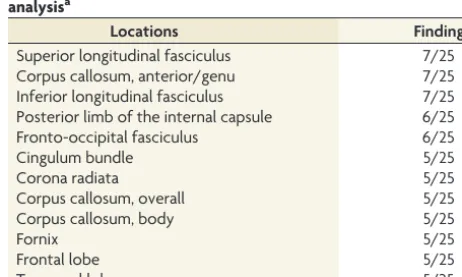
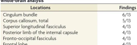
Tables 1–3 show the most commonly identified areas of abnormal FA in region-of-interest, tractography, and whole-brain analyses; the most commonly implicated regions are generally similar across approaches. However, they are not entirely consistent. For example, the centrum semiovale and the brain stem are locations where abnormal FA has commonly been identified by using re-gion-of-interest analysis, but these locations are not commonly identified by using either tractography or whole-brain analysis. Additionally, the superior longitudinal fasciculus is the most commonly identified location with abnormal FA by using whole-Table 1: Most common locations of abnormal FA by ROI analysisa
Locations Findings
Corpus callosum, anterior/genu 22b/30
Corpus callosum, posterior/splenium 21b/32
Posterior limb of the internal capsule 11/22
Corpus callosum, body 10/18
Frontal lobe 7/10
Corona radiata 6b/10
Cingulum bundle 7/8
Centrum semiovale 6/11
Brain stem 5/8
Cerebral peduncle 5/7
a
Values indicate the number of articles reporting abnormally low FA. Denominators represent the number of studies that assessed FA at these locations, including those that did not find abnormal FA.
b
[image:5.594.300.532.55.171.2]Includes articles reporting abnormally high FA. A fully referenced version of this Table is available in On-line Table 2.
Table 2: Most common locations of abnormal FA by tractographya
Locations Findings
Corpus callosum, total 10b/11
Corpus callosum, anterior/genu 8/8
Corpus callosum, posterior/splenium 7/8
Cingulum bundle 6/10
Fornix 5/7
Corpus callosum, body 4/6
Fronto-occipital fasciculus 4/5
Inferior longitudinal fasciculus 4/5
Uncinate fasciculus 4/5
Hippocampus 3/3
a
Values indicate the number of articles reporting abnormally low FA. Denominators represent the number of studies that assessed FA at these locations, including those that did not find abnormal FA.
bIncludes articles reporting abnormally high FA. A fully referenced version of this
[image:5.594.301.532.243.360.2]Table is available in On-line Table 3.
Table 3: Most common locations of abnormal FA by whole-brain analysisa
Locations Findings
Superior longitudinal fasciculus 7/25
Corpus callosum, anterior/genu 7/25
Inferior longitudinal fasciculus 7/25
Posterior limb of the internal capsule 6/25
Fronto-occipital fasciculus 6/25
Cingulum bundle 5/25
Corona radiata 5/25
Corpus callosum, overall 5/25
Corpus callosum, body 5/25
Fornix 5/25
Frontal lobe 5/25
Temporal lobe 5/25
a
[image:5.594.301.532.431.569.2]brain analysis, but this region does not appear as a common loca-tion by using either region-of-interest analysis or tractography. This discrepancy may be due to the fact that the superior longitu-dinal fasciculus was examined infrequently and does not neces-sarily indicate that it is an uncommon site of TAI. Tables 4 – 6 show the most commonly identified areas of abnormal MD in each of the region-of-interest, tractography, and whole-brain analyses. The corpus callosum is the most commonly identified region of abnormal FA and MD, perhaps because it is the largest white matter tract in the brain and an important site of TBI pa-thology. For both reasons, the corpus callosum is commonly cho-sen as a target of regional analyses, a choice that may bias the net findings of the literature.
The reliability of regional analyses depends on accurate and reproducible spatial localization of ROIs or tracts across subjects. Approaches to ensuring reliability may include specific reliability and reproducibility testing when expert observers perform place-ment of ROIs. Alternatively, subject images may be spatially nor-malized to a standard template, with placement of ROIs based on the template. This latter approach ensures that region-of-interest placement is consistent among subjects; however, it depends on the robustness of the registration of the subject brains to a
stan-dard space. Fourteen articles used spatial normalization to ensure standardization of region-of-interest/tract location.5,13,14,35,54, 61-63,72,73,77,86,92,100Sixty-three articles used expert manual
place-ment of regions of interest.3,4,6,9-11,15,16,19,22-28,30,31,33,34,37-43,46-51, 55,57,59,60,63-67,69,72,75,76,80-85,87-89,91,93-95,97-99,102However, only 35
of 63 studies reported that they performed reliability assess-ments, such as inter-/intraobserver reliability.6,9-11,19,25,40-43, 46,47,49,53,55,59,60, 64-67,75,76,81,83-85,87-89,91,93,94,98,99The studies
that did report reliability testing included 63% (18/30) of arti-cles using tractography9,11,19,41,42,46,47,55,67,75,83-85,87-89,91,94
but only 31% (19/61) of articles using region-of-interest analyses.6,10,19,25,40,41,43,46,55,59,60,64-66,76,81,93,98,99
In comparing articles using whole-brain approaches, 5/8 of the TBSS studies found significant abnormalities,14,32,36,68,78
whereas all of the articles using voxelwise approaches identified significant differences. The greater likelihood of identifying ab-normalities through voxelwise approaches as opposed to TBSS might be indicative of spurious group differences between pa-tients and controls due to misalignment between subjects and the standard template, an issue that may be minimized by the TBSS approach.107On the other hand, TBSS may be inherently less
sensitive because the analyses are restricted to a limited white matter skeleton.
SPECIFIC DIFFUSION MEASURES STUDIED
Diffusion tensor imaging yields multiple measures at each voxel. FA describes the directional coherence of water diffusion in tissue. MD is the scalar measure of the total direction-independent dif-fusion within a voxel. Axial diffusivity describes difdif-fusion along the principal axis of the diffusion ellipsoid, while radial diffusivity is an average of diffusion along its 2 minor axes. FA was the most commonly studied parameter across the studies we reviewed. Ab-normally low FA is widely held to represent alterations of white matter microstructure consistent with TAI.108Elevations of FA
have been much less frequently reported (see below). Although some authors have hypothesized that abnormally high FA repre-sents cytotoxic edema,54the mechanistic basis of abnormally high
FA remains uncertain.
FA was examined in all studies. All 100 articles, except for 46,54,89,96(which showed elevated FA), described findings of low
FA in subjects with TBI, regardless of the time of injury and across the spectrum of injury severity. MD is the next most commonly reported parameter (n ⫽ 51).3,9,10,13-17,19,21,22,25,28,29,33-40,42, 43,48-50,55,56,59-63,66,67,69,71,72,76,77,81,83,90,91,93,95,96,100-102 Articles
that examined parameters other than FA almost always identified areas of abnormal FA and then assessed other parameters within these regions. One article initially identified loci of abnormal MD and then assessed FA in the regions of abnormal MD.20Axial diffusivity
and radial diffusivity were much less commonly assessed (n ⫽ 18).3,8,11,13,14,20,22,36-38,49,54,56,63,66,76,81,89Only 2 articles reported
radial diffusivity and not axial diffusivity.29,66 One article
used a predictive model in which all 4 measures were incorporated.65
[image:6.594.53.285.64.129.2]While FA is a useful summary measure, more detailed infor-mation regarding diffusional uniformity, potentially obtainable through study of eigenvalue measures, may be important for di-agnosis and outcome prediction and especially for understanding Table 4: Most common locations of abnormal mean diffusivity by
ROI analysisa
Locations Findings
Corpus callosum, posterior/splenium 10b/20
Corpus callosum, anterior/genu 10/16
Frontal lobe 9/10
White matter 7/7
Thalamus 4/6
a
Values indicate the number of articles reporting abnormally low MD. Denominators represent the number of studies that assessed MD at these locations, including those that did not find abnormal MD.
b
[image:6.594.53.285.203.268.2]Includes articles reporting abnormally high MD. A fully referenced version of this Table is available in On-line Table 5.
Table 5: Most common locations of abnormal mean diffusivity by tractography analysisa
Locations Findings
Corpus callosum, anterior/genu 4/4
Fronto-occipital fasciculus 4/5
Inferior longitudinal fasciculus 4/5
Uncinate fasciculus 4/4
Cingulum bundle 3b/7
a
Values indicate the number of articles reporting abnormally low MD. Denominators represent the number of studies that assessed MD at these locations, including those that did not find abnormal MD.
bIncludes articles reporting abnormally high MD. A fully referenced version of this
[image:6.594.53.287.340.417.2]Table is available in On-line Table 6.
Table 6: Most common locations of abnormal mean diffusivity by whole-brain analysisa
Locations Findings
Cingulum bundle 6/13
Corpus callosum, total 5/13
Superior longitudinal fasciculus 4/13
Posterior limb of the internal capsule 4/13
Fronto-occipital fasciculus 4/13
Frontal lobe 4/13
a
pathologic mechanisms of TBI in humans. Eigenvalue measures implicate specific pathologic mechanisms in animal models of TBI and other types of brain injury.2Including eigenvalue measures
in a future study would facilitate a transitional bridge from animal to human studies and enable more informed targeting of novel inter-ventions in a patient group with few therapeutic options.
BRAIN REGIONS
Brain regions examined varied greatly among the articles. We systematically tabulated the location of abnormal DTI measures (FA and MD separately) across all articles (Tables 1– 6). To effec-tively summarize a large number of regions, we list only the top 10 most commonly identified regions of abnormal FA and the top 5 most commonly identified regions of abnormal MD via region-of-interest, tractography, and whole-brain analyses. When tabu-lating abnormal regions, it is important to recognize the relation-ship between study design and detection—that is, regional analyses can only detect a region as abnormal if the study design specifically examines that region. Whole-brain analyses, on the other hand, are positioned to detect all abnormal areas in the brain. Thus, the number of studies reporting an area as abnormal is influenced by the study design. Nonetheless, overall we found a large degree of consistency between studies regardless of the method of analysis. The corpus callosum, frontal lobe, internal capsule, and cingulum are among the most commonly identified regions of abnormality in DTI studies of TBI, perhaps because these structures are particularly vulnerable to injury due to their anatomic relationship to the skull and other structures such as the falx cerebri. These findings are almost entirely based on group comparisons and, therefore, do not necessarily reflect the distri-bution of injuries in the individual.
FUNCTIONAL OUTCOMES AFTER TBI
Seventy-two of 100 articles examined outcomes associated with TBI in addition to the imaging findings,4-9,11,13,14,16-23,25,27,30,33, 36-43,45-47,49-59,61,62,64-68,70,71,74-76,78,80-82,84,87-94,96,98,101,102 and
almost all of these 72 articles reported specifically on the asso-ciation of DTI findings with the outcomes. Most surprising however, 3 articles, though they reported outcomes, did not report analysis of the relationship between the outcome mea-sures and DTI.27,35,56Fifty-one of the 72 articles assessed
neu-ropsychological outcomes.5-7,9-14,16-19,22,23,25,27,30,36-39,41-43, 45,46,50-52,54,57-60,64,65,67,68,74,75,80,81,88,90-94,96,102 Eighteen of
72 used a global outcome measure (eg, Glasgow Outcomes
Scale, Coma Recovery Scale, Mini-Mental Status Examina-tion),4,16,22,33,42,50,57-59,63,66,70,76,80-82,84,8814 articles reported
correlation with the GCS,4,5,7,8,22,33,38,42,61,62,66,87,91,988
arti-cles correlated FA with intelligence quotient ,5,14,16,22,54,58,80,92
and 12 articles examined the relationship between DTI and postconcussive symptoms (eg, headaches, visual distur-bances, cognitive symptoms) and/or mood disor-ders.6,20,23,40,53,54,78,80,81,89,92,99 Several articles reported the
association of DTI with other patient assessments as surrogate outcome measures. These measures included electroencephalog-raphy,79 fMRI,10,63,69,75,90,100 MR spectroscopy,5,82 regional
brain volumes,85and motor-evoked potentials.97
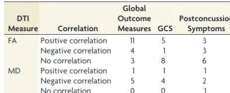
Of the articles that correlated DTI metrics with outcome, we found considerable heterogeneity among studies with respect to the specific outcome measures used. Choice of specific neuropsy-chological tests was most variable, with only a few articles report-ing the same measure (range, 1–19; mean, 1.8; median, 1.5). To summarize the data from these studies, we divided the various outcome measures into domains, including the following: atten-tion, executive funcatten-tion, memory, motor funcatten-tion, psychomotor/ processing speed, visuospatial function, global outcome, GCS, in-telligence quotient, and postconcussive symptoms. Tables 7 and 8 summarize the significant associations reported between DTI metrics and these outcome categories.
In addition to heterogeneity of the outcome measures used, additional variability (eg, brain region examined, analysis type, and DTI metric assessed) among studies examining the same out-Table 7: Relationship of DTI metrics to cognitive outcome measuresa
DTI
Measure Correlation Attention
Executive
Function Memory Motor
Psychomotor/
Processing Speed Visuospatial IQ
FA Positive correlation 11 9 14 4 5 4 2
Negative correlation 6 5 2 0 2 0 0
No correlation 2 6 6 1 1 0 6
MD Positive correlation 3 2 2 0 0 1 0
Negative correlation 4 6 7 0 1 3 0
No correlation 1 4 3 1 2 0 0
Note:—IQ indicates intelligence quotient.
a
[image:7.594.53.537.56.143.2]Total number of articles assessing relationships between DTI measures and cognitive outcomes. Cognitive-outcome measures have been categorized as 7 domains (top row). Articles are classified as reporting positive correlation, negative correlation, or no correlation. Positive correlation indicates a correlation coefficient greater than zero. Negative correlation indicates a correlation coefficient less than zero. No correlation includes articles that reported analyzing relationships between the DTI measures and cognitive outcomes within a domain but either reported finding no correlation (correlation coefficient equal to zero) or a correlation with aPvalue⬎.05. A fully referenced version of this Table is available in On-line Table 8.
Table 8: Relationship of DTI metrics to general clinical assessmentsa
DTI
Measure Correlation
Global Outcome Measures GCS
Postconcussion Symptoms
FA Positive correlation 11 5 3
Negative correlation 4 1 3
No correlation 3 8 6
MD Positive correlation 1 1 1
Negative correlation 5 4 2
No correlation 0 0 1
aTotal number of articles assessing relationships between DTI measures and global
[image:7.594.301.533.225.319.2]come domain further complicated summary of the literature. The average number of studies examining the same outcome domain was 15 (range, 6 –29; Tables 7 and 8). However, even among arti-cles examining the same domain, results are inconsistent.
Discordance in the results of outcome studies can, perhaps, be attributed to several issues. Although studies may examine the same domain, they typically vary in the choice of the specific mea-sure/instrument used. Because the sensitivity and specificity of any 2 measures, though designed to test the same cognitive domain, for instance, will differ,109results of studies relating imaging to these
differing outcome measures will potentially differ between the 2 studies as well. Moreover, the severity of TBI might determine the extent to which a patient experiences impairment in cognition or other adverse outcomes. As a result, comparison between studies including different injury severities might not be appropriate. Fi-nally, impairment secondary to TBI, particularly mild TBI, can be subtle and, therefore, escape detection by using some formal testing tools. Perhaps the most salient message to be derived from this seg-ment of the literature is that standardization of outcome measures and study design is essential to future meaningful study of TBI.110
ASSESSMENT OF INDIVIDUAL PATIENTS WITH TBI
The heterogeneity of injury mechanisms that cause TBI is likely best captured in studies assessing individual patients with TBI; group comparisons are inherently insensitive to interindividual variation, which is a hallmark of TBI. All of the articles included in this review report group analyses of patients with TBI. However,⬎35 additional articles report the use of DTI in individual TBI cases (eg, Gold and Lipton111). Several of the articles included in
this review reported assessment of individual patients in addition to their group analyses. Two articles examined whole-brain white matter histograms of individual patients,8,50but only 1 reported
results at the single-subject level, finding the distribution of FA in patients with TBI to be skewed toward lower FA in comparison with controls.8Three articles applied a tractography approach to
individual subjects,31,58,63while 2 articles applied the whole-brain
approach to individuals.44,50Assessment of individual patients
with TBI is important to the characterization of outliers, who might differ from the group in terms of extent and spatial distri-bution of injury, and is a prerequisite to clinical use of DTI in evaluating patients with TBI.
IMPLICATIONS, LIMITATIONS, AND POSSIBILITIES
DTI has been studied extensively as a tool for identification of brain abnormalities related to TBI and to understand the relation-ship of these brain abnormalities to other clinical features of the disorder. During the past decade, the number of such studies has risen exponentially and continues to increase with no sign of abatement. A unifying theme can be deduced from this large body of research: DTI is an extremely useful and robust tool for the detection of TBI-related brain abnormalities. The overwhelming consensus of these studies is that low white matter FA is charac-teristic of TBI. This finding is consistent across almost all the articles we reviewed, despite significant variability in patient demographics, modest differences in data acquisition parameters, and a multiplicity of data analysis techniques. This consistency across studies attests to the robustness of DTI as a measure of brain injury in TBI. The findingof significant differences in FA histograms that pool all white matter voxels across the whole brain is particularly compelling, indicating that a substantial portion of the hundreds of thousands of voxels in the image datasets are abnormal.
We also found an overwhelming consensus that imaging ab-normalities detected with DTI are associated with important clin-ical outcomes. This further validates DTI as a meaningful measure of clinically important brain injury. However, heterogeneity among the outcome measures that have been reported limits our ability to draw direct generalizable connections between DTI ab-normalities at specific brain locations and specific outcomes. The greatest degree of variability among the studies we reviewed was in the choice of outcome measures. As others have suggested, an impor-tant priority for future studies of TBI should be the use of standard-ized approaches, particularly standardization of outcome mea-sures.112Additionally, more high-quality longitudinal studies are
needed to extend the power of DTI, from identifying patients with TBI at cross-section to accurately predicting future clinical status.
By far, FA was the DTI measure used most commonly across the studies we reviewed. Too few articles reported analyses of eigenvalues to permit meaningful inferences regarding the role of eigenvalue findings in the assessment of TBI at this time. This is an important area of deficiency because preclinical studies indicate that differential effects on eigenvalue measures can separate pathologic mechanisms. Thus, more detailed study of the full pal-ette of metrics available from DTI is a great area for future study. The variety of data analysis approaches applied across the studies we reviewed presents a significant obstacle to summary of the data and limits the inferences that can be made on the basis of the literature as a whole. It is primarily on the basis of this factor that we determined that meta-analysis methods would not be ap-propriate for assessment of this literature. Studies reporting re-gional analyses might be considered comparable on the basis of similarity of brain regions tested across multiple studies. How-ever, the precise spatial location represented by a given region-of-interest, tract, or brain structure descriptors may vary signifi-cantly among studies on the basis of the methods, criteria, and raters used to define ROIs. Intra- and inter-rater variability can confound inferences based on these studies. While reliability test-ing can and should be performed to verify the reproducibility of region-of-interest placement, only a minority of studies did so.
Whole-brain analyses perhaps offer the greatest promise for pooling of results across studies, provided that the studies nor-malize their image data to the same brain template. Variability in the brain atlases used for spatial normalization and inconsistent reporting of coordinates for abnormalities in the studies we re-viewed limit such cross-study comparison at this time.
such as head and body composition make it highly probable that despite some commonalities, many areas of injury will differ among patients. Further application of individualized assess-ments of regional brain injury is thus needed to realize the full potential of DTI as a research and clinical tool.
On the basis of our analysis of the current literature, we suggest that important focus areas for future study should include larger longitudinal studies, incorporation of multiple outcome mea-sures in statistical models that account for the complexity inher-ent in TBI populations, and assessminher-ent of interindividual differ-ences. Standardization across centers, specifically with regard to data acquisition parameters, data analysis techniques, and the specific outcome measures assessed, promises to greatly increase the yield of such studies.
In summary, DTI provides a robust measure of clinically im-portant TAI at cross-section, despite the variability inherent in characteristics of patients with TBI and injury mechanisms as well as study differences in data acquisition and analysis methods. Larger longitudinal studies will be essential for the evaluation of DTI as a prognostic biomarker in TBI. More detailed assessment of DTI metrics and translational imaging studies should be un-dertaken to link pathophysiologic mechanisms in animal models to important clinical outcomes in patients. Together, these ap-proaches promise to realize the full potential of DTI to improve diagnosis and treatment of patients with TBI.
ACKNOWLEDGMENTS
We thank Tova M. Gardin for graphic arts assistance.
Disclosures: Molly E. Zimmerman—UNRELATED:Grants/Grants Pending: National Institutes of Health (NIH)/National Institute on Aging,* Alzheimer’s Association,* Bristol-Myers Squibb.* Michael L. Lipton—UNRELATED:Grants/Grants Pending: NIH,* Dana Foundation,* Resurrecting Lives Foundation,*Patents (planned, pending, or issued): Einstein patent pending,*Comments: for data analysis techniques. *Money paid to the institution.
REFERENCES
1. Niogi SN, Mukherjee P.Diffusion tensor imaging of mild trau-matic brain injury.J Head Trauma Rehabil2010;25:241–55 2. Mac Donald CL, Dikranian K, Bayly P, et al.Diffusion tensor
im-aging reliably detects experimental traumatic axonal injury and indicates approximate time of injury.J Neurosci2007;27:11869 –76 3. Arfanakis K, Haughton VM, Carew JD, et al.Diffusion tensor MR imaging in diffuse axonal injury.AJNR Am J Neuroradiol2002;23: 794 – 802
4. Akpinar E, Koroglu M, Ptak T.Diffusion tensor MR imaging in pediatric head trauma.J Comput Assist Tomogr2007;31:657– 61 5. Babikian T, Marion SD, Copeland S, et al.Metabolic levels in the
corpus callosum and their structural and behavioral correlates af-ter moderate to severe pediatric TBI.J Neurotrauma2010;27: 473– 81
6. Bazarian JJ, Zhong J, Blyth B, et al.Diffusion tensor imaging detects clinically important axonal damage after mild traumatic brain injury: a pilot study.J Neurotrauma2007;24:1447–59
7. Bendlin BB, Ries ML, Lazar M, et al.Longitudinal changes in pa-tients with traumatic brain injury assessed with diffusion-tensor and volumetric imaging.Neuroimage2008;42:503–14
8. Benson RR, Meda SA, Vasudevan S, et al.Global white matter anal-ysis of diffusion tensor images is predictive of injury severity in traumatic brain injury.J Neurotrauma2007;24:446 –59
9. Bigler ED, McCauley SR, Wu TC, et al.The temporal stem in trau-matic brain injury: preliminary findings.Brain Imaging Behav
2010;4:270 – 82
10. Bonnelle V, Leech R, Kinnunen KM, et al.Default mode network connectivity predicts sustained attention deficits after traumatic brain injury.J Neurosci2011;31:13442–51
11. Caeyenberghs K, Leemans A, Coxon J, et al.Bimanual coordination and corpus callosum microstructure in young adults with trau-matic brain injury: a diffusion tensor imaging study. J Neu-rotrauma2011;28:897–913
12. Caeyenberghs K, Leemans A, Geurts M, et al.Correlations between white matter integrity and motor function in traumatic brain in-jury patients.Neurorehabil Neural Repair2011;25:492–502 13. Caeyenberghs K, Leemans A, Geurts M, et al.Brain-behavior
rela-tionships in young traumatic brain injury patients: DTI metrics are highly correlated with postural control.Hum Brain Mapp2010; 31:992–1002
14. Caeyenberghs K, Leemans A, Geurts M, et al.Brain-behavior rela-tionships in young traumatic brain injury patients: fractional an-isotropy measures are highly correlated with dynamic visuomotor tracking performance.Neuropsychologia2010;48:1472– 82 15. Chang MC, Jang SH.Corpus callosum injury in patients with
dif-fuse axonal injury: a diffusion tensor imaging study. NeuroReha-bilitation2010;26:339 – 45
16. Chang MC, Kim SH, Kim OL, et al.The relation between fornix injury and memory impairment in patients with diffuse axonal injury: a diffusion tensor imaging study.NeuroRehabilitation2010; 26:347–53
17. Cho HK, Hong JH, Kim SH, et al.Clinical usefulness of diffusion tensor imaging in patients with transtentorial herniation follow-ing traumatic brain injury.Brain Inj2011;25:1005– 09
18. Choi GS, Kim OL, Kim SH, et al.Classification of cause of motor weakness in traumatic brain injury using diffusion tensor imag-ing.Arch Neurol2012;69:363– 67
19. Christidi F, Bigler ED, McCauley SR, et al.Diffusion tensor imaging of the perforant pathway zone and its relation to memory function in patients with severe traumatic brain injury.J Neurotrauma
2011;28:711–25
20. Chu Z, Wilde EA, Hunter JV, et al.Voxel-based analysis of diffu-sion tensor imaging in mild traumatic brain injury in adolescents.
AJNR Am J Neuroradiol2010;31:340 – 46
21. Cubon VA, Putukian M, Boyer C, et al.A diffusion tensor imaging study on the white matter skeleton in individuals with sports-re-lated concussion.J Neurotrauma2011;28:189 –201
22. Ewing-Cobbs L, Prasad MR, Swank P, et al.Arrested development and disrupted callosal microstructure following pediatric trau-matic brain injury: relation to neurobehavioral outcomes. Neuro-image2008;42:1305–15
23. Geary EK, Kraus MF, Pliskin NH, et al.Verbal learning differences in chronic mild traumatic brain injury.J Int Neuropsychol Soc2010; 16:506 –16
24. Greenberg G, Mikulis DJ, Ng K, et al.Use of diffusion tensor imag-ing to examine subacute white matter injury progression in mod-erate to severe traumatic brain injury.Arch Phys Med Rehabil2008; 89:S45–50
25. Grossman EJ, Ge Y, Jensen JH, et al.Thalamus and cognitive im-pairment in mild traumatic brain injury: a diffusional kurtosis imaging study.J Neurotrauma2012;29:2318 –27
26. Gupta RK, Saksena S, Agarwal A, et al.Diffusion tensor imaging in late posttraumatic epilepsy.Epilepsia2005;46:1465–71
27. Hanten G, Wilde EA, Menefee DS, et al.Correlates of social prob-lem solving during the first year after traumatic brain injury in children.Neuropsychology2008;22:357–70
28. Hartikainen KM, Waljas M, Isoviita T, et al.Persistent symptoms in mild to moderate traumatic brain injury associated with executive dysfunction.J Clin Exp Neuropsychol2010;32:767–74
29. Henry LC, Tremblay J, Tremblay S, et al.Acute and chronic changes in diffusivity measures after sports concussion.J Neurotrauma
2011;28:2049 –59
tissue texture analysis correlated to neuropsychological and DTI findings.Acad Radiol2010;17:1096 –102
31. Hong JH, Kim OL, Kim SH, et al.Cerebellar peduncle injury in patients with ataxia following diffuse axonal injury.Brain Res Bull
2009;80:30 –35
32. Huang MX, Theilmann RJ, Robb A, et al.Integrated imaging ap-proach with MEG and DTI to detect mild traumatic brain injury in military and civilian patients.J Neurotrauma2009;26:1213–26 33. Huisman TA, Schwamm LH, Schaefer PW, et al.Diffusion tensor
imaging as potential biomarker of white matter injury in diffuse axonal injury.AJNR Am J Neuroradiol2004;25:370 –76
34. Inglese M, Makani S, Johnson G, et al.Diffuse axonal injury in mild traumatic brain injury: a diffusion tensor imaging study.J Neuro-surg2005;103:298 –303
35. Kennedy MR, Wozniak JR, Muetzel RL, et al.White matter and neurocognitive changes in adults with chronic traumatic brain in-jury.J Int Neuropsychol Soc2009;15:130 –36
36. Kinnunen KM, Greenwood R, Powell JH, et al.White matter dam-age and cognitive impairment after traumatic brain injury.Brain
2011;134:449 – 63
37. Kraus MF, Susmaras T, Caughlin BP, et al.White matter integrity and cognition in chronic traumatic brain injury: a diffusion ten-sor imaging study.Brain2007;130:2508 –19
38. Kumar R, Gupta RK, Husain M, et al.Comparative evaluation of corpus callosum DTI metrics in acute mild and moderate trau-matic brain injury: its correlation with neuropsychometric tests.
Brain Inj2009;23:675– 85
39. Kumar R, Husain M, Gupta RK, et al.Serial changes in the white matter diffusion tensor imaging metrics in moderate traumatic brain injury and correlation with neuro-cognitive function.J Neu-rotrauma2009;26:481–95
40. Lange RT, Iverson GL, Brubacher JR, et al.Diffusion tensor imag-ing findimag-ings are not strongly associated with postconcussional disorder 2 months following mild traumatic brain injury.J Head Trauma Rehabil2012;27:188 –98
41. Levin HS, Wilde E, Troyanskaya M, et al.Diffusion tensor imaging of mild to moderate blast-related traumatic brain injury and its sequelae.J Neurotrauma2010;27:683–94
42. Levin HS, Wilde EA, Chu Z, et al.Diffusion tensor imaging in re-lation to cognitive and functional outcome of traumatic brain in-jury in children.J Head Trauma Rehabil2008;23:197–208 43. Levin HS, Wilde EA, Hanten G, et al.Mental state attributions and
diffusion tensor imaging after traumatic brain injury in children.
Dev Neuropsychol2011;36:273– 87
44. Lipton ML, Gellella E, Lo C, et al.Multifocal white matter ultra-structural abnormalities in mild traumatic brain injury with cog-nitive disability: a voxel-wise analysis of diffusion tensor imaging.
J Neurotrauma2008;25:1335– 42
45. Lipton ML, Gulko E, Zimmerman ME, et al.Diffusion tensor im-aging implicates prefrontal axonal injury in executive function impairment following mild traumatic brain injury.Radiology
2009;252:816 –24
46. Little DM, Kraus MF, Joseph J, et al.Thalamic integrity underlies executive dysfunction in traumatic brain injury.Neurology2010; 74:558 – 64
47. Ljungqvist J, Nilsson D, Ljungberg M, et al.Longitudinal study of the diffusion tensor imaging properties of the corpus callosum in acute and chronic diffuse axonal injury.Brain Inj2011;25:370 –78 48. Lo C, Shifteh K, Gold T, et al.Diffusion tensor imaging abnormal-ities in patients with mild traumatic brain injury and neurocogni-tive impairment.J Comput Assist Tomogr2009;33:293–97 49. Mac Donald CL, Johnson AM, Cooper D, et al.Detection of
blast-related traumatic brain injury in U.S. military personnel.N Engl J Med2011;364:2091–100
50. Marquez de la Plata CD, Yang FG, Wang JY, et al.Diffusion tensor imaging biomarkers for traumatic axonal injury: analysis of three analytic methods.J Int Neuropsychol Soc2011;17:24 –35
51. Maruta J, Suh M, Niogi SN, et al.Visual tracking synchronization
as a metric for concussion screening.J Head Trauma Rehabil2010; 25:293–305
52. Matsushita M, Hosoda K, Naitoh Y, et al.Utility of diffusion tensor imaging in the acute stage of mild to moderate traumatic brain injury for detecting white matter lesions and predicting long-term cognitive function in adults.J Neurosurg2011;115:130 –39 53. Matthews SC, Strigo IA, Simmons AN, et al.A multimodal imaging
study in U.S. veterans of Operations Iraqi and Enduring Freedom with and without major depression after blast-related concussion.
Neuroimage2011;54(suppl 1):S69 –75
54. Mayer AR, Ling J, Mannell MV, et al.A prospective diffusion tensor imaging study in mild traumatic brain injury.Neurology2010;74: 643–50
55. McCauley SR, Wilde EA, Bigler ED, et al.Diffusion tensor imaging of incentive effects in prospective memory after pediatric trau-matic brain injury.J Neurotrauma2011;28:503–16
56. Messe´ A, Caplain S, Paradot G, et al.Diffusion tensor imaging and white matter lesions at the subacute stage in mild traumatic brain injury with persistent neurobehavioral impairment.Hum Brain Mapp2011;32:999 –1011
57. Miles L, Grossman RI, Johnson G, et al.Short-term DTI predictors of cognitive dysfunction in mild traumatic brain injury.Brain Inj
2008;22:115–22
58. Nakayama N, Okumura A, Shinoda J, et al.Evidence for white mat-ter disruption in traumatic brain injury without macroscopic le-sions.J Neurol Neurosurg Psychiatry2006;77:850 –55
59. Newcombe V, Chatfield D, Outtrim J, et al.Mapping traumatic axonal injury using diffusion tensor imaging: correlations with functional outcome.PloS One2011;6:e19214
60. Newcombe VF, Outtrim JG, Chatfield DA, et al.Parcellating the neuroanatomical basis of impaired decision-making in traumatic brain injury.Brain2011;134:759 – 68
61. Newcombe VF, Williams GB, Nortje J, et al.Concordant biology underlies discordant imaging findings: diffusivity behaves differ-ently in grey and white matter post acute neurotrauma.Acta Neu-rochir Suppl2008;102:247–51
62. Newcombe VF, Williams GB, Nortje J, et al.Analysis of acute trau-matic axonal injury using diffusion tensor imaging.Br J Neurosurg
2007;21:340 – 48
63. Newcombe VF, Williams GB, Scoffings D, et al.Aetiological differ-ences in neuroanatomy of the vegetative state: insights from dif-fusion tensor imaging and functional implications.J Neurol Neu-rosurg Psychiatry2010;81:552– 61
64. Niogi SN, Mukherjee P, Ghajar J, et al.Extent of microstructural white matter injury in postconcussive syndrome correlates with impaired cognitive reaction time: a 3T diffusion tensor imaging study of mild traumatic brain injury.AJNR Am J Neuroradiol2008; 29:967–73
65. Niogi SN, Mukherjee P, Ghajar J, et al.Structural dissociation of attentional control and memory in adults with and without mild traumatic brain injury.Brain2008;131:3209 –21
66. Oni MB, Wilde EA, Bigler ED, et al.Diffusion tensor imaging anal-ysis of frontal lobes in pediatric traumatic brain injury.J Child Neurol2010;25:976 – 84
67. Pal D, Gupta RK, Agarwal S, et al.Diffusion tensor tractography indices in patients with frontal lobe injury and its correlation with neuropsychological tests.Clin Neurol Neurosurg2012;114:564 –71 68. Palacios EM, Fernandez-Espejo D, Junque C, et al.Diffusion tensor imaging differences relate to memory deficits in diffuse traumatic brain injury.BMC Neurol2011;11:24
69. Palmer HS, Garzon B, Xu J, et al.Reduced fractional anisotropy does not change the shape of the hemodynamic response in survi-vors of severe traumatic brain injury.J Neurotrauma 2010;27: 853– 62
71. Porto L, Jurcoane A, Margerkurth J, et al.Morphometry and diffu-sion MR imaging years after childhood traumatic brain injury.Eur J Paediatr Neurol2011;15:493–501
72. Rutgers DR, Fillard P, Paradot G, et al.Diffusion tensor imaging char-acteristics of the corpus callosum in mild, moderate, and severe trau-matic brain injury.AJNR Am J Neuroradiol2008;29:1730 –35 73. Rutgers DR, Toulgoat F, Cazejust J, et al.White matter
abnormal-ities in mild traumatic brain injury: a diffusion tensor imaging study.AJNR Am J Neuroradiol2008;29:514 –19
74. Salmond CH, Menon DK, Chatfield DA, et al.Diffusion tensor imaging in chronic head injury survivors: correlations with learn-ing and memory indices.Neuroimage2006;29:117–24
75. Scheibel RS, Newsome MR, Wilde EA, et al.Brain activation during a social attribution task in adolescents with moderate to severe traumatic brain injury.Soc Neurosci2011;6:582–98
76. Sidaros A, Engberg AW, Sidaros K, et al.Diffusion tensor imaging during recovery from severe traumatic brain injury and relation to clinical outcome: a longitudinal study.Brain2008;131:559 –72 77. Singh M, Jeong J, Hwang D, et al.Novel diffusion tensor imaging
methodology to detect and quantify injured regions and affected brain pathways in traumatic brain injury.Magn Reson Imaging
2010;28:22– 40
78. Smits M, Houston GC, Dippel DW, et al.Microstructural brain injury in post-concussion syndrome after minor head injury. Neu-roradiology2011;53:553– 63
79. Sponheim SR, McGuire KA, Kang SS, et al.Evidence of disrupted functional connectivity in the brain after combat-related blast in-jury.Neuroimage2011;54(suppl 1):S21–29
80. Sugiyama K, Kondo T, Oouchida Y, et al.Clinical utility of diffu-sion tensor imaging for evaluating patients with diffuse axonal injury and cognitive disorders in the chronic stage.J Neurotrauma
2009;26:1879 –90
81. Tasker RC, Gunn Westland A, White DK, et al.Corpus callosum and inferior forebrain white matter microstructure are related to functional outcome from raised intracranial pressure in child traumatic brain injury.Dev Neurosci2010;32:374 – 84
82. Tollard E, Galanaud D, Perlbarg V, et al.Experience of diffusion tensor imaging and 1H spectroscopy for outcome prediction in severe traumatic brain injury: preliminary results.Crit Care Med
2009;37:1448 –55
83. Wang JY, Bakhadirov K, Abdi H, et al.Longitudinal changes of structural connectivity in traumatic axonal injury. Neurology
2011;77:818 –26
84. Wang JY, Bakhadirov K, Devous MD, Sr., et al.Diffusion tensor trac-tography of traumatic diffuse axonal injury.Arch Neurol2008;65: 619 –26
85. Warner MA, Marquez de la Plata C, Spence J, et al.Assessing spatial relationships between axonal integrity, regional brain volumes, and neuropsychological outcomes after traumatic axonal injury.
J Neurotrauma2010;27:2121–30
86. Wei CW, Tharmakulasingam J, Crawley A, et al.Use of diffusion-ten-sor imaging in traumatic spinal cord injury to identify concomitant traumatic brain injury.Arch Phys Med Rehabil2008;89:S85–91 87. Wilde EA, Bigler ED, Haider JM, et al.Vulnerability of the anterior
commissure in moderate to severe pediatric traumatic brain in-jury.J Child Neurol2006;21:769 –76
88. Wilde EA, Chu Z, Bigler ED, et al.Diffusion tensor imaging in the corpus callosum in children after moderate to severe traumatic brain injury.J Neurotrauma2006;23:1412–26
89. Wilde EA, McCauley SR, Hunter JV, et al.Diffusion tensor imaging of acute mild traumatic brain injury in adolescents.Neurology
2008;70:948 –55
90. Wilde EA, Newsome MR, Bigler ED, et al.Brain imaging correlates of verbal working memory in children following traumatic brain injury.Int J Psychophysiol2011;82:86 –96
91. Wilde EA, Ramos MA, Yallampalli R, et al.Diffusion tensor imag-ing of the cimag-ingulum bundle in children after traumatic brain in-jury.Dev Neuropsychol2010;35:333–51
92. Wozniak JR, Krach L, Ward E, et al.Neurocognitive and neuroimag-ing correlates of pediatric traumatic brain injury: a diffusion tensor imaging (DTI) study.Arch Clin Neuropsychol2007;22:555– 68 93. Wu TC, Wilde EA, Bigler ED, et al.Longitudinal changes in the
corpus callosum following pediatric traumatic brain injury.Dev Neurosci2010;32:361–73
94. Wu TC, Wilde EA, Bigler ED, et al.Evaluating the relationship between memory functioning and cingulum bundles in acute mild traumatic brain injury using diffusion tensor imaging.J Neu-rotrauma2010;27:303– 07
95. Xu J, Rasmussen IA, Lagopoulos J, et al.Diffuse axonal injury in severe traumatic brain injury visualized using high-resolution diffusion tensor imaging.J Neurotrauma2007;24:753– 65 96. Yallampalli R, Wilde EA, Bigler ED, et al.Acute white matter
differ-ences in the fornix following mild traumatic brain injury using diffu-sion tensor imaging.J Neuroimaging2013;23:224–27
97. Yasokawa YT, Shinoda J, Okumura A, et al.Correlation between diffusion-tensor magnetic resonance imaging and motor-evoked potential in chronic severe diffuse axonal injury.J Neurotrauma
2007;24:163–73
98. Yuan W, Holland SK, Schmithorst VJ, et al.Diffusion tensor MR imaging reveals persistent white matter alteration after traumatic brain injury experienced during early childhood.AJNR Am J Neu-roradiol2007;28:1919 –25
99. Yurgelun-Todd DA, Bueler CE, McGlade EC, et al.Neuroimaging correlates of traumatic brain injury and suicidal behavior.J Head Trauma Rehabil2011;26:276 – 89
100. Zhang K, Johnson B, Pennell D, et al.Are functional deficits in concussed individuals consistent with white matter structural alterations: com-bined FMRI & DTI study.Exp Brain Res2010;204:57–70
101. Hong JH, Jang SH, Kim OL, et al.Neuronal loss in the medial cho-linergic pathway from the nucleus basalis of Meynert in patients with traumatic axonal injury: a preliminary diffusion tensor im-aging study.J Head Trauma Rehabil2012;27:172–76
102. Kumar R, Saksena S, Husain M, et al.Serial changes in diffusion tensor imaging metrics of corpus callosum in moderate traumatic brain injury patients and their correlation with neuropsychometric tests: a 2-year follow-up study.J Head Trauma Rehabil2010;25:31– 42 103. Rosenbaum SB, Lipton ML.Embracing chaos: the scope and im-portance of clinical and pathological heterogeneity in mTBI.Brain Imaging Behav2012;6:255– 82
104. Kashluba S, Hanks RA, Casey JE, et al.Neuropsychologic and func-tional outcome after complicated mild traumatic brain injury.
Arch Phys Med Rehabil2008;89:904 –11
105. Burdette JH, Durden DD, Elster AD, et al.High b-value diffu-sion-weighted MRI of normal brain.J Comput Assist Tomogr
2001;25:515–19
106. Dudink J, Larkman DJ, Kapellou O, et al.High b-value diffusion tensor imaging of the neonatal brain at 3T.AJNR Am J Neuroradiol
2008;29:1966 –72
107. Smith SM, Jenkinson M, Johansen-Berg H, et al.Tract-based spatial statistics: voxelwise analysis of multi-subject diffusion data. Neu-roimage2006;31:1487–505
108. Shenton ME, Hamoda HM, Schneiderman JS, et al.A review of mag-netic resonance imaging and diffusion tensor imaging findings in mild traumatic brain injury.Brain Imaging Behav2012;6:137–92 109. Cicerone KD, Azulay J.Diagnostic utility of attention measures in
postconcussion syndrome.Clin Neuropsychol2002;16:280 – 89 110. Duhaime AC, Holshouser B, Hunter JV, et al.Common data
ele-ments for neuroimaging of traumatic brain injury: pediatric con-siderations.J Neurotrauma2012;29:629 –33
111. Gold MM, Lipton ML.Neurological picture: diffusion tractogra-phy of axonal degeneration following shear injury.J Neurol Neu-rosurg Psychiatry2008;79:1374 –75