• No results found

Review Article Angiotensin-converting enzyme (insertion/deletion) gene polymorphism does not contribute to sporadic Alzheimer’s disease risk in caucasian individuals: a meta-analysis

N/A
N/A
Protected

Academic year: 2020

Share "Review Article Angiotensin-converting enzyme (insertion/deletion) gene polymorphism does not contribute to sporadic Alzheimer’s disease risk in caucasian individuals: a meta-analysis"

Copied!
14
0
0

Loading.... (view fulltext now)

Full text

(1)

Review Article

Angiotensin-converting enzyme (insertion/deletion) gene polymorphism does not contribute to sporadic Alzheimer’s disease risk in caucasian

individuals: a meta-analysis

Hai Yuan

1

, Juanjuan Cao

2

, Liping Ding

1

, Pingping Ge

1

, Yuyu Fang

1

, Qing Xia

1

, Xiaotong Wang

3

1

Department of Rehabilitation Medicine, The Second People’s Hospital of Hefei City, Hefei, China;

2

Department of Rehabilitation Medicine, The Second People’s Hospital of Nantong City, Nantong, China;

3

Department of Neurology, The Second Affiliated Hospital, Wenzhou Medical University, Wenzhou, China

Received November 30, 2016; Accepted December 23, 2016; Epub July 15, 2017; Published July 30, 2017 Abstract: Despite extensive research on the role of ACE in the development of sporadic Alzheimer’s disease (SAD) in Caucasian individuals, studies about the influence of ACE Insertion/Deletion (I/D) polymorphism on the risk of SAD were inconsistent. To explore whether the ACE I/D polymorphism confers susceptibility to SAD in Caucasian in- dividuals, a meta-analysis comprising 12,108 SAD cases and 6180 controls from 34 case-control comparisons was performed by searching electronic databases. The results showed no significant differences in five models involving genotypes II, ID, and DD: II versus (ID + DD) (odds ratio (OR) = 1.02, 95% confidence interval (CI) = 0.94-1.12, P

= 0.59), II versus DD (OR = 1.07, 95% CI = 0.97-1.19, P = 0.16), II versus ID (OR = 1.00, 95% CI = 0.91-1.09, P = 0.93), ID versus DD (OR = 1.10, 95% CI = 0.98-1.23, P = 0.11), and (II + ID) versus DD (OR = 1.09, 95% CI = 0.98- 1.22, P = 0.12) on the basis of all studies. When a subgroup analysis was performed based on the age at onset or the epsilon 4 allele of apolipoprotein E (APOEε4) carrier status, significant correlations were demonstrated, but they were not consistent with the overall results. The pooled results suggested that ACE I/D polymorphism might not be a risk factor for SAD in Caucasians, and genetic interactions with the age at onset or APOEε4 carrier status might affect these correlations. Large-sample studies are needed to confirm these findings in Caucasians.

Keywords: Alzheimer’s disease, angiotensin-converting enzyme, gene, polymorphism, caucasians, meta-analysis

Introduction

Alzheimer’s disease (AD), a progressive neuro- degenerative disorder, accounts for impairment in cognitive function. The essential pathologi- cal features of AD are characterized by a selec- tive neuronal loss associated with neurofibril- lary tangles and deposition of amyloid in the medial temporal lobe structures and cortical areas of the brain [1, 2]. Although the contribu- tion of major factors to the pathogenesis of the sporadic AD (SAD) is incompletely understood, increasing attention has been focused on the association between angiotensin-converting enzyme (ACE) and SAD.

ACE prompts the formation of angiotensin II in the renin-angiotensin system and plays an

important role in blood pressure and sodium homeostasis [3]. Neurons, which are important elements for memory and cognition in the hip- pocampus and amygdala, are excited by angio- tensin II [4, 5]. The levels of amyloid b-protein (Ab) have been found to be lowered by ACE via promoting its degradation [6, 7], and the activi- ty or serum levels of ACE increased in the brain of patients with SAD [8, 9]. These studies sug- gested the contribution of ACE to the pathogen- esis of AD.

An insertion/deletion (I/D) polymorphism in

intron 16 of the ACE gene on chromosome

17q23 has been identified and constitutes the

genotypes II, ID, and DD [10]. ACE I/D polymor-

phism was found to be associated with the lev-

(2)

Angiotensin-converting enzyme gene and Alzheimer’s disease

els and activity of ACE [11]. A potential correla- tion between ACE I/D polymorphism and SAD risk was reported [12] in Caucasians. However, this association is still controversial.

Previously published meta-analyses reported a significant correlation between ACE I/D poly- morphism and risk of SAD [13, 14]. However, it remains unclear whether ethnicity could affect this correlation. Since then, several studies on this correlation using a large sample size have been reported. The present study investigated the possible association of ACE I/D polymor- phism with the risk of SAD through a meta-anal- ysis in Caucasian individuals. Subgroup analy- ses were performed on the basis of the age at onset and the epsilon 4 allele of apolipoprotein E (APOEε4) carrier status.

Materials and methods Search strategy

Studies were identified by searching the stan- dard databases: MEDLINE, EMBASE, and HuGEnet, without language restriction, and the search focused on studies conducted on human subjects only. The following Medical Subject Headings and text words were used:

Alzheimer’s disease, Alzheimer disease, AD in combination with ACE, angiotensin-converting enzyme, polymorphism, genotype, gene, or mutation. Two investigators (Juanjuan Cao and Liping Ding) independently reviewed abstracts or full text of all citations to identify eligible studies. The identified articles had to meet the following criteria: (1) SAD was diagnosed clini-

specified selection criteria. The relevant data of eligible studies were extracted and entered separate databases. The discrepancy was re- solved by discussion. The following informa- tion was extracted from eligible studies: first author, year of publication, ethnicity, clinical characteristics, number of genotype (II, ID, DD) of cases and controls, and genotyping me- thods.

Statistical analysis

For dichotomous outcomes, the odds ratios (ORs) and their 95% confidence intervals (CIs) were calculated using the Review Manager software, version 5.2. Five different ORs were calculated in the present study: dominant model (II + ID vs DD), recessive model [II vs (ID + DD)], homozygote comparison (II vs DD), and heterozygote comparison (ID vs DD; II vs ID).

The statistical significance was determined by the Z test. A P value ≤ 0.05 was considered sta- tistically significant. Subgroup analyses were conducted on the basis of patients with the APOEε4 carrier status and age at onset (the cri- terion for the age at onset ≥ 65 years was defined as late-onset AD).

The genotype distribution of the control popula- tion in eligible studies was tested for deviation from the Hardy-Weinberg equilibrium (HWE) using the chi-square test (with P ≤ 0.1 consid- ered as significant). If the genotype distribution was not in accordance with HWE, the study was excluded. The test for heterogeneity between studies was performed with Cochran’s Q statis- tic (P > 0.10 was considered the representative Figure 1. Flow chart of literature search and study selection.

cally; (2) it was a case-control study; (3) the frequency of people and individual ACE I/D genotype (II, ID, DD) in cases and controls was reported;

and (4) both cases and con- trols were Caucasian indivi- duals. The exclusion criteria were as follows: (1) a family history of dementia in cases and (2) case reports, editori- als, and review articles.

Data extraction

All studies were checked by

two investigators (Juanjuan

Cao and Liping Ding) indepen-

dently according to the pre-

(3)

9795 Int J Clin Exp Med 2017;10(7):9793-9806 Table 1. Characteristics of inclusive studies evaluating ACE I/D polymorphism and SAD risk

Author Year Country (Ethnicity) Genotyping Method Diagnosis Criteria Matching

Characteristics Specimen HWE

χ

2

P

Alvarez et al. [51] 1999 Spain (European) PCR-RFLP NINCDS-ADRDA - - 1.2633 0.26102

Bowirrat et al. [48] 2005 Israel (European) PCR-RFLP NINCDS-ADRDA - - 2.3939 0.12181

Buss et al. [56] 2002 Germany et al. (European) PCR-RFLP NINCDS-ADRDA Age and Gender Blood 0.2491 0.61773

Camelo et al. [25] 2004 Colombia (European) PCR-RFLP NINCDS/ADRDA Age and Gender Blood 0.0393 0.84285

Carbonell et al. [35] 2003 UK (European) NINCDS-ADRDA - Blood 0.0149 0.90278

Chapman et al. [49] 1998 Israel (European) PCR-RFLP NINCDS-ADRDA DSM-III-R Age Blood 0.0985 0.75368

Crawford et al. [28] 2000 USA (American) PCR-RFLP CERAD Blood 5.5274 0.01872

Farrer et al. [54] a 2000 Russia (European) PCR-RFLP NINCDS-ADRDA Age Blood 1.4962 0.22126

Farrer et al. [54] b 2000 Canada (American) PCR-RFLP NINCDS-ADRDA Age - 6.1123 0.01342

Helbecque et al. [32] 2009 France et al. (European) DSM-III-R NINCDS-ADRDA Age and Gender Blood 0.0013 0.9707

Isbir et al. [41] 2001 Turkey (European) PCR-RFLP NINCDS-ADRDA Age and Gender Blood 0.8522 0.35592

Kehoe et al. [11] a 1999 UK (European) - - - - 0.0518 0.81989

Kehoe et al. [11] b 1999 UK (European) - - - - 1.3195 0.25068

Kehoe et al. [11] c 1999 UK (European) - - - - 0.4877 0.48493

Kehoe et al. [60] 2003 Sweden (European) - NINCDS-ADRDA CERAD - - 0.3027 0.58222

Keikhaee et al. [42] 2006 Iran (European) PCR-RFLP NINCDS-ADRDA Age and Gender Blood 0.06 0.80649

Kǒlsch et al. [31] 2005 Germany (European) PCR-RFLP DSM IV Age and Gender Blood 1.8477 0.17405

Lendon et al. [46] 2002 UK (European) PCR-RFLP NINCDS-ADRDA DSM-III-R - Blood, Brain 0.8202 0.36513

Mattila et al. [55] 2000 Finland (European) PCR-RFLP NINCDS-ADRDA CERAD Age and Gender Blood, Brain 0.0233 0.87859

Miners et al. [7] 2009 Sweden (European) Fluorescent peptide assay CERAD - - 0.4828 0.48715

Monastero et al. [30] 2002 Italy (European) NINCDS-ADRDA Age and Gender 0.5853 0.44426

Myllykangas et al. [47] 2000 Finland (European) PCR-RFLP NINCDS-ADRDA DSM-III-R Age and Gender 2.1167 0.1457

Nacmias et al. [27] 2007 Italy (European) PCR-RFLP DSM-IV Age and Gender Blood 0.3657 0.54535

Narain et al. [44] 2000 UK (European) PCR-RFLP CERAD Age and Gender Blood 12.452 0.00042

Nirmal et al. [36] 2011 India (Asian) - DSM-IV - Blood 0.0062 0.93709

Palumbo et al. [33] 1999 Italy (European) - NINCDS-ADRDA Age and Gender Blood 2.5671 0.10911

Panza et al. [53] 2002 Italy (European) - - - - 2.4875 0.11475

Perry et al. [62] 2001 USA (American) - - - - 10.855 0.00099

Prince et al. [63] 2001 Sweden (European) - - - - 1.7971 0.18006

Richard et al. [64] 2001 French (European) PCR-RFLP NINCDS-ADRDA DSM-III-R Age and Gender - 2.2219 0.13606

Scacchi et al. [52] 1998 Italy (European) - NINCDS-ADRDA DSM-III-R Gender Blood 0.09 0.76415

Seripa et al. [26] a 2003 USA (European) PCR-RFLP NINCDS-ADRDA Age and Gender Brain 2.7409 0.09781

Seripa et al. [26] b 2003 Italy (European) PCR-RFLP NINCDS-ADRDA Age and Gender Blood 0.1402 0.70812

Shcherbatykhet al. [34] 2001 USA (American) - NINCDS-ADRDA Age and Gender Blood 1.9297 0.16479

Sleegers et al. [19] 2005 Netherlands (European) PCR-RFLP DSM-IIIR - - 0.0086 0.92606

Trebunova et al. [50] 2008 Slovakia (European) PCR-RFLP NINCDS-ADRDA Age and Gender Blood 0.7991 0.37136

Vardy et al. [61] 2009 UK (European) Fluorescent peptide assay NINCDS-ADRDA Age and Gender Blood 0.0006 0.98016

Schǎchter 1994 France (European) PCR-RFLP - - Blood 2.1603 0.14162

Zuliani et al. [45] 2001 Italy (European) PCR-RFLP NINCDS-ADRDA Age and Gender Blood 0.9994 0.31745

Neurological and Communicative Disorders and Stroke (NINCDS) and the Diagnostic and Statistical Manual of Mental Disorders, third edition, revised (DSM-III-R) and the Alzheimer’s Disease and Related Disorders Association (ADRDA).

CERAD: The Consortium to Establish a Registry for Alzheimer’s disease. Part I. Clinical and neuropsychological assessment of Alzheimer’s disease. PCR-RFLP: polymerase chain reaction (PCR)-restriction fragment length polymorphism.

(4)

Angiotensin-converting enzyme gene and Alzheimer’s disease

of homogeneity). A pooled OR was calculated using the fixed-effects model (Mantel-Haenszel method) in the case of homogeneity; otherwise, the random-effects model (Der Simonian-Laird) was adopted.

The stability of the results was detected by per- forming a sensitivity analysis. The higher het- erogeneity studies involved in the meta-analy- sis were deleted to reflect the influence of the related data on the pooled ORs. The Begg’s funnel plot was used to explore publication bias, and the Egger’s linear regression test was used to quantitatively assess the publication bias (P ≤ 0.10 was considered statistically significant) (version 12.0, Stata Corp).

Results

Identification of eligible studies

The 183 potentially relevant studies were

retrieved using the search criteria, and 126 of

these articles were excluded as irrelevant to

SAD risk and ACE I/D polymorphism. Abstracts

from 57 articles were reviewed, and 5 studies

were excluded (3 reviews [15-17] and 2 meta-

analyses [13, 14]); 2 studies with unidentified

allele frequency [18, 19] and 6 studies with no

controls [20-25] were excluded. Also, 13 arti-

cles on Asian individuals were excluded [26-

38]. No additional relevant studies were found

from the references reviewed. Thus, 31 articles

Figure 2. Forest plot for ACE I/D polymorphism (ID versus DD) and SAD risk.

(5)

[8, 12, 20, 39-65, 31] met the inclusion criteria (Figure 1). Different comparisons were made based on population distribution in the three articles [12, 40, 57]. The genotype distribution for the control group in four comparisons did not follow HWE [42, 49, 57, 62], and these com- parisons were exclusive in the present meta- analysis. The characteristics of included stud- ies are presented in Table 1.

A total of 34 comparisons consisting of 12,108 patients with SAD and 6180 controls were included in this meta-analysis. For most stud- ies, the polymerase chain reaction (PCR)- restriction fragment length polymorphism was performed. Also, the diagnosis of definite or probable SAD was established according to the criteria of the National Institute of Neurological

and Communicative Disorders and Stroke and the Alzheimer’s Disease and Related Disorders Association (NINCDS-ADRDA), the age- or sex- matched controls to the cases were found, and genomic DNA was isolated from peripheral tis- sues according to the standard procedure (Table 1).

Meta-analysis database

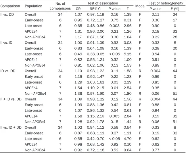
The combined results showed no significant dif- ference in II versus (ID + DD) (OR = 1.02, 95%

CI = 0.94-1.12, P = 0.59), II versus DD (OR =

1.07, 95% CI = 0.97-1.19, P = 0.16), and II ver-

sus ID (OR = 1.00, 95% CI = 0.91-1.09, P =

0.93) under the fixed-effects model, and ID ver-

sus DD (OR = 1.10, 95% CI = 0.98-1.23, P =

0.11) and II + ID versus DD (OR = 1.09, 95% CI

Figure 3. Forest plot for ACE I/D polymorphism (II+ID versus DD) and SAD risk.

(6)

Angiotensin-converting enzyme gene and Alzheimer’s disease

= 0.98-1.22, P = 0.12) (Figures 2-6) under the random-effects model. All results for genetic models and the test for heterogeneity are sum- marized in Table 2.

Subgroup analysis

A statistically significant difference was ob- served in late-onset AD (II vs DD: OR = 0.65, 95% CI = 0.48-0.86, P = 0.003 < 0.05; II vs ID:

OR = 0.49, 95% CI = 0.38-0.65, P < 0.05; ID vs DD: OR = 1.29, 95% CI = 1.03-1.61, P = 0.03 <

0.05; or II versus ID + DD: OR = 0.55, 95% CI = 0.42-0.70, P < 0.05) (Table 2), but no statistical significance in early-onset AD was found. When stratified by the APOEε4 carrier status, a higher risk for SAD was also observed for the APOEε4 carrier status (ID vs DD: OR = 1.54, 95% CI =

1.10-2.15, P = 0.01 < 0.05; II + ID vs DD: OR = 1.58, 95% CI = 1.15-2.16, P = 0.005 < 0.05);

however, the results were not pronounced for the non-APOEε4 carrier status (Table 2).

Sensitivity analysis

The sensitivity analysis indicated that three

independent comparisons [12, 58] were the

main origin of heterogeneity. The heterogeneity

was removed after the exclusion of these three

comparisons [test for heterogeneity for ID vs

DD (I

2

= 12%, P = 0.28) and (II + ID) vs DD (I

2

=

14%, P = 0.25)], and the corresponding pooled

ORs were not materially altered [II vs (ID + DD)

(OR = 1.02, 95% CI = 0.94-1.12, P = 0.59), II vs

DD (OR = 1.07, 95% CI = 0.97-1.19, P = 0.16), II

vs ID (OR = 1.00, 95% CI = 0.91-1.09, P = 0.93),

Figure 4. Forest plot for ACE I/D polymorphism (II versus ID + DD) and SAD risk.

(7)

ID vs DD (OR = 1.10, 95% CI = 0.98-1.23, P = 0.11), and II + ID vs DD (OR = 1.09, 95% CI = 0.98-1.22, P = 0.12) under the fixed-effects model]. No effect of any study on the pooled results in the aforementioned five models was found. The results of sensitivity analysis indi- cated the stability of the results.

Publication bias

The shape of the funnel plots in genetic models seemed symmetrical, indicating no evidence for obvious publication bias (Figures 7-11).

Further, the Egger’s test showed no significant publication bias in the genetic model [II vs (ID + DD), P = 0.249; II vs DD, P = 0.873; II vs ID, P = 0.141; ID vs DD, P = 0.22, and (II + ID) vs DD, P

= 0.326)]. Therefore, the potential publication bias did not materially alter the combined risk estimate.

Discussion

Epidemiological and pathogenetic evidences strongly suggested a correlation between genetic factors and the risk for SAD. On the basis of this hypothesis, the contribution of various candidate genes to the risk for SAD was investigated, and one of the candidate genes that have been analyzed as a risk factor for SAD was found to be the ACE gene. ACE modu- lates the progression of AD via degradation of Ab or components of renin-angiotensin system in the brain [6, 7].

Figure 5. Forest plot for ACE I/D polymorphism (II versus ID) and SAD risk.

(8)

Angiotensin-converting enzyme gene and Alzheimer’s disease

A collaborative study demonstrated a possible correlation between ACE gene I allele and the risk for SAD [12]. Since then a considerable number of studies were performed to replicate these results. In Colombian patients, ACE I/D polymorphism does not appear to confer an added risk for SAD [39]. However, ambiguous results have been presented. The ACE I/D gen- otypes are associated with an increased risk for SAD in patients with German origin [44]. A possible protective role for the II genotype of ACE was observed by Shcherbatykh et al. [47].

The failure to reproduce replicated studies might be due to the small sample size used. In inclusive 34 comparisons, the pooled results confirmed that ACE I/D polymorphism had no effect on the risk for SAD. However, the results

of I-square and Cochran’s Q statistic showed heterogeneity in the two models (Table 2). The most plausible explanation for the heterogene- ity may be the presence of an interaction between age at onset, APOEε4 carrier status, and ACE I/D polymorphism for the risk of SAD.

The association with genetic risk factors might

be dependent on the heterogeneity of age at

onset. Helbecque et al. showed that the ACE D

allele was at a reduced risk for SAD for the old-

est patients with SAD [45]. However, no correla-

tion of ACE with the age at onset was found for

patients with SAD in Italy [40]. On the basis of

age at onset, statistically significant difference

was found in late-onset AD. The present study

further supported the previous reports of an

Figure 6. Forest plot for ACE I/D polymorphism (II versus DD) and SAD risk.

(9)

Table 2. Meta-analysis of the association between ACE I/D polymorphism and SAD risk Comparison Population No. of

comparisons

Test of association Mode Test of heterogeneity

OR 95% CI P value Z P value I

2

(%)

II vs. DD Overall 34 1.07 0.97, 1.19 0.16 1.39 F 0.11 24

Early-onset 6 0.95 0.72, 1.27 0.75 0.31 F 0.30 17

Late-onset 6 0.65 0.48, 0.86 0.003 2.96 F 0.90 0

APOEε4 7 1.31 0.86, 2.00 0.21 1.26 F 0.18 33

Non-APOEε4 7 1.17 0.87, 1.56 0.30 1.04 F 0.22 28

II vs. ID Overall 34 1.00 0.91, 1.09 0.93 0.08 F 0.33 8

Early-onset 6 0.83 0.64, 1.08 0.16 1.39 F 0.28 20

Late-onset 6 0.49 0.38, 0.65 < 0.05 5.15 F 0.54 0

APOEε4 7 0.82 0.55, 1.21 0.32 1.00 F 0.91 0

Non-APOEε4 7 0.81 0.62, 1.06 0.13 1.53 F 0.89 0

ID vs. DD Overall 34 1.10 0.98, 1.23 0.11 1.58 R 0.004 44

Early-onset 6 1.16 0.92, 1.47 0.22 1.23 F 0.99 0

Late-onset 6 1.29 1.03, 1.61 0.03 2.19 F 0.85 0

APOEε4 7 1.54 1.10, 2.15 0.01 2.54 F 0.35 0

Non- APOEε4 7 1.36 0.97, 1.90 0.07 1.80 R 0.06 51

II + ID vs. DD Overall 34 1.09 0.98, 1.22 0.12 1.56 R 0.004 44

Early-onset 6 1.09 0.88, 1.36 0.42 0.81 F 0.88 0

Late-onset 6 1.07 0.86, 1.32 0.54 0.61 F 0.94 0

APOEε4 7 1.58 1.15, 2.16 0.005 2.84 F 0.19 31

Non-APOEε4 7 1.28 0.92, 1.78 0.15 1.44 R 0.06 51

II vs. ID + DD Overall 34 1.02 0.94, 1.12 0.59 0.54 F 0.33 8

Early-onset 6 0.87 0.68, 1.11 0.27 1.11 F 0.19 32

Late-onset 6 0.55 0.42, 0.70 < 0.05 4.70 F 0.62 0

APOEε4 7 0.98 0.68, 1.42 0.92 0.10 F 0.62 0

Non-APOEε4 7 0.92 0.72, 1.18 0.52 0.64 F 0.77 0

R: random-effects model; F: fixed-effect model.

interaction between age at onset and ACE I/D polymorphism for the risk for SAD [26, 29, 45].

gistic interaction existed between ACE I/D poly- morphism and APOEε4 carrier status for the risk of SAD, and ACE I/D polymorphism might A gene-gene interaction analy- sis should be explored. When cases and controls were strat- ified according to the APOEε4 carrier status, no significant difference was revealed [42].

Otherwise, an increase in the

risk of developing SAD was

found in subjects carrying both

the ACE DD genotype and the

APOEε4 allele (OR = 10.32,

95% CI = 2.67-39.81) [53]. A

statistically significant differ-

ence in the APOEε4 carrier sta-

tus was observed between

ACE I/D polymorphism and the

risk for SAD in the present

meta-analysis. Hence, a syner-

Figure 7. Begg’s funnel plot for ID versus DD.

(10)

Angiotensin-converting enzyme gene and Alzheimer’s disease

Figure 8. Begg’s funnel plot for (II+ID) versus DD.

Figure 9. Begg’s funnel plot for II versus (ID+DD).

be a genetic risk for patients with SAD having the APOEε4 carrier status.

The present meta-analysis of observational studies had some limitations. First, the effects of clinical heterogene- ity (age, gender, and so on) on the conclusions were explored.

However, the small sample size might have yielded false- positive results due to the lack of statistical power. Therefore, it is critical that larger-scale, well-designed studies should be performed to identify the- se effects. On the contrary, the samples (blood and br- ain) were selected, and differ- ent genotyping methods were used with different sensitivity and specificity, which might have also resulted in selection bias and clinical heterogene- ity. If the heterogeneity was removed by sensitivity analy- sis, the overall results were not materially altered, sug- gesting the stability of the results. Third, an important issue that is often raised in a methodological meta-analysis is publication bias. Publication bias was not detected by the Begg’s funnel plot and Egger’s test in this meta-analysis. How- ever, publication bias might not be ruled out completely. It is possible that relevant un- published articles with null results might not have been included, leading to publica- tion bias.

Despite the aforementioned

limitations, this meta-analysis

demonstrated that the ACE

I/D polymorphism might not

be associated with the risk for

SAD. It was suggested that the

interaction between ACE I/D

polymorphism and age at on-

set or APOEε4 carrier status

might account for the risk for

SAD. On the basis of SAD with

(11)

Figure 11. Begg’s funnel plot for II versus DD.

sults of the present meta-analysis should be properly replicated in future prospective cohort studies, taking into consideration the in- teractions.

Disclosure of conflict of interest None.

Address correspondence to: Dr. Qing Xia, Depart- ment of Rehabilitation Medicine, The Second People’s Hospital of Hefei City, 246 Heping Road, Hefei 230011, Anhui Province, China. Tel: +86 551 62965302; Fax: +86 551 62965302; E-mail:

345464516@qq.com; Dr. Xiaotong Wang, De- partment of Neurology, The Second Affiliated Hos- pital, Wenzhou Medical University, 109 Xueyuan Road, Wenzhou 325027, Zhejiang Province, China.

Tel: +86 577 8669 9362; Fax: +86 577 8669 9362;

E-mail: wangxt805@126.com References

[1] Hu J, Igarashi A, Kamata M and Nakagawa H. Angiotensin-converting enzyme degrades Alzheimer amyloid beta-peptide (A beta); re- tards A beta aggregation, deposition, fibril for- mation; and inhibits cytotoxicity. J Biol Chem 2001; 276: 47863-47868.

[2] Selkoe DJ. The molecular pathology of al- zheimer’s disease. Neurobiol Aging 1991; 6:

487-498.

[3] Reid IA. Interactions between ANG II, sympa- thetic nervous system, and baroreceptor re- flexes in regulation of blood pressure. Am J Physiol 1992; 262: E763-778.

[4] von Bohlen und Halbach O. Angiotensin IV in the central nervous system. Cell Tissue Res 2003; 311: 1-9.

[5] Skoog I, Wallin A, Fredman P, Hesse C, Aevarsson O, Karlsson I, Gottfries CG and Blennow K. A population study on blood-brain barrier function in 85-year-olds: re- lation to Alzheimer’s dis- ease and vascular demen- tia. Neurology 1998; 50:

966-971.

[6] Karch CM and Goate AM.

Alzheimer’s disease risk genes and mechanisms of disease pathogenesis. Biol Psychiatry 2015; 77: 43-51.

[7] Hemming ML and Selkoe DJ. Amyloid beta-protein is degraded by cellular an- giotensin-converting enzy- me (ACE) and elevated by an ACE inhibitor. J Biol Chem 2005; 280: 37644- 37650.

[8] Miners S, Ashby E, Baig S, Harrison R, Tayler H, Speedy E, Prince JA, Love S and Kehoe PG.

Angiotensin-converting enzyme levels and ac- tivity in Alzheimer’s disease: differences in brain and CSF ACE andassociation with ACE1 genotypes. Am J Transl Res 2009; 1: 163-177.

[9] Savaskan E, Hock C, Olivieri G, Bruttel S, Rosenberg C, Hulette C and Müller-Spahn F.

Cortical alterations of angiotensin converting enzyme, angiotensin II and AT1 receptor in Alzheimer’s dementia. Neurobiol Aging 2001;

22: 541-546.

[10] Arbustini E, Grasso M, Fasani R, Klersy C, Diegoli M, Porcu E, Banchieri N, Fortina P, Danesino C and Specchia G. Angiotensin con- verting enzyme gene deletion allele is indepen- dently and strongly associated with coronary atherosclerosis and myocardial infarction. Br Heart J 1995; 74: 584-591.

[11] Kluijtmans LA, van den Heuvel LP, Boers GH, Frosst P, Stevens EM, van Oost BA, den Heijer M, Trijbels FJ, Rozen R and Blom HJ. Molecular genetic analysis in mild hyperhomocystein- emia: a common mutation in the methy lene- tetra hydrofolate reductase gene is a genetic risk factor for cardiovascular disease. Am J Hum Genet 1996; 58: 35-41.

[12] Kehoe PG, Russ C, McIlory S, Williams H, Holmans P, Holmes C, Liolitsa D, Vahidassr D, Powell J, McGleenon B, Liddell M, Plomin R, Dynan K, Williams N, Neal J, Cairns NJ, Wilcock G, Passmore P, Lovestone S, Williams J and Owen MJ. Variation in DCP1, encoding ACE, is associated with susceptibility to Alzheimer dis- ease. Nat Genet 1999; 21: 71-72.

[13] Narain Y, Yip A, Murphy T, Brayne C, Easton D,

Evans JG, Xuereb J, Cairns N, Esiri MM, Furlong

RA and Rubinsztein DC. The ACE gene and

(12)

Angiotensin-converting enzyme gene and Alzheimer’s disease

Alzheimer’s disease susceptibility. J Med Genet 2000; 37: 695-697.

[14] Lehmann DJ, Cortina-Borja M, Warden DR, Smith AD, Sleegers K, Prince JA, van Duijn CM and Kehoe PG. Large meta-analysis establish- es the ACE insertion-deletion polymorphism as a marker of Alzheimer’s disease. Am J Epidemiol 2005; 162: 305-317.

[15] Richard F and Amouyel P. Genetic susceptibili- ty factors for Alzheimer’s disease. Eur J Pharmacol 2001; 412: 1-12.

[16] Panza F, Solfrizzi V, D’ Introno A, Colacicco AM, Capurso C, Capurso A and Kehoe PG. Shifts in angiotensin I converting enzyme insertion al- lele frequency across Alzheimer: implications for Alzheimer’s disease risk. J Neurol Neurosurg Psychiatry 2003; 74: 1159-1161.

[17] Elias-Sonnenschein LS, Bertram L and Visser PJ. Relationship between genetic risk factors and markers for Alzheimer’s disease patholo- gy. Biomark Med 2012; 6: 477-495.

[18] Lucatelli JF, Barros AC, Silva VK, Machado Fda S, Constantin PC, Dias AA, Hutz MH and de Andrade FM. Genetic influences on Alzheimer’s disease: evidence of interactions between the genes APOE, APOC1 and ACE in a sample pop- ulation from the south of Brazil. Neuro- chem Res 2011; 36: 1533-1539.

[19] Qiu WQ, Mwamburi M, Besser LM, Zhu H, Li H, Wallack M, Phillips L, Qiao L, Budson AE, Stern R and Kowall N. Angiotensin converting en- zyme inhibitors and the reduced risk of Alzheimer’s disease in the absence of apolipo- protein E4 allele. J Alzheimers Dis 2013; 37:

421-428.

[20] Sleegers K, den Heijer T, van Dijk EJ, Hofman A, Bertoli-Avella AM, Koudstaal PJ, Breteler MM and van Duijn CM. ACE gene is associated with Alzheimer’s disease and atrophy of hippocam- pus and amygdala. Neurobiol Aging 2005; 26:

1153-1159.

[21] Vardy ER, Brown K, Stopford CL, Thompson JC, Richardson AM, Neary D, Kalsheker N, Morgan K, Mann DM and Snowden JS. Cognitive phe- notypes in Alzheimer’s disease and genetic variants in ACE and IDE. Neurobiol Aging 2012;

33: 1486.e1-2.

[22] Tian J, Shi J, Bailey K, Harris JM, Pritchard A, Lambert JC, Chartier-Harlin MC, Pickering- Brown SM, Lendon CL and Mann DM. A poly- morphism in the angiotensin 1-converting en- zyme gene is associated with damage to cere- bral cortical white matter in Alzheimer’s dis- ease. Neurosci Lett 2004; 354: 103-106.

[23] Purandare N, Oude Voshaar RC, Davidson Y, Gibbons L, Hardicre J, Byrne J, McCollum C, Jackson A, Burns A and Mann DM. Deletion/

insertion polymorphism of the angiotensin- converting enzyme gene and white matter hy-

perintensities in dementia: a pilot study. J Am Geriatr Soc 2006; 54: 1395-1400.

[24] Kehoe PG, Katzov H, Andreasen N, Gatz M, Wilcock GK, Cairns NJ, Palmgren J, de Faire U, Brookes AJ, Pedersen NL, Blennow K and Prince JA. Common variants of ACE contribute to variable age-at-onset of Alzheimer’s dis- ease. Hum Genet 2004; 114: 478-483.

[25] Miners JS, van Helmond Z, Raiker M, Love S and Kehoe PG. ACE variants and association with brain Aβ levels in Alzheimer’s disease. Am J Transl Res 2010; 3: 73-80.

[26] Yang JD, Feng G, Zhang J, Lin ZX, Shen T, Breen G, St Clair D and He L. Association between angiotensin-converting enzyme gene and late onset Alzheimer’s disease in han Chinese.

Neurosci Lett 2000; 295: 41-44.

[27] Nirmal S, Tripathi M, Shastri SS and Sagar R, SV. Association of angiotensin-converting en- zyme insertion (I)/deletion (D) genotype in Alzheimer’s disease patients of north Indian population. Int J Neurosci 2011; 121: 557- 561.

[28] Cheng CY, Hong CJ, Liu HC, Liu TY and Tsai SJ.

Study of the association between Alzheimer’s disease and angiotensin-converting enzyme gene polymorphism using DNA from lympho- cytes. Eur Neurol 2002; 47: 26-29.

[29] Zhang JW, Li XQ, Zhang ZX, Chen D, Zhao HL, Wu YN and Qu QM. Association between angio- tensin-converting enzyme gene polymorphism and Alzheimer’s disease in a Chinese popula- tion. Dement Geriatr Cogn Disord 2005; 20:

52-56.

[30] Wang HK, Fung HC, Hsu WC, Wu YR, Lin JC, Ro LS, Chang KH, Hwu FJ, Hsu Y, Huang SY, Lee- Chen GJ and Chen CM. Apolipoprotein E, angio- tensin-converting enzyme and kallikrein gene polymorphisms and the risk of Alzheimer’s dis- ease and vascular dementia. J Neural Transm (Vienna) 2006; 113: 1499-1509.

[31] Isbir T, Agaçhan B, Yilmaz H, Aydin M, Kara I, Eker D and Eker E. Interaction between apoli- poprotein-E and angiotensin-converting en- zyme genotype in Alzheimer’s disease. Am J Alzheimers Dis Other Demen 2001; 16: 205- 210.

[32] Keikhaee MR, Hashemi SB, Najmabadi H and Noroozian M. C677T methy lentetra hydrofu- late reductase and angiotensin converting en- zyme gene polymorphisms in patients with Alzheimer’s disease in iranian population.

Neurochem Res 2006; 31: 1079-1083.

[33] Wang B, Jin F, Yang Z, Lu Z, Kan R, Li S, Zheng

C and Wang L. The insertion polymorphism in

angiotensin-converting enzyme gene associat-

ed with the APOE epsilon 4 allele increases the

risk of late-onset Alzheimer disease. J Mol

Neurosci 2006; 30: 267-271.

(13)

[34] Bowirrat A, Cui J, Waraska K, Friedland RP, Oscar-Berman M, Farrer LA, Korczyn A and Baldwin CT. Lack of association between an- giotensin-converting enzyme and dementia of the Alzheimer’s type in an elderly arab popula- tion in Wadi ara, lsrael. Neuropsychiatr Dis Treat 2005; 1: 73-76.

[35] Chapman J, Wang N, Treves TA, Korczyn AD and Bornstein NM. ACE, MTHFR, factor V Leiden, and APOE polymorphisms in patients with vascular and Alzheimer’s dementia. Stro- ke 1998; 29: 1401-1404.

[36] Hu J, Miyatake F, Aizu Y, Nakagawa H, Na- kamura S, Tamaoka A, Takahash R, Urakami K and Shoji M. Angiotensin-converting enzyme genotype is associated with Alzheimer disease in the Japanese population. Neurosci Lett 1999; 277: 65-67.

[37] Wu C, Zhou D, Guan Z, Fan J and Qiao Y. The association between angiotensin I converting enzyme gene polymorphism and Chinese late onset Alzheimer disease. Zhonghua Yi Xue Yi Chuan Xue Za Zhi 2002; 19: 401-404.

[38] Ning M, Yang Y, Zhang Z, Chen Z, Zhao T, Zhang D, Zhou D, Xu J, Liu Z, Wang Y, Liu Y, Zhao X, Li W, Li S and He L. Amyloid-β-related genes SORL1 and ACE are genetically associated with risk for late-onset Alzheimer disease in the Chinese population. Alzheimer Dis Assoc Disord 2010; 24: 390-396.

[39] Camelo D, Arboleda G, Yunis JJ, Pardo R, Arango G, Solano E, López L, Hedmont D and Arboleda H. Angiotensin-converting enzyme and alpha-2-macroglobulin gene polymor- phisms are not associated with Alzheimer’s disease in colombian patients. J Neurol Sci 2004; 218: 47-51.

[40] Seripa D, Forno GD, Matera MG, Gravina C, Margaglione M, Palermo MT, Wekstein DR, Antuono P, Davis DG, Daniele A, Masullo C, Bizzarro A, Gennarelli M and Fazio VM. Methy lenetetra hydrofolate reductase and angioten- sin converting enzyme gene polymorphisms in two genetically and diagnostically distinct co- hort of Alzheimer patients. Neurobiol Aging 2003; 24: 933-939.

[41] Nacmias B, Bagnoli S, Tedde A, Cellini E, Bessi V, Guarnieri B, Ortensi L, Piacentini S, Bracco L and Sorbi S. Angiotensin converting enzyme insertion/deletion polymorphism in sporadic and familial Alzheimer’s disease and longevity.

Arch Gerontol Geriatr 2007; 45: 201-206.

[42] Crawford F, Abdullah L, Schinka J, Suo Z, Gold M, Duara R and Mullan M. Gender-specific as- sociation of the angiotensin converting en- zyme gene with Alzheimer’s disease. Neurosci Lett 2000; 280: 215-219.

[43] Monastero R, Caldarella R, Mannino M, Cefalù AB, Lopez G, Noto D, Camarda C, Camarda LK, Notarbartolo A, Averna MR and Camarda R.

Lack of association between angiotensin con-

verting enzyme polymorphism and sporadic Alzheimer’s disease. Neurosci Lett 2002; 335:

147-149.

[44] Kölsch H, Jessen F, Freymann N, Kreis M, Hentschel F, Maier W and Heun R. ACE I/D polymorphism is a risk factor of Alzheimer’s disease but not of vascular dementia. Neurosci Lett 2005; 377: 37-39.

[45] Helbecque N, Codron V, Cottel D and Amouyel P. An age effect on the association of common variants of ACE with Alzheimer’s disease.

Neurosci Lett 2009; 461: 181-184.

[46] Palumbo B, Cadini D, Nocentini G, Filipponi E, Fravolini ML and Senin U. Angiotensin convert- ing enzyme deletion allele in different kinds of dementia disorders. Neurosci Lett 1999; 267:

97-100.

[47] Shcherbatykh TV, Kiryanov SA, Korovaitseva GI, Selezneva ND, Voskresenskaya NI, Golim- bet VE, Farrer L, Gavrilova SI and Rogaev EI.

The angiotensin-converting enzyme gene as a possible risk or protective factor in Alzheimer’s disease. Neurosci Behav Physiol 2001; 31:

179-181.

[48] Carbonell J, Allen R, Kalsi G, McQuillin A, Livingston G, Katona C, Walker Z, Katz A, Rands G, Stevens T, Crossan I, Curtis D and Gurling H. Variation in the DCP1 gene, encod- ing the angiotensin converting enzyme ACE, is not associated with increased susceptibility to Alzheimer’s disease. Psychiatr Genet 2003;

13: 47-50.

[49] Narain Y, Yip A, Murphy T, Brayne C, Easton D, Evans JG, Xuereb J, Cairns N, Esiri MM, Furlong RA and Rubinsztein DC. The ACE gene and Alzheimer’s disease susceptibility. J Med Genet 2000; 37: 695-697.

[50] Zuliani G, Ble’ A, Zanca R, Munari MR, Zurlo A, Vavalle C, Atti AR and Fellin R. Genetic poly- morphisms in older subjects with vascular or Alzheimer’s dementia. Acta Neurol Scand 2001; 103: 304-308.

[51] Lendon CL, Thaker U, Harris JM, McDonagh AM, Lambert JC, Chartier-Harlin MC, Iwatsubo T, Pickering-Brown SM and Mann DM. The an- giotensin 1-converting enzyme insertion (I)/

deletion (D) polymorphism does not influence the extent of amyloid or tau pathology in pa- tients with sporadic Alzheimer’s disease.

Neurosci Lett 2002; 328: 314-318.

[52] Myllykangas L, Polvikoski T, Sulkava R, Verkkoniemi A, Tienari P, Niinistö L, Kontula K, Hardy J, Haltia M and Pérez-Tur J. Cardiovascular risk factors and Alzheimer’s disease: a genetic association study in a population aged 85 or over. Neurosci Lett 2000; 292: 195-198.

[53] Trebunova M, Slaba E, Habalova V and Gdovinova Z. ACE I/D polymorphism in Alzheimer’s disease. Cent Eur J Biol 2008; 3:

49-54.

(14)

Angiotensin-converting enzyme gene and Alzheimer’s disease

[54] Alvarez R, Alvarez V, Lahoz CH, Martínez C, Peña J, Sánchez JM, Guisasola LM, Salas-Puig J, Morís G, Vidal JA, Ribacoba R, Menes BB, Uría D and Coto E. Angiotensin converting en- zyme and endothelial nitric oxide synthase DNA polymorphisms and late onset Alzheimer’s disease. J Neurol Neurosurg Psychiatry 1999;

67: 733-736.

[55] Scacchi R, De Bernardini L, Mantuano E, Vilardo T, Donini LM, Ruggeri M, Gemma AT, Pascone R and Corbo RM. DNA polymorphisms of apolipoprotein B and angiotensin I-con- verting enzyme genes and relationships with lipid levels in italian patients with vascular de- mentia or Alzheimer’s disease. Dement Geriatr Cogn Disord 1998; 9: 186-190.

[56] Panza F, Solfrizzi V, D’ introno A, Capurso C, Colacicco AM, Argentieri G and Capurso A.

Lack of association between ace polymor- phism and Alzheimer’s disease in southern Italy. Arch Gerontol Geriatr Suppl 2002; 8:

239-245.

[57] Farrer LA, Sherbatich T, Keryanov SA, Korovai- tseva GI, Rogaeva EA, Petruk S, Premkumar S, Moliaka Y, Song YQ, Pei Y, Sato C, Selezneva ND, Voskresenskaya S, Golimbet V, Sorbi S, Duara R, Gavrilova S, St George-Hyslop PH and Rogaev EI. Association between angiotensin- converting enzyme and Alzheimer disease.

Arch Neurol 2000; 57: 210-214.

[58] Mattila KM, Rinne JO, Röyttä M, Laippala P, Pietilä T, Kalimo H, Koivula T, Frey H and Lehtimäki T. Dipeptidyl carboxypeptidase 1 (DCP1) and butyrylcholinesterase (BCHE) gene interactions with the apolipoprotein E epsilon4 allele as risk factors in Alzheimer’s disease and in Parkinson’s disease with coexisting Alzheimer pathology. J Med Genet 2000; 37:

766-770.

[59] Buss S, Müller-Thomsen T, Hock C, Alberici A, Binetti G, Nitsch RM, Gal A and Finckh U. No association between DCP1 genotype and late- onset Alzheimer disease. Am J Med Genet 2002; 114: 440-445.

[60] Kehoe PG, Katzov H, Feuk L, Bennet AM, Johansson B, Wiman B, de Faire U, Cairns NJ, Wilcock GK, Brookes AJ, Blennow K and Prince JA. Haplotypes extending across ACE are asso- ciated with Alzheimer’s disease. Hum Mol Genet 2003; 12: 859-867.

[61] Vardy ER, Rice PJ, Bowie PC, Holmes JD, Catto AJ and Hooper NM. Plasma angiotensin-con- verting enzyme in Alzheimer’s disease. J Alzheimers Dis 2009; 16: 609-618.

[62] Perry RT, Collins JS, Harrell LE, Acton RT and Go RC. Investigation of association of 13 poly- morphisms in eight genes in southeastern African American Alzheimer disease patients as compared to age-matched controls. Am J Med Genet 2001; 105: 332-342.

[63] Prince JA, Feuk L, Sawyer SL, Gottfries J, Ricksten A, Nägga K, Bogdanovic N, Blennow K and Brookes AJ. Lack of replication of associa- tion findings in complex disease: an analysis of 15 polymorphisms in prior candidate genes for sporadic Alzheimer’s disease. Eur J Hum Genet 2001; 9: 437-444.

[64] Richard F, Fromentin-David I, Ricolfi F, Ducime- tière P, Di Menza C, Amouyel P and Helbecque N. The angiotensin I converting enzyme gene as a susceptibility factor for dementia.

Neurology 2001; 56: 1593-1595.

[65] Schächter F, Faure-Delanef L, Guénot F, Rouger H, Froguel P, Lesueur-Ginot L and Cohen D.

Genetic associations with human longevity at

the APOE and ACE loci. Nat Genet 1994; 6: 29-

32.

Figure

Figure 5. Forest plot for ACE I/D polymorphism (II versus ID) and SAD risk.
Table 2. Meta-analysis of the association between ACE I/D polymorphism and SAD risk Comparison Population No
Figure 8. Begg’s funnel plot for (II+ID) versus DD.
Figure 11. Begg’s funnel plot for II versus DD.

References

Related documents

Neurodevelopmental problems, including neurologic soft signs, learning problems, and ADHD, were also associated with behavior disorders and subclinical psychotic spectrum symptoms,

In addition 12th and Maraka, Badhaka significator Saturn is conjoined with 2nd Maraka lord Jupiter (expansion of growth) in 10 (ineffective medication) in Jupiter sign Sagitta- rius;

like the characters in the Dead or Alive series who wear school girl outfits in a fight, it can give men unrealistic expectations on what they want a mate because both men and

The harvested iliac block bones were divided in the following groups: autogenous iliac block bone with preservation of the periosteum (the periosteum group), autogenous iliac block

Abstract: The purpose of this study is to examine and analyze the online marketing in Madurai with special reference to fruits and vegetables (especially in Madurai website and

Abstract – This paper comprises with different methods of facial feature extraction of Face Recognition using Gabor, Local Binary Pattern and its fusion.. In

After comparing methods, the following measure- ments were adopted for each site: line intercept canopy cover by species, line intercept plant height by life form, line intercept

As the optimal buyback no-lending policy is at odds with the data (i.e the first and third lines in Table 4 are very different) several normative recommendations emerge: