R E S E A R C H
Open Access
Postoperative intubation time is associated with
acute kidney injury in cardiac surgical patients
Matthias Heringlake
1*, Yvonne Nowak
1, Julika Schön
1, Jens Trautmann
1, Astrid Ellen Berggreen
1,
Efstratios I Charitos
2and Hauke Paarmann
1Abstract
Introduction:Acute kidney injury (AKI) is a frequent complication after cardiac surgery and is associated with a poor prognosis. Mechanical ventilation is an important risk factor for developing AKI in critically ill patients. Ventilation with high tidal volumes has been associated with postoperative organ dysfunction in cardiac surgical patients. No data are available about the effects of the duration of postoperative respiratory support in the immediate postoperative period on the incidence of AKI in patients after cardiac surgery.
Method:We performed a secondary analysis of 584 elective cardiac surgical patients enrolled in an observational trial on the association between preoperative cerebral oxygen saturation and postoperative organ dysfunction and analyzed the incidence of AKI in patients with different times to extubation. The latter variable was graded in 4 h intervals (if below 16 h) or equal to or greater than 16 h. AKI was staged according to the AKI Network criteria. Results:Overall, 165 (28.3%) patients developed AKI (any stage), 43 (7.4%) patients needed renal replacement therapy. Patients developing AKI had a significantly (P<0.001) lower renal perfusion pressure (RPP) in the first 8 hours after surgery (57.4 mmHg (95% CI: 56.0 to 59.0 mmHg)) than patients with a postoperatively preserved renal function (60.5 mmHg ((95% CI: 59.9 to 61.4 mmHg). The rate of AKI increased from 17.0% in patients extubated within 4 h postoperatively to 62.3% in patients ventilated for more than 16 h (P<0.001). Multivariate logistic regression analysis of variables significantly associated with AKI in the univariate analysis revealed that the time to the first extubation (OR: 1.024/hour, 95% CI: 1.011 to 1.044/hour;P<0.001) and RPP (OR: 0.963/mmHg; 95% CI: 0.934 to 0.992;P<0.001) were independently associated with AKI.
Conclusion:Without taking into account potentially unmeasured confounders, these findings are suggestive that the duration of postoperative positive pressure ventilation is an important and previously unrecognized risk factor for AKI in cardiac surgical patients, independent from low RPP as an established AKI trigger, and that even a moderate delay of extubation increases AKI risk. If replicated independently, these findings may have relevant implications for clinical care and for further studies aiming at the prevention of cardiac surgery associated AKI.
Introduction
Cardiac surgery-associated acute kidney injury (CSA-AKI) is an important complication and an independent mortality factor in patients undergoing cardiac surgery [1] and even minor deteriorations in postoperative renal function have been associated with an increased mortality risk [2].
The pathophysiological factors mediating CSA-AKI may be summarized as a complex interplay between a
decrease in renal blood flow (induced either directly by changes in systemic perfusion or indirectly by activation of the renin-angiotensin system and/or the renal sym-pathetic nervous system), hyperglycemia and increased inflammation as well as intrinsic and extrinsic toxins (that is myoglobin and hemoglobin, radiocontrast agents) (for overview see: [1,3,4]). No therapeutic concept to prevent or ameliorate this complication has been proved to be successful in adequately powered trials [3,4].
Cardiac surgical procedures are typically performed during general anesthesia and most patients are mechan-ically ventilated in the postoperative period until normo-thermia and hemodynamic stability have been achieved. * Correspondence:Heringlake@t-online.de
1
Department of Anesthesiology and Intensive Care Medicine, University of Lübeck, Ratzeburger Allee 160, 23538 Lübeck, Germany
Full list of author information is available at the end of the article
Interestingly, the duration of postoperative positive pres-sure ventilation has been largely ignored as a factor poten-tially mediating CSA-AKI in almost all interventional studies as well as in scientific reviews of this field, despite the fact that it has long been known that especially mech-anical ventilation with positive end-expiratory pressure (PEEP), but also continuous positive airway pressure (CPAP) breathing may have profound effects on renal function [5,6].
Very recently, van den Akker and coworkers have shown that mechanical ventilation for more than 24 h is associated with a threefold increase in the risk for devel-oping acute kidney injury (AKI) in critically ill patients [7]. However, only one small study including cardiac sur-gical patients was included in this meta-analysis.
Lellouche and coworkers analyzed the effects of venti-lation with low (<10 ml/kg), traditional (10 to 12 ml/kg) or high (>12 ml/kg) tidal volumes immediately after car-diac surgery on the incidence of postoperative organ dysfunction and observed a clear association between non-protective ventilation and organ dysfunction includ-ing AKI. However, and despite patients with low tidal volumes being extubated earlier after surgery than patients with traditional or high, that is injurious tidal volumes, the authors did not address the effects of short-term ventilation (below 24 h) on organ dysfunction in this trial [8].
The present study thus explores the influence of the duration of continuous postoperative positive pressure ventilation within the immediate postoperative period on the incidence of CSA-AKI in a heterogeneous cohort of cardiac surgical patients.
Materials and methods
The present study is a secondary analysis of data from a large observational trial on the relationship between pre-operative cerebral oxygen saturation (ScO2) determined by near-infrared spectroscopy (NIRS) and postoperative organ dysfunction. This data set has already been used for other analyses and the details of recruitment as well as consenting have been presented elsewhere [9,10]. Following approval from the local ethics committee (Ethikkommission der Universität zu Lübeck, Lübeck, Germany), all patients scheduled for cardiac surgery at the University of Lübeck from 1 April 2009 to 29 December 2009 were screened for participation in this trial. Written informed consent was obtained preoperatively.
The only exclusion criterion for the observational trial was an age of less than 18 years. The present analysis was restricted to elective patients and additionally excluded patients preoperatively treated with dialysis (Figure 1).
Anesthesiological, surgical, and intensive care treatment followed standardized algorithms established at the Department of Anesthesiology and the Department of
Thoracic Vascular and Cardiac Surgery of the Univer-sity of Lübeck and have also been presented in detail elsewhere [9,10]. Hemodynamic treatment was partially adapted from a German guideline for the hemodynamic treatment of cardiac surgical patients [11]. Accordingly, intra- and postoperative fluid therapy was performed with balanced Ringer’s solution, 6% hydroxyethyl starch 130, and gelatinesuccinate at the discretion of the attending physicians. Starting in July 2009, all patients were add-itionally treated perioperatively with a continuous infusion of 4 mmol/kg sodium bicarbonate [9].
Surgery was performed during mild hypothermia using antegrade blood cardioplegia. After surgery, patients were transferred to the ICU while being sedated with a combination of remifentanil and propofol adjusted to their clinical needs. In absence of contraindications (glu-cose-6-phosphate dehydrogenase deficiency, porphyria), a continuous infusion of metamizole 3 g/24 h was started. After normal body temperature had been achieved, propo-fol was stopped, analgesics (pethidine and/or piritramide) were applied, and the remifentanil infusion was continu-ously reduced. Further piritramide was applied according to patients’analgesic needs.
Postoperative ventilation was performed with biphasic intermittent positive airway pressure (BIPAP) and pressure support ventilation (PSV) with a minimum PEEP of 5 mbar and inspiratory pressures/pressure support-adjusted to achieve a tidal volume around 6 to 8 ml/kg. Postoperative adjustments in ventilatory support as well as weaning from the respirator and extubation were performed at the discretion of the attending physicians of the ICU.
The severity of postoperative kidney dysfunction was quantified by the Acute Kidney Injury Network (AKIN) criteria [12] from perioperative changes in plasma creatin-ine (relative increase in relation to the preoperative base-line) and urine flow (within the first 24 h after surgery).
Statistical analysis
Analyses were performed with MedCalc 12.7.7.0 for Windows and R version 3.1.0 [13]. For comparisons of the descriptive characteristics, simple statistical tests (chi-square test, Wilcoxon, Mann-Whitney test, Kruskal-Wallis test) were used as appropriate. AP<0.05 was con-sidered to indicate statistical significance. If not specified otherwise, data are presented as median and the 95% con-fidence interval (CI) of the median.
Patients were grouped according to the postoperative time until first extubation into five groups: <4 h; ≥4 h to <8 h;≥8 h to <12 h;≥12 h to <16 h;≥16 h. The cut-off value of 16 h for ‘prolonged’ventilation was chosen due to the clinical observation that a cardiac surgical patient ventilated for more than 16 h after surgery usually cannot be discharged routinely on the morning of the first postoperative day.
Heringlakeet al. Critical Care2014,18:547 Page 2 of 13
Renal perfusion pressure (RPP) was calculated as the difference between mean arterial (MAP) and central venous pressure (CVP).
Univariate analyses were used to identify variables with significant statistical influence on the development of postoperative AKI. The following variables that showed a highly significant statistical difference (P<0.001) in the univariate analyses were entered a stepwise backward logistic regression model: age; Euroscore; preoperative plasma creatinine level; ScO2minox; type of surgery; duration of surgery; duration of cardiopulmonary bypass (CPB); lowest hematocrit during CPB; intra- and postop-erative use of inotropic/vasoactive agents (noradrenaline, dobutamine, vasopressine); cumulative use of blood pro-ducts; mean RPP within the first 8 hours after ICU admis-sion, time to first extubation (hours), and reintubation. RPP was used as a surrogate parameter of MAP and CVP to avoid overfitting of the model.
Results
Risk factors and treatments in patients with and without AKI
Overall, 165 (28.3%) patients developed AKI of any stage (AKI stage 1: 85 (14.6%), AKI stage 2: 7 (1.2%), AKI stage 3: 73 (12.5%)). Forty-three (7.4%) patients needed renal replacement therapy (RRT). Of these pa-tients, 33 (20%), 7 (4.2%), and 1 (0.6%) fulfilled the AKI urine output criteria for the AKI stages 1 to 3, respectively, while the other 75.1% primarily met the AKI creatin-ine criteria or were treated with RRT upon clinical indications (inadequate response to diuretics, acidosis, and so on).
With the exception of 10 patients, in which RRT was started later than 48 h postoperatively, (range 49 h to 139 h after surgery), the majority of patients developed AKI in the immediate period after surgery (median time to the start of RRT: 26.5 h (95% CI: 14 h to 40 h).
No consent to participate (n=3) Surgery cancelled (n=28) Incomplete data (n = 3)
Off-pump or interventional surgery (n = 26) Patients on dialysis (n = 20)
Isolated/ combined
thoracic vasc. surgery (n=56) Isolated
CABG surgery
(n=222)
Any other surgery
(n=11) Isolated/
combined valve surgery
(n=174)
30-day follow-up Alive (n=573) Dead (n=11) Patients included in analysis
(n= 584) 1.4.2009 to 31.12.2009,
796 patients undergoing cardiac surgery screened
CABG + valve surgery
(n=120)
[image:3.595.61.539.88.488.2]Exclusion of urgent and emergency cases (n= 132)
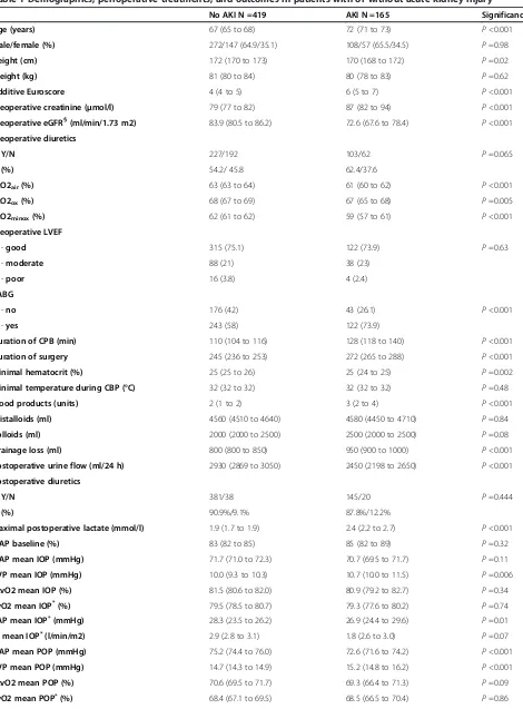
Patient demographics, cardiovascular risk factors, and additive Euroscore were significantly different in patients with and without postoperative AKI (Table 1). Addition-ally, surgical core markers (duration of surgery and dur-ation of CPB) pointed to a significantly higher operative risk. Patients with AKI were treated more frequently and with higher doses of inotropes and vasopressors, needed more blood products and showed a trend toward a higher use of synthetic colloids. Specific analysis of the doses of hydroxyethyl starch and gelatine showed that patients with AKI were treated with a significantly higher amount of gelatine (no AKI: 1,500 (1,500 to 2,000) ml, AKI: 2,000 (1,500 to 2,000) ml;P=0.009).
Analysis of intra- and postoperative hemodynamics showed minor, but at several time points statistically sig-nificant differences in mean arterial and central venous pressures, central venous oxygen saturation (ScvO2) and, in invasively monitored patients, differences in mean pulmonary artery pressure (PAP) and cardiac index (CI) between patients with postoperative preserved renal func-tion and patients with AKI (Table S1 in Addifunc-tional file 1 and Table 1). Average renal perfusion pressure during the first postoperative 8 h was significantly lower in patients with AKI in comparison with patients without this com-plication (Table 1).
Time to extubation and renal outcomes
Median time to the first extubation was 7 h (6.8 to 7.0 h). Forty-seven patients (8.1%) were reintubated for at least one time. Total postoperative ventilation time in rein-tubated patients was 190 h (109 to 365 h). Seventeen patients were treated with tracheostomy for long-term respiratory support.
The rate of AKI increased from 17.0% in patients extu-bated within 4 h postoperatively to 62.3% in patients ventilated for equal or more than 16 h (P<0.001) in the total cohort (Figure 2). Exclusion of the 47 patients needing reintubation revealed a lower incidence of AKI (24.6%), but a comparable pattern, with an AKI rate of 12.4% in patients with less than 4 h to first extubation and 53.8% in patients with prolonged (≥16 h) ventilator support (P<0.001).
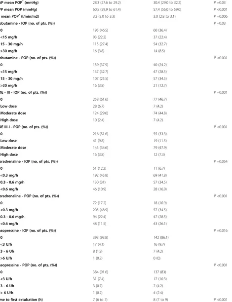
Potential risk factors for longer mechanical ventilation after cardiothoracic surgery in the groups with different extubation times are summarized in Table 2, showing that - for the total cohort - patients that were venti-lated longer were older, had a higher Euroscore, a lower estimated glomerular filtration rate (eGFR), a longer duration of surgery, a higher postoperative drainage loss, a higher use of blood products, and were post-operatively treated with higher doses of noradrenaline and dobutamine.
Analyses of hemodynamics within the first 8 h after surgery in patients with different times to extubation
revealed that patients ventilated for more than 16 h had a lower postoperative MAP, a higher CVP and, consecu-tively, a lower RPP than patients ventilated for a shorter periods of time. There was no significant differences in RPP between patients ventilated for ≤4 h and >4 h to ≤8 h after surgery (Table 2).
Logistic regression in the total cohort showed that time to the first extubation was a predictor of AKI taking into account variables significantly associated with AKI in the univariate analysis and the need for reintubation. In the total cohort, the multivariate logistic regression showed that the duration of ventilation (odds ratio (OR): 1.024/ hour, 95% CI: 1.011 to 1.044/hour; P <0.001), age (OR: 1.035/year, 95% CI: 1.015 to 1.057/hour; P <0.001), dur-ation of surgery (OR: 1.003, 95% CI: 1.001 to 1.006;
P =0.02), RPP (OR: 0.963/mmHg; 95% CI: 0.934 to
0.992/mmHg; P <0.001), and reintubation (OR: 5.27;
95% CI: 2.545 to 11.499; P <0.001) were independently associated with AKI.
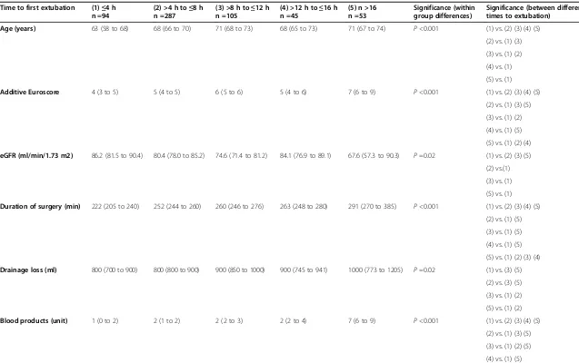
Figures 3 and 4 present the results of the multivariate logistic regression model for prototypical input values (25thand 75thpercentile) of the model’s variables.
Discussion
The present study aims to determine the effects of the duration of postoperative ventilation on the inci-dence of AKI in a heterogeneous cohort of elective car-diac surgical patients. The results of this analysis are highly suggestive that - among other well-accepted risk factors for AKI like age and renal hypoperfusion - a delay of extubation substantially increases the risk for develop-ing AKI.
AKI is an important complication in patients undergo-ing cardiac surgery [1,4]. Several mechanisms mediatundergo-ing a perioperative decrease in renal function have been identified within the last years. Among these factors are: exogeneous and endogeneous toxins, anemia, atheroem-bolic events, systemic and renal inflammation, hypergly-cemia, and neurohumoral activation [1,3,4,14]. However, a decrease in RPP and renal blood flow leading to a reduction in oxygen delivery remains a common and important pathway in these patients. The latter me-chanism has frequently been attributed to perioperative periods of overt low systemic blood flow, anemia, and/or hypotension [15,16], especially during CPB. It has been largely ignored that almost all patients undergoing cardiac surgery are mechanically ventilated during and after surgery, and that positive pressure ventilation itself impacts renal function via humoral and neurohumoral pathways ultimately leading to a decrease in glomerular filtration rate and renal blood flow [5,6]. It is of note that the complex neurohumoral effects of mechanical ventilation on the kidney are - at least partially - inde-pendent from overt changes in hemodynamics and may
Heringlakeet al. Critical Care2014,18:547 Page 4 of 13
Table 1 Demographics, perioperative treatments, and outcomes in patients with or without acute kidney injury
No AKI N =419 AKI N =165 Significance
Age (years) 67 (65 to 68) 72 (71 to 73) P<0.001
Male/female (%) 272/147 (64.9/35.1) 108/57 (65.5/34.5) P=0.98
Height (cm) 172 (170 to 173) 170 (168 to 172) P=0.02
Weight (kg) 81 (80 to 84) 80 (78 to 83) P=0.62
Additive Euroscore 4 (4 to 5) 6 (5 to 7) P<0.001
Preoperative creatinine (μmol/l) 79 (77 to 82) 87 (82 to 94) P<0.001
Preoperative eGFR§(ml/min/1.73 m2) 83.9 (80.5 to 86.2) 72.6 (67.6 to 78.4) P<0.001
Preoperative diuretics
Y/N 227/192 103/62 P=0.065
(%) 54.2/ 45.8 62.4/37.6
ScO2air(%) 63 (63 to 64) 61 (60 to 62) P<0.001
ScO2ox(%) 68 (67 to 69) 67 (65 to 68) P=0.005
ScO2minox(%) 62 (61 to 62) 59 (57 to 61) P<0.001
Preoperative LVEF
-good 315 (75.1) 122 (73.9) P=0.63
-moderate 88 (21) 38 (23)
-poor 16 (3.8) 4 (2.4)
CABG
-no 176 (42) 43 (26.1) P<0.001
-yes 243 (58) 122 (73.9)
Duration of CPB (min) 110 (104 to 116) 128 (118 to 140) P<0.001
Duration of surgery 245 (236 to 253) 272 (265 to 288) P<0.001
Minimal hematocrit (%) 25 (25 to 26) 25 (24 to 25) P=0.002
Minimal temperature during CBP (°C) 32 (32 to 32) 32 (32 to 32) P=0.48
Blood products (units) 2 (1 to 2) 3 (2 to 4) P<0.001
Cristalloids (ml) 4560 (4510 to 4640) 4580 (4450 to 4710) P=0.84
Colloids (ml) 2000 (2000 to 2500) 2500 (2000 to 2500) P=0.08
Drainage loss (ml) 800 (800 to 850) 950 (900 to 1000) P<0.001
Postoperative urine flow (ml/24 h) 2930 (2869 to 3050) 2450 (2198 to 2650) P<0.001
Postoperative diuretics
Y/N 381/38 145/20 P=0.444
(%) 90.9%/9.1% 87.8%/12.2%
Maximal postoperative lactate (mmol/l) 1.9 (1.7 to 1.9) 2.4 (2.2 to 2.7) P<0.001
MAP baseline (%) 83 (82 to 85) 85 (82 to 89) P=0.32
MAP mean IOP (mmHg) 71.7 (71.0 to 72.3) 70.7 (69.5 to 71.7) P=0.11
CVP mean IOP (mmHg) 10.0 (9.3 to 10.3) 10.7 (10.0 to 11.5) P=0.006
ScvO2 mean IOP (%) 81.5 (80.6 to 82.0) 80.9 (79.2 to 82.7) P=0.34
SvO2 mean IOP*(%) 79.5 (78.5 to 80.7) 79.3 (77.6 to 80.2) P=0.74
PAP mean IOP*(mmHg) 28.3 (23.5 to 26.2) 26.9 (24.4 to 29.6) P=0.01
CI mean IOP*(l/min/m2) 2.9 (2.8 to 3.1) 1.8 (2.6 to 3.0) P=0.07
MAP mean POP (mmHg) 75.2 (74.4 to 76.0) 72.6 (71.6 to 74.2) P<0.001
CVP mean POP (mmHg) 14.7 (14.3 to 14.9) 15.2 (14.8 to 16.2) P<0.001
ScvO2 mean POP (%) 70.6 (69.5 to 71.7) 69.3 (66.4 to 71.3) P=0.09
Table 1 Demographics, perioperative treatments, and outcomes in patients with or without acute kidney injury (Continued)
PAP mean POP*(mmHg) 28.3 (27.6 to 29.2) 30.4 (29.0 to 32.2) P=0.03
RPP mean POP (mmHg) 60.5 (59.9 to 61.4) 57.4 (56.0 to 59.0) P<0.001
CI mean POP*(l/min/m2) 3.2 (3.0 to 3.3) 3.0 (2.8 to 3.1) P=0.006
Dobutamine - IOP (no. of pts. (%)) P=0.03
0 195 (46.5) 60 (36.4)
<15 mg/h 93 (22.2) 37 (22.4)
15 - 30 mg/h 115 (27.4) 54 (32.7)
>30 mg/h 16 (3.8) 14 (8.5)
Dobutamine - POP (no. of pts. (%)) P<0.001
0 159 (37.9) 40 (24.2)
<15 mg/h 137 (32.7) 47 (28.5)
15 - 30 mg/h 107 (25.5) 57 (34.5)
>30 mg/h 16 (3.8) 21 (12.7)
PDE - III - IOP (no. of pts. (%)) P=0.001
0 258 (61.6) 77 (46.7)
Low dose 28 (6.7) 7 (4.2)
Moderate dose 124 (29.6) 74 (44.8)
High dose 10 (2.4) 7 (4.2)
PDE III-I - POP (no. of pts. (%)) P<0.001
0 216 (51.6) 55 (33.3)
Low dose 41 (9.8) 19 (11.5)
Moderate dose 145 (34.6) 79 (47.9)
High dose 16 (3.8) 12 (7.3)
Noradrenaline - IOP (no. of pts. (%)) P=0.054
0 51 (12.2) 11 (6.7)
<0.3 mg/h 192 (45.8) 69 (41.8)
0.3 - 0.6 mg/h 130 (31) 57 (34.5)
<0.6 mg/h 46 (10.9) 28 (16.9)
Noradrenaline - POP (no. of pts. (%)) P<0.001
0 72 (17.2) 18 (10.9)
<0.3 mg/h 205 (48.9) 57 (34.5)
0.3 - 0.6 mg/h 94 (22.4) 47 (28.5)
<0.6 mg/h 48 (11.5) 43 (26.1)
Vasopressine - IOP (no. of pts. (%)) P=0.016
0 393 (93.8) 142 (86.1)
<3 U/h 17 (4.1) 16 (9.7)
3 - 6 Uh 8 (1.9) 7 (4.2)
>6 U/h 1 (0.2) 0 (0)
Vasopressine - POP (no. of pts. (%)) P<0.001
0 384 (91.6) 137 (83)
<3 U/h 31 (7.4) 17 (10.3)
3 - 6 Uh 3 (0.7) 7 (4.2)
> 6 U/h 1 (0.2) 4 (2.4)
Time to first extubation (h) 7 (6 to 7) 8 (7 to 9) P<0.001
Heringlakeet al. Critical Care2014,18:547 Page 6 of 13
be difficult to detect using conventional clinical moni-toring [17].
Thus, despite the interactions between established mediators of CSA-AKI and mechanical ventilation have not been formally analyzed, it is rather plausible that mechanical ventilation may act synergistically with estab-lished AKI - triggers to further reduce renal function in the perioperative setting.
The findings of the present study clearly support this concept: according to the results of the logistic regres-sion analysis every hour of postoperative mechanical ventilation independently increases the odds of develop-ing AKI by a factor of 1.024. It is of note that the time to the first extubation was more important as a risk
factor for AKI than conventional risk stratification tools like additive Euroscore [18] and cerebral oxygen satur-ation [10] and remained statistically significant in the multivariate logistic regression after adjustment for the fact that patients developing AKI also had a lower RPP in the immediate time period after surgery. Interestingly, the average MAP in patients that developed AKI in the postoperative course was in a range that is convention-ally regarded as safe, the CVP values were only slightly above normal, and only the calculation of the effective RPP revealed that patients developing AKI suffered from (still numerically moderate) renal hypoperfusion.
[image:7.595.59.539.113.211.2]Despite the effects of positive airway pressure on kidney function have been the focus of several experimental Table 1 Demographics, perioperative treatments, and outcomes in patients with or without acute kidney injury (Continued)
Reintubation P<0.001
No 409 (97.6) 133 (80.6)
Yes 9 (2.1) 32 (19.4)
Total ventilation time (h) 7 (6 to 7) 9 (7.4 to 11) P<0.001
Duration of ICU treatment (h) 23 (22 to 23) 66 (38 to 81) P<0.001
Duration of HDU treatment (h) 70 (65 to 77) 124 (108 to 144) P<0.001
Mortality 30 d (no. of pts. (%)) 1 (0.2) 10 (6.1) P<0.001
Continuous data are given as median and 95% confidence interval of the median; categorical data as absolute number with percentages in parenthesis. §
Estimated glomerular filtration rate determined by the modifications of diet in renal disease equation;*
data derived from patients monitored with pulmonary artery catheter. AKI: acute kidney injury; eGFR: estimated glomerular filtration rate; ScO2: cerebral oxygen saturation determined by near-infrared spectroscopy (ScO2air: bihemispheric mean values during room air; ScO2ox: bihemispheric mean values during oxygen supplementation; ScO2minox: lowest value of either side
during oxygen supplementation); LVEF: left ventricular ejection fraction; CABG: coronary artery bypass grafting; CPB: cardiopulmonary bypass; IOP: intraoperatively; POP: postoperatively; MAP: mean arterial pressure; CVP: central venous pressure; PAP: mean pulmonary arterial pressure; ScvO2: central venous oxygen saturation; SvO2: mixed venous oxygen saturation; CI: cardiac index; RPP: renal perfusion pressure (MAP - CVP); PDE-III: phosphodiesterase III inhibitors (enoximone or milrinone), ICU: intensive care unit; HDU: high dependency unit. The amount of fluids, blood products (packed red cells and fresh frozen plasma) represents the volumes or units applied during surgery and within the first day on the ICU.
[image:7.595.56.540.501.705.2]Table 2 Demographics, risk factors, and treatments in patients with different time to extubation after cardiac surgery
Time to first extubation (1)≤4 h n =94
(2) >4 h to≤8 h n =287
(3) >8 h to≤12 h n =105
(4) >12 h to≤16 h n =45
(5) n >16 n =53
Significance (within group differences)
Significance (between different times to extubation)
Age (years) 63 (58 to 68) 68 (66 to 70) 71 (68 to 73) 68 (65 to 73) 71 (67 to 74) P<0.001 (1) vs. (2) (3) (4) (5)
(2) vs. (1) (3)
(3) vs. (1) (2)
(4) vs. (1)
(5) vs. (1)
Additive Euroscore 4 (3 to 5) 5 (4 to 5) 6 (5 to 6) 5 (4 to 6) 7 (6 to 9) P<0.001 (1) vs. (2) (3) (4) (5)
(2) vs. (1) (3) (5)
(3) vs. (1) (2)
(4) vs. (1) (5)
(5) vs. (1) (2) (4)
eGFR (ml/min/1.73 m2) 86.2 (81.5 to 90.4) 80.4 (78.0 to 85.2) 74.6 (71.4 to 81.2) 84.1 (76.9 to 89.1) 67.6 (57.3 to 90.3) P=0.02 (1) vs. (2) (3) (5)
(2) vs.(1)
(3) vs. (1)
(5) vs. (1)
Duration of surgery (min) 222 (205 to 240) 252 (244 to 260) 260 (246 to 276) 263 (248 to 280) 291 (270 to 385) P<0.001 (1) vs. (2) (3) (4) (5)
(2) vs. (1) (5)
(3) vs. (1) (5)
(4) vs. (1) (5)
(5) vs. (1) (2) (3) (4)
Drainage loss (ml) 800 (700 to 900) 800 (800 to 900) 900 (850 to 1000) 900 (745 to 941) 1000 (773 to 1205) P=0.02 (1) vs. (3) (5)
(2) vs. (3) (5)
(3) vs. (1) (2)
(5) vs. (1) (2)
Blood products (unit) 1 (0 to 2) 2 (1 to 2) 2 (2 to 3) 2 (2 to 4) 7 (6 to 9) P<0.001 (1) vs. (2) (3) (4) (5)
(2) vs. (1) (3) (5)
(3) vs. (1) (2) (5)
(4) vs. (1) (5)
(5) vs. (1) (2) (3) (4)
Heringlake
et
al.
Critical
Care
2014,
18
:547
Page
8
o
f
1
3
http://ccfo
rum.com/con
(3) vs. (1) (4)
(4) vs. (3)
(5) vs. (1)
CVP POP (mmHg) 13.9 (13.2 to 14.7) 14.4 (14.0 to 14.8) 15.2 (14.9 to 15.9) 15.6 (14.6 to 16.1) 16.9 (15.6 to 18.0) P<0.001 (1) vs. (3) (4) (5)
(2) vs. (3) (4) (5)
(3) vs. (1) (2) (5)
(4) vs. (1) (2)
(5) vs. (1) (2) (3)
RPP POP (mmHg) 62.4 (60.0 to 65.1) 60.1 (59.5 to 61.3) 57.9 (56.8 to 59.6) 60.6 (58.3 to 63.4) 57.8 (53.6 to 59.5) P<0.001 (1) vs. (3) (5)
(2) vs. (3) (5)
(3) vs. (1) (2)
(4) vs. (5)
(5) vs. (1) (2) (4)
Dobutamine - POP P<0.001
0 39 (41.5%) 108 (37.6%) 26 (13.3%) 12 (26.7%) 14 (26.4%)
<15 mg/h 28 (29.8%) 94 (32.8%) 32 (16.4%) 14 (31.1%) 16 (30.2%)
15 - 30 mg/h 23 (24.5%) 72 (25.1%) 41 (21%) 17 (37.8%) 12 (22.6%)
>30 mg/h 4 (4.2%) 14 (4.9%) 6 (3.1%) 2 (4.4%) 11 (20.8%)
Noradrenaline - POP P<0.001
0 18 (19.1%) 57 (19.9%) 9 (4.6%) 3 (6.7%) 3 (5.7%)
<0.3 mg/h 50 (53.2%) 136 (47.4%) 44 (22.6%) 23 (51.1%) 9 (17%)
0.3 - 0.6 mg/h 18 (19.1%) 68 (23.7%) 33 (16.9%) 13 (28.9%) 9 (17%)
>0.6 mg/h 8 (8.5%) 26 (9.1%) 19 (9.7%) 6 (13.3%) 32 (60.4%)
AKI stage P<0.001
0 78 (83%) 212 (73.9%) 75 (71.5%) 34 (75.5%) 20 (37.7%)
1 8 (8.5%) 45 (15.7%) 18 (17,1%) 8 (17.8%) 6 (11.3%)
2 1 (1.1%) 5 (1.7%) 0 (0%) 0 (0%) 1 (1.9%)
3 7 (7.4%) 25 (8.7%) 12 (11.4%) 3 (6.7%) 26 (49.1%)
Differences in variables strongly associated with acute kidney injury in univariate analysis in groups of cardiac surgical patients stratified according to the times of first extubation after surgery. Data are given as median and 95% confidence interval of the median or absolute numbers with percentages in parenthesis. ScO2ox: preoperative oxygen-supplemented cerebral oxygen saturation determined by near-infrared spectroscopy. eGFR: estimated
glomerular filtration rate by the Modifications of Diet in Renal Disease (MDRD) formula; POP: postoperative; MAP: average mean arterial pressure within 8 h after surgery; CVP: average central venous pressure within 8 h after surgery; RPP: average renal perfusion pressure (MAP - CVP) within 8 h after surgery; AKI: acute kidney injury. Kruskal-Wallis and chi-square test.
et
al.
Critical
Care
2014,
18
:547
Page
9
o
f
1
3
[image:9.794.63.691.67.491.2]Figure 4Graphical depiction of the effect of intubation time and renal perfusion pressure on the probability of developing acute kidney injury (y-axis) for a prototypical patient (age 66 years; duration of surgery 266 min).
Figure 3Graphical depiction of the results of the multivariate logistic regression model in the total cohort using prototypical input values (25th and 75th percentiles of the respective variables).Intubation time (x-axis) independently increased the probability of developing acute kidney injury (y-axis).Left:Duration of surgery 266 min; mean renal perfusion pressure 60 mmHg.Right:Age 64 years old, mean renal perfusion pressure 60 mmHg. Intubation time (x-axis) independently increased the probability of developing acute kidney injury (y-axis).
Heringlakeet al. Critical Care2014,18:547 Page 10 of 13
studies [6], clinical data on this topic from cardiac surgical patients are sparse. Van den Akker and coworkers have shown in a meta-analysis, that mechanical ventilation for more than 24 h is associated with a threefold increase in the risk for developing AKI in critically ill patients [7]. However, only one study in cardiac surgical patients was included in this analysis. Brito and coworkers studied 186 patients during a three-year period and observed, that in the univariate analysis, mechanical ventilation for more than 24 h was a risk factor of AKI (defined as a 50% increase in creatinine or need for dialysis). Probably as an effect of the relatively small sample size, this variable was no longer significant in multivariate analysis [19].
Koning and coworkers analyzed the effects of intermit-tent positive airway pressure (IPPV) and spontaneous ventilation on cardiac output, renal blood flow, creatinine clearance, and urinary electrolyte excretion and observed decreased renal blood flow and creatinine clearance during IPPV conditions that partially improved during spontaneous ventilation. However, no data on the effects of mechanical ventilation on incidence of AKI were pre-sented in this study [20].
Within the last years, further interactions between mechanical ventilation and AKI have been observed that may also have implications for cardiac surgical patients. A wealth of experimental [21] and an in-creasing number of clinical studies are suggestive that non-protective ventilation may induce pulmonary bio-trauma, inflammation, and subsequent non-pulmonary organ dysfunction (including kidney dysfunction) [22,23]. In line with this, Lellouche and coworkers observed that car-diac surgical patients ventilated with traditional (10 ml/kg) or higher tidal volume are at increased risk for extrapulmon-ary organ dysfunction as well as prolonged ICU stay and that these complications translate into poor long-term outcomes [8]. And despite some other studies failing to show an effect of the ventilation mode on systemic inflam-mation [24], a recent meta-analysis - including several studies in cardiac surgical patients - came to the con-clusion that protective ventilation with lower volumes may lead to better clinical outcomes [25].
Limitations
Despite the sound pathophysiological background de-scribed above, it has to be acknowledged that patients in the present study who were not extubated immediately after surgery were older, had a higher risk profile, had a lower RPP, and were more frequently treated with vaso-active or inotropic drugs than patients extubated early. However, the results of the logistic regression analysis are clearly indicative that the duration of postoperative positive pressure ventilation is an independent risk fac-tor of AKI, taking into account all the above-mentioned factors. Nonetheless, some uncertainty remains, since it
cannot definitively be ruled out that the adverse effects of longer ventilation after surgery may be mediated by unrecognized or undetermined confounders like the nephrotoxic effects of synthetic colloids [26] that were more frequently used in patients with AKI or short-term variations in hemodynamics that remained undetectable during hourly measurements.
Unfortunately, we have no data on individual patient ventilation strategies in the present study. Tidal volumes during mechanical ventilation or while breathing spon-taneously with pressure support were adjusted by nurses and physicians according to standard hospital practice in the range between 6 and 8 ml/kg. However, it cannot, of course, be completely ruled out that periods of non-protective ventilation may have occurred in these patients and have induced a proinflammatory state amplifying the development of AKI.
With respect to the observational nature of the present study we did not perform a power analysis a priori for this specific research question. The relatively high number of events (that is patients developing AKI) strengthens our findings, even if the total sample size was only moderate. However, our findings present only a single-center experi-ence and thus may need to be replicated in a prospective and multicenter fashion.
Conclusions
In conclusion, the results of the present study suggest that mechanical ventilation is an important trigger for AKI in cardiac surgical patients and that even a moder-ate delay of extubation after surgery may substantially increase the risk of developing AKI. If replicated inde-pendently, this observation may not only have relevant implications for clinical management (supporting the concept of fast-track cardiac anesthesia) but also for AKI trials.
Key messages
Acute kidney injury (AKI) is a serious and frequent complication in patients undergoing cardiac surgery.
Positive pressure ventilation may influence renal hemodynamics via neurohumoral pathways.
The present observational study shows that the time to the first extubation after cardiac surgery is independently associated with the risk of developing AKI.
Additional file
Additional file 1: Table S1.Perioperative hemodynamics in cardiac surgical patients with or without acute kidney injury.
Abbreviations
AKI:acute kidney injury; AKIN: Acute Kidney Injury Network; BIPAP: biphasic intermittent positive airway pressure; CABG: coronary artery bypass grafting; CI: cardiac index; CPAP: continuous positive airway pressure; CPB: cardiopulmonary bypass; CSA-AKI: cardiac surgery-associated acute kidney injury; CVP: central venous pressure; eGFR: estimated glomerular filtration rate; HDU: high dependency unit; ICU: intensive care unit; IPAP: intermittent positive airway pressure; LVEF: left ventricular ejection fraction; MAP: mean arterial blood pressure; NIRS: near-infrared spectroscopy; PAP: mean pulmonary arterial pressure; PEEP: positive end-expiratory pressure; PSV: pressure support ventilation; RPP: renal perfusion pressure; RRT: renal replacement therapy; ScO2: cerebral oxygen saturation; ScvO2: central venous oxygen saturation.
Competing interests
The authors JS and MH receive honoraria for lectures from Covidien Germany GmbH, Neustadt, Germany, the manufacturer of the cerebral oximeter used in this study. The other authors declare that they have no competing interests.
Authors’contributions
MH designed the study, participated in data acquisition and analyses, and wrote the manuscript. YN participated in data acquisition, data analyses, and preparation of the manuscript. JS, AEB, JT, EIC, and HP participated in the data analyses and drafting of the manuscript. All authors read and approved the manuscript.
Funding
The present work was funded by the Department of Anesthesiology, University of Lübeck.
Presented in abstracted form during the 36thAnnual Meeting of the Society of Cardiovascular Anesthesiologists 29 March 2014 to 2 April 2014, New Orleans, LA, USA.
Author details
1
Department of Anesthesiology and Intensive Care Medicine, University of Lübeck, Ratzeburger Allee 160, 23538 Lübeck, Germany.2Department of Cardiac and Thoracic Vascular Surgery, University of Lübeck, Ratzeburger Allee 160, 23538 Lübeck, Germany.
Received: 16 May 2014 Accepted: 17 September 2014
References
1. Stafford-Smith M, Shaw A, Swaminathan M:Cardiac surgery and acute kidney injury: emerging concepts.Curr Opin Crit Care2009,15:498–502. 2. Lassnigg A, Schmidlin D, Mouhieddine M, Bachmann LM, Druml W, Bauer P,
Hiesmayr M:Impact of minimal increases in serum creatinine on outcome in patients after cardiothoracic surgery: do we have to revise current definitions of acute renal failure?Crit Care Med2008,
36:1129–1137.
3. Bellomo R, Auriemma S, Fabbri A, D’Onofrio A, Katz N, McCullough PA, Ricci Z, Shaw A, Ronco C:The pathophysiology of cardiac surgery-associated acute kidney injury (CSA-AKI).Int J Artif Organs2008,31:166–178.
4. Mao H, Katz N, Ariyanon W, Blanca-Martos L, Adýbelli Z, Giuliani A, Danesi TH, Kim JC, Nayak A, Neri M, Virzi GM, Brocca A, Scalzotto E, Salvador L, Ronco C:Cardiac surgery-associated acute kidney injury.Blood Purif2014,
37:34–50.
5. Marquez JM, Douglas ME, Downs JB, Wu WH, Mantini EL, Kuck EJ, Calderwood HW:Renal function and cardiovascular responses during positive airway pressure.Anesthesiology1979,50:393–398.
6. Kaczmarczyk G:Pulmonary-renal axis during positive-pressure ventilation. New Horiz1994,2:512–517.
7. Van den Akker JP, Egal M, Groeneveld JA:Invasive mechanical ventilation as a risk factor for acute kidney injury in the critically ill: a systematic review and meta-analysis.Crit Care2013,17:R98.
8. Lellouche F, Dionne S, Simard S, Bussières J, Dagenais F:High tidal volumes in mechanically ventilated patients increase organ dysfunction after cardiac surgery.Anesthesiology2012,116:1072–1082.
9. Heringlake M, Heinze H, Schubert M, Nowak Y, Guder J, Kleinebrahm M, Paarmann H, Hanke T, Schön J:A perioperative infusion of sodium bicarbonate does not improve renal function in cardiac surgery patients: a prospective observational cohort study.Crit Care2012,16:R156. 10. Heringlake M, Garbers C, Kabler JH, Anderson I, Heinze H, Schön J, Berger
KU, Dibbelt L, Sievers HH, Hanke T:Preoperative cerebral oxygen saturation and clinical outcomes in cardiac surgery.Anesthesiology2011,
114:58–69.
11. Carl M, Alms A, Braun J, Dongas A, Erb J, Goetz A, Goepfert M, Gogarten W, Grosse J, Heller AR, Heringlake M, Kastrup M, Kroener A, Loer SA, Marggraf G, Markewitz A, Reuter D, Schmitt DV, Schirmer U, Wiesenack C, Zwissler B, Spies C:S3 guidelines for intensive care in cardiac surgery patients: hemodynamic monitoring and cardiocirculary system.Ger Med Sci2010,
8:Doc 12.
12. Mehta RL, Kellum JA, Shah SV, Molitoris BA, Ronco C, Warnock DG, Levin A, Acute Kidney Injury Network:Acute Kidney Injury Network: report of an initiative to improve outcomes in acute kidney injury.Crit Care2007,
11:R31.
13. Development Core Team:R: a language and environment for statistical computing.Vienna, Austria: R Foundation for Statistical Computing; 2014. ISBN 3-900051-07-0.
14. Mao H, Katz N, Ariyanon W, Blanca-Martos L, Adýbelli Z, Giuliani A, Danesi TH, Kim JC, Nayak A, Neri M, Virzi GM, Brocca A, Scalzotto E, Salvador L, Ronco C:Cardiac surgery-associated acute kidney injury.Cardiorenal Med
2013,3:178–199.
15. Sickeler R, Phillips-Bute B, Kertai MD, Schroder J, Mathew JP, Swaminathan M, Stafford-Smith M:The risk of acute kidney injury with co-occurrence of anemia and hypotension during cardiopulmonary bypass relative to anemia alone.Ann Thorac Surg2014,97:865–871.
16. Haase M, Bellomo R, Story D, Letis A, Klemz K, Matalanis G, Seevanayagam S, Dragun D, Seeliger E, Mertens PR, Haase-Fielitz A:Effect of mean arterial pressure, haemoglobin and blood transfusion during cardiopulmonary bypass on post-operative acute kidney injury.Nephrol Dial Transplant
2012,27:153–160.
17. Braam B, Cupples WA, Joles JA, Gaillard C:Systemic arterial and venous determinants of renal hemodynamics in congestive heart failure.Heart Fail Rev2012,17:161–175.
18. Ho J, Reslerova M, Gali B, Nickerson PW, Rush DN, Sood MM, Bueti J, Komenda P, Pascoe E, Arora RC, Rigatto C:Serum creatinine measurement immediately after cardiac surgery and prediction of acute kidney injury. Am J Kidney Dis2012,59:196–201.
19. Brito DJ, Nina VJ, Nina RV, Figueiredo Neto JA, Oliveira MI, Salgado JV, Lages JS, Salgado FN:Prevalence and risk factors for acute renal failure in the postoperative of coronary artery bypass grafting.Rev Bras Cir Cardiovasc2009,24:297–304.
20. Koning HM, Leusink JA, Nas AA, van Scheyen EJ, van Urk P, Haas FJ, Koning AJ:Renal function following open heart surgery: the influence of postoperative artificial ventilation.Thorac Cardiovasc Surg1988,36:1–4. 21. Dreyfuss D, Saumon G:Ventilator-induced lung injury: lessons from
experimental studies.Am J Respir Crit Care Med1998,157:294–323. 22. Ranieri VM, Suter PM, Tortorella C, De Tullio R, Dayer JM, Brienza A, Bruno F,
Slutsky AS:Effect of mechanical ventilation on inflammatory mediators in patients with acute respiratory distress syndrome: A randomized controlled trial.JAMA1999,282:54–61.
23. Reis Miranda D, Gommers D, Struijs A, Dekker R, Mekel J, Feelders R, Lachmann B, Bogers AJ:Ventilation according to the open lung concept attenuates pulmonary inflammatory response in cardiac surgery. Eur J Cardiothorac Surg2005,28:889–895.
24. Wrigge H, Zinserling J, Stüber F, von Spiegel T, Hering R, Wetegrove S, Hoeft A, Putensen C:Effects of mechanical ventilation on release of cytokines into systemic circulation in patients with normal pulmonary function.Anesthesiology2000,93:1413–1417.
25. Serpa Neto A, Cardoso SO, Manetta JA, Pereira VG, Espósito DC, Pasqualucci Mde O, Damasceno MC, Schultz MJ:Association between use of lung-protective ventilation with lower tidal volumes and clinical outcomes
Heringlakeet al. Critical Care2014,18:547 Page 12 of 13
among patients without acute respiratory distress syndrome: a meta-analysis.JAMA2012,308:1651–1659.
26. Zarychanski R, Abou-Setta AM, Turgeon AF, Houston BL, McIntyre L, Marshall JC, Fergusson DA:Association of hydroxyethyl starch administration with mortality and acute kidney injury in critically ill patients requiring volume resuscitation: a systematic review and meta-analysis.JAMA2013,309:678–688.
doi:10.1186/s13054-014-0547-4
Cite this article as:Heringlakeet al.:Postoperative intubation time is associated with acute kidney injury in cardiac surgical patients.Critical Care201418:547.
Submit your next manuscript to BioMed Central and take full advantage of:
• Convenient online submission
• Thorough peer review
• No space constraints or color figure charges
• Immediate publication on acceptance
• Inclusion in PubMed, CAS, Scopus and Google Scholar
• Research which is freely available for redistribution