ISSN Online: 2168-1597 ISSN Print: 2168-1589
DOI: 10.4236/abcr.2018.72010 Apr. 30, 2018 134 Advances in Breast Cancer Research
Role of ESR Pathway Genes in Breast Cancer: A
Review
Deepak Kumar
1*, Marilyn Rae Myers
2, Ussama Al Homsi
3, Valentin Ilyin
11Computational Biology, Carnegie Mellon University in Qatar (CMU-Q), Doha, Qatar 2Pennsylvania State University, University Park, USA
3Hematology and Oncology Department, NCCCR, Hamad Medical Corporation, Doha, Qatar
Abstract
Breast cancer is the leading cause of death in women. Prognosis of breast cancer is often pessimistic because the tumors are prone to metastasizing to the bone, brain, and lung. The estrogen signaling receptor (ESR) pathway contains 39 main genes and proteins which makes it one of the larger signal-ing pathways. Predominately this pathway and the proteins within are in-volved in breast growth and development, making it a prospective area of study for breast cancer. While the healthy ESR pathway has been constructed and is well established, a mechanistic model of mutated genes of ESR pathway has not been delved upon. Such mutated models could be utilized for selecting combinational targets for drug therapies, as well as elucidating crosstalk be-tween other pathways and feedback mechanisms. To construct the mutated models of the ESR pathway it is imperative to assess what is currently unders-tood in the literature and what inconsistencies exist in order to resolve them. Without this information, a model of the ESR pathway will be unreliable and likely unproductive. This review is the detailed literature survey of the biolog-ical studies performed on ESR pathways genes, and their respective roles in breast cancer. Furthermore, the details mentioned in the review can be bene-ficial for the integrated study of the ESR pathway genes, which includes, structural and dynamics study of the genes products, to have a holistic under-standing of the cancer mechanism.
Keywords
Estrogen Signaling Receptor (ESR) Pathway, Breast Cancer, ESR Genes, Mechanistic Modeling, Integrated Study, Kyoto Encyclopedia of Genes and Genomes (KEGG), PubMed, Literature Survey
How to cite this paper: Kumar, D., Myers, M.R., Al Homsi, U. and Ilyin, V. (2018) Role of ESR Pathway Genes in Breast Can-cer: A Review. Advances in Breast Cancer Research, 7, 134-186.
https://doi.org/10.4236/abcr.2018.72010
Received: March 20, 2018 Accepted: April 27, 2018 Published: April 30, 2018
Copyright © 2018 by authors and Scientific Research Publishing Inc. This work is licensed under the Creative Commons Attribution International License (CC BY 4.0).
DOI: 10.4236/abcr.2018.72010 135 Advances in Breast Cancer Research
1. Introduction
Breast cancer is an aggressive and invasive disease that produces a malig-nant tumor due to multiple mutations in the genome. It is known that breast cancer is the leading cause of mortality and morbidity in women by cancer worldwide, although in some rare cases the disease has been diagnosed in male patients. In 2016, it is estimated that there will be 246,660 new cases of invasive breast cancer
and 40,450 deaths attributed to breast cancer in the United States [1].
Addition-ally, it is alarming to observe that the incidence rate has been rising in many countries [2]. A major factor in breast cancer fatality is that the disease fre-quently metastasizes and invades multiple regions in the body, including the bone, lung, and brain. Consequently, it is pertinent to research how breast can-cer develops in order to design an appropriate method of treatment.
To develop such a treatment, it is critical to have a comprehensive under-standing of the mutated genes that cause breast cancer and the mechanistic changes that occur. Whereas there is much research on individual genes and smaller cascade models, there has been little done to obtain a more holis-tic comprehension of these effects. In order to facilitate the development of mutated pathway models, it is the aim of this report to compile and incorporate sources of research to be used for designing this larger pathway model. However, it must be noted that although this would be a useful endeavor it would not be specific to individuals. This is a significant distinction as there are multiple subtypes of breast cancer often containing different mutations which subsequently can cause varied reactions to specific treatments. While it would be advantageous to do this for all pathways that are known to mutate in the development of breast can-cer, this report will specifically look at the estrogen signaling receptor (ESR) pathway, using the Kyoto Encyclopedia of Genes and Genomes (KEGG) ESR
pathway as reference (Figure 1).
The ESR pathway is a prospective starting point to unravel the mechanisms of breast cancer. Estrogen, an integral component of the ESR pathway, is a steroidal hormone which is involved in multiple functions including the development and maturation of breasts. Due to its involvement in the growth of breasts it is a likely candidate in providing information regarding tumor formation. Conse-quently, study of the major ESR pathway estrogen is involved in, will improve our comprehension of the breast tumor development and progression. Research already shows that the ESR pathway is implicated in breast cancer progression,
and the majority of human breast cancers start out as estrogen dependent [3]. It
DOI: 10.4236/abcr.2018.72010 136 Advances in Breast Cancer Research
Figure 1. Representation of the ESR pathway from Kyoto Encyclopedia of Genes and Genomes (KEGG-PATHWAY: map04915).
By collecting information on what is currently known about the proteins and genes involved in the ESR pathway this can direct future studies, such as com-putational studies involving structural analysis of the molecules. Molecular dy-namic studies along with other computational methods could overcome the li-mitations of crystallography and provide useful structural and functional
in-sights [4]. Such studies can be useful in understanding the network dynam-ics of
the genes and protein molecules involved in breast cancer. Moreover, structural level study can provide insight into the architectural and conformational changes of the molecules occurring due to point mutations or single nucleotide polymorphism (SNP). While computational studies have been utilized in many areas of research, there are many avenues in the breast cancer field that could still benefit from such studies. Molecular dynamic studies have already benefit-ted some research, specifically by revealing a detailed consequence of the muta-tions on the p53 DNA-binding core domain that may now provide insight for
therapeutic approaches in breast cancer [5]. By laying out what is currently
DOI: 10.4236/abcr.2018.72010 137 Advances in Breast Cancer Research In Table 1, depending on the literature study, genes in varied correlation cat-egory were found to act as both tumor suppressor and promoter. Genes in the consensus correlation category are the genes that do not deviate from suppress-ing or promotsuppress-ing tumor progression accordsuppress-ing to current literature.
2. Literature Consensus on Correlations to Breast Cancer
This section contains information on genes where there is agreement across the literature on the correlation the gene has to breast cancer. However, the litera-ture may propose different mechanisms by which the gene induces tumor
for-mation. Overview of all the genes assessed and evaluated is illustrated in Table
2.
2.1. HBEGF Induces Invadopodia and Metastasis
[image:4.595.186.539.385.735.2]Heparin binding EGF like growth factor (HBEGF) can be found either bound to the cell membrane or as a solute in the ECM. Although the molecule must un-dergo proteolytic cleavage to become a solute in the ECM, the molecule’s function
Table 1. Shows whether in current literature a particular gene correlation to breast cancer is agreed upon or whether a gene is observed to act both as a tumor suppressor and pro-moter, as determined by this review. The genes are from ESR pathway of KEGG (PATHWAY: map04915).
Varied Correlations Consensus Correlation
E2 HBEGF
MMP EGFR
GABA GABAB
OPRM1 GIRK
AC mGluR
Shc Src
cAMP Gα
PI3K Grb2
PKC Akt
PKA Ras
PLC SOS
IP3R DAG
IP3 CAM
eNOS Raf
MAPK MEK
AP1 FKBP
NO CREB
DOI: 10.4236/abcr.2018.72010 138 Advances in Breast Cancer Research
Table 2. Genes and their cognate variants found in the healthy ESR pathway compiled using the ESR reference pathway from Kyoto Encyclopedia of Genes and Genomes (KEGG).
Genes found in the healthy ESR pathway Gene variants involved in the healthy ESR pathway
Estradiol-17 beta (E2)
Matrix metalloproteinase (MMP) MMP2, MMP9 Heparin-binding EGF-like growth factor (HBEGF)
Gamma-aminobutyric acid (GABA) Beta-Endorphin (βEnd)
Adenylate Cyclase (AC) ADCY5, ADCY6, ADCY7, ADCY8, ADCY9 ADCY1, ADCY2, ADCY3, ADCY4,
Epidermal growth factor receptor (EGFR)
Gamma-aminobutyric acid B receptor (GABAB) GABBR
GIRK KCNJ3, KCNJ5, KCNJ6, KCNJ9
Opioid receptor µ (OPRM1) Membrane estrogen receptor (mER)
Metabotropic glutamate receptors (mGluR1a) GRM1
Estrogen receptor (ER) NR3A1, NR3A2
Proto-oncogene, non-receptor tyrosine kinase (c-Src) Src Tyrosine-protein kinase (Shc) Shc1, Shc2, Shc3, Shc4 Cyclic adenosine monophosphate (cAMP)
Phosphatidylinositol-4,5-bisphosphate 3-kinase
(PI3K) PIK3C, PIK3R
Guanine nucleotide-binding protein G(i) subunit
alpha (Gi/o) GNAI, GNAO
Novel protein kinase C delta type (PKC delta) PRKCD Guanine Nucleotide-Binding Protein Alpha-Q (Gq) GNAQ Adenylate Cyclase-Stimulating G Alpha Protein (Gs) GNAS Protein kinase cAMP-activated catalytic subunit alpha
(PKA)
Growth factor receptor bound protein 2 (Grb2) Rac serine/threonine-protein kinase (Akt)
Phospholipase C (PLC) PLGB
GTPaseHras (Ras) HRAS, KRAS, NRAS
G protein-coupled estrogen receptor (GPER) GPR30 Son of Sevenless (SOS)
Diaclyglycerol (DAG)
Inositol 1,4,5-trisphosphate receptor (IP3R) ITPR1, ITPR2, ITPR3
Calmodulin (CAM) CALM
DOI: 10.4236/abcr.2018.72010 139 Advances in Breast Cancer Research
Continued
Raf proto-oncogene serine/threonine-protein kinase
(Raf) RAF1
Mitogen-activated protein kinase kinase (MEK) MAP2K1, MAP2K2 D-myo-inositol 1,4,5 triphosphate (IP3)
FK506 binding protein 4 (FKBP52) FKBP4_5 Mitogen-activated protein kinase (ERK1/2) MAPK1_3
Nitric ocide (NO)
FBJ murine osteosarcoma viral oncogene homolog
(TF) Fos, Jun, SP1
remains unchanged. HBEGF is implicated as a participant in various normal physiological and pathologic processes such as cell attachments, chemotaxis,
mitosis, and inhibition of cellular apoptosis [6]. It is a microenvironment
orga-nizer which contributes to niches where normal stem and progenitor cells are maintained and released into differentiation and where oncogenic processes take
place and keep developing [7]. Most importantly HBEGF is not only observed in
breast cancer but in a wide variety of cancers, such as ovarian and gastric cancer. There is little disagreement in the medical community over the expression of HBEGF in breast cancer; however the particular mechanism by which HBEGF acts is still under investigation. One report suggests that its subcellular localiza-tion and release of N- and C-terminal fragments are involved in oncogenic
be-haviors [8]. Additionally, the report observes that HBEGF is a potent inducer of
angiogenesis in vivo. It has been speculated that HBEGF and epidermal growth factor receptors (EGFRs) enhances invadopodia in breast cancer, which then acts as a mechanism for cell autonomous invasion that is mediated by matrix
metalloproteinase (MMP) 2 and 9 [9]. This is particularly interesting
consider-ing that some reports do not observe MMP 2 in breast cancer samples, and con-sequently acts as evidence for there to be additional research on MMPs presence in breast cancer and their associations with HBEGF.
2.2. EGFR Amplified in Triple Negative Breast Cancer
EGFR comprises of two domains, the extracellular ligand binding domain and the intracellular receptor tyrosine kinase (RTK) domain. When bound to a li-gand at the cellular level, the complex can induce cell proliferation but also alter adhesion and motility and prevent apoptosis; at the physiological level, the
com-plex promotes invasion and angiogenesis [10]. EGFR is involved in regulating
gefiti-DOI: 10.4236/abcr.2018.72010 140 Advances in Breast Cancer Research nib, cetuximab, and lapatinib, are often unsuccessful, and perhaps would be more effective by applying personalized medicine methods in order to predict patients who would most benefit from inhibition of EGFR. Though EGFR over-expression is observed in all subtypes of breast cancer, EGFR is more frequently overexpressed in triple-negative breast cancer (TNBC) and inflammatory breast
cancer, which are especially aggressive [11]. Therefore, it can be inferred that
personalized medicine could be more efficacious in EGFR treatment since some patients are more likely to have overexpression of EGFR. While EGFR is typical-ly more common in particular subsets of breast cancer, the gene is more liketypical-ly amplified than mutated. One study reports that out of 47 TNBC tumor samples there were no EGFR mutations but amplification of EGFR was observed in
well-characterized TNBCs (up to 92%) [12]. In addition to being able to predict
which patients will respond best to EGFR drug treatments, it is critical to reveal why EGFR treatments are not always effective. There is evidence for significant interactions between EGFR family members and other RTKs, such as the recep-tors for hepatocyte growth factor and insulin-like growth factor, and it is possi-ble that such alternative signaling pathways are linked to resistance to
EGFR-targeted therapies [13]. However, these speculations will require further
investigations in order to utilize EGFR treatments appropriately.
2.3. Tumor Suppressing Qualities of Beta-Endorphin
β-Endorphin (BEP) shares the µ-opioid receptor (OPRM) with opium and
con-sequently parallels morphine effects. These effects in clude euphoria through in-hibition of stress production, stress behavior, and pain. It has been shown that BEP not only inhibits the stress response of hypothalamicpituitary-adrenal axis through interaction with corticotrophin-releasing hormone neurons in the pa-raventricular nucleus (PVN), but also inhibits the sympathetic nervous system
through innervations of the PVN where these BEP molecules bind to δ- and
µ-opioid receptors to modulate the neuro-transmission in neurons of the
auto-nomic nervous system [14].
Unlike many of the other genes/proteins examined in the ESR pathway, this gene does not seem to be directly related to cell survival or growth which would seem pertinent to cancer cells, however a correlation between the two has been observed. It is now believed that stress can be a detriment to cancer treatments and therefore BEP is an important molecule to examine in cancer therapy. It has been shown that while stress-induced neuroendocrine activation has a negligible impact on growth of the primary breast cancer tumor, it induces a 30-fold
in-crease in metastasis to distant tissues including lymph nodes and lung [15].
Taking this into consideration, it is not unexpected that BEP transplantation, due to its stress-inhibiting qualities, has strongly been linked to the inhibition of tumor progression in breast cancer and lung metastasis of mammary
adenocar-cinoma cells [16]. This same report speculates that the plausible molecular
re-DOI: 10.4236/abcr.2018.72010 141 Advances in Breast Cancer Research sponse. However, these results were observed in rats and further research should be conducted on BEP, and clinical trials should observe the effects of BEP in ad-dition to standard treatment.
2.4. Lack of Contemporary Research on GABAB and GIRK
Gamma-Aminobutyric Acid type B Receptor (GABAB) is made of two sub-units. Both subunits contain a large extracellular N-terminal domain, sev-en-transmembrane domains, and a short intracellular C-terminal domain. The C-terminal domains of both subunits contain a coiled-coil structure involved in
the heterodimerization of GABAB [17]. It is interesting to note that whereas
io-notropic GABA receptors are ligand-gated chloride channels that mediate fast GABA response, GABAB, belonging to the C family of G-protein-coupled re-ceptors (GCPRs), mediate slow GABA response by acti-vating G-proteins and
their downstream effectors [18]. One of these G-proteins is the G protein-coupled
inwardly-rectifying potassium channel (GIRK). Upon activation, by GABA or other neurotransmitters involved in pain transmission such as dopamine,
sero-tonin, and opioids, GIRK induces postsynaptic inhibition [19]. Unlike GIRK, an
increasing number of studies have demonstrated the potential roles of the
neu-rotransmitter receptor GABAB as tumor suppressors in various cancers [20]. For
instance, in colorectal cancer, upregulation of GABAB significantly inhibits the
growth of the tumor by halting cell proliferation [21]. GIRK can induce varied
responses, although it is known that GIRKs function is highly susceptible to the concentration of GIRK sub-unit present. Most literature that investigates GIRKs has been obtained using animal models that lack one or two of the GIRK chan-nel subunits and consequently much information about GIRK function and
mechanisms is unknown in humans [22].
Although activation of GABAB seems to have tumor suppressing effects in some cancers, there has been little investigation on its presence in breast cancer. One report observes that GABAB 1 expression was significantly higher in
ma-lignant tissues than in nonmama-lignant tissues in ductal breast cancer [23].
Unfor-tunately, this observation is hardly reliable since this was only observed in 3 out of 6 samples, and consequently to have more accurate and reliable analyses, it is pertinent to study GABAB in more breast cancer samples. While its role in breast cancer is not thoroughly explored, there is information on the mechanism of activation. It is suggested that GABAB agonist-induced conformational changes may lead to a rearrangement of the transmembrane domain
heterodi-mer for signal transduction across the membrane [24]. This particular activation
DOI: 10.4236/abcr.2018.72010 142 Advances in Breast Cancer Research also prove to be a fruitful endeavor.
There is even less data on GIRKs involvement in breast cancer. Regardless, GIRK has been observed and analyzed in human breast cancer. Specifically, GIRK variants, GIRK1a, GIRK1c and GIRK1d, were not only overexpressed in some breast cancer cell lines but also increased velocity, motility, and invasion of
the cells [25]. However, other sources are outdated and therefore more modern
techniques and methods could be utilized and applied to analyze GIRK variants in breast cancer and their mechanisms by which they govern breast cancer cells in humans.
2.5. mGluRs Mediators of Angiogenesis Causing Tumor Growth
Metabotropic glutamate receptors (mGluRs) typically bind to glutamate, a non-essential amino acid. However, glutamate can also bind to ionotropic glutamate receptors. The major difference between the two receptors is their mechanism of activation. While ionotropic glutamate receptors are voltage-gated channel that change ion concentration, mGluRs are GPCRs that stimulate second messengers. Not all mGluRs activate the same secondary messengers. However, all mGluR isoforms contain the typical seven transmembrane domains as well as an ami-noterminal ligand-binding domain and a carboxy-terminal cytoplasmic domain
[26]. In comparison to ionotropic glutamate receptors, many studies indicate
that mGluRs are the predominant mediators of glutamatergic signaling in cancer
[27]. Moreover, mGluRs have been implicated as novel drivers of oncogenesis in
melanoma and other tumor types, with somatic mutations that altered
down-stream mGluR1 intracellular localization signaling [28][29].
Unsurprisingly, mGluRs mutations and amplifications have been found in breast cancer. One report found that mGluR1 was present in 5 TNBC cell lines tested and in normal mammary epithelial cells, however their analysis suggested that mGluR1 might be functional in breast cancer cells but not in normal
mam-mary epithelium [30]. Additionally, they observed that the inhibition of mGluR1
induces apoptosis in vivo by xenografts in mice. This concurs with another study which states that mGluR1 activity is increased in breast cancer cell lines,
specifi-cally TNBC by promotingmigration and invasion [31]. The same report notes
that, consistent with the concept that multiple genetic changes are usually re-quired to transform a normal cell to a malignant phenotype, mGluR1 appears to function in the background of genetic changes that “prime” the cell (in MCF10AT1, c-Ha-ras). This response to the activity of mGluR1 by transforma-tion appears to be a dose requirement, given that MCF10AT1 cells already ex-press more mGluR1 than MCF10A. The way these genes interact and cooperate have not been investigated, and this provides an avenue for further research. Another analysis shows that mGluR1 is a mediator of angiogenesis and since the angiogenic process is highly dependent on VEGF, and PKC is a downstream mediator of VEGF activity, it is possible that PKC acts as a coincidence detector,
DOI: 10.4236/abcr.2018.72010 143 Advances in Breast Cancer Research While these suggestions are useful in understanding the changes that occur in the ESR signaling pathway during breast cancer, these mechanisms need further analysis to be accurate and reliable. Especially considering the small amount of cancer cell lines used for these specific reports.
2.6. Crosstalk Effecting Src Inhibition
Proliferation, motility, invasion are just a few of the functions the Src family contributes to through its interaction with mediators such as integrins and GPCRs. It is then logical to infer from these functions that Src may participate in cancer cell formation. Src kinase activation is common in various types of can-cers although activating mutations and genomic amplifications are very rare
[33]. In cancer, the overexpression of Src activation could be caused by the
mu-tation of upstream proteins. These proteins are typically phosphatases or kinases as they control the phosphorylation of Src and consequently its activity. Src con-sists of two well defined protein-protein interaction sequences named Src
ho-mology domain 2 and 3 (SH2 and SH3) [34].
Current literature has linked and associated Src expression with the progres-sion of breast cancer. One such report suggests that Src expresprogres-sion is most criti-cal for TNBC as it was most sensitive to an inhibitor of Src, while ER+ breast
cancer was not [35]. The study shows that by the inhibition of Src the levels of
MAPK phosphorylation and/or Akt phosphorylation were reduced. It must be noted that because the definition of TNBC is not always precise, other factors should be accounted for to determine the effectiveness of Src targeted treatments for patients. This is one of the factors that causes Src inhibitors to not always be an effective treatment on its own. One report suggests that by combining the in-hibitors of focal adhesion kinases and Srcs can reduce invasion, migration and
mammosphere formation more efficiently than individual inhibitions [36].
The study suggests that Src inhibition reduced the expression of MMP9, which confirms other reports suggesting that MMP9 promotes tumor formation and progression. While it would be easy to assume that Src has one role and function in tumor formation, it has been determined that Src has multiple func-tions leading to the malignant phenotype of breast cancer. This is illustrated in HER2+ tumors, where it was observed that targeting Src signaling significantly
sensitized resistant tumors to anti-HER2 therapies [37] [38]. Similarly, the
ob-served detachment of the antiestrogen resistant cells after dasatinib treatment, a kinase inhibitor, may be the result of Src-mediated signaling to the focal
adhe-sion kinase [39]. This explains why inhibitors that target Src need to be in
con-junction with other inhibitors such as to focal adhesion kinase proteins. As men-tioned previously, Src is a protein that cooperates with numerous proteins in breast cancer to promote tumor progression. This conclusion concurs with the implication that there is a functional crosstalk between EGFR and Src in the
on-set of lapatinib, an inhibitor of EGFR and HER 2, resistance [40]. Although
inhibi-DOI: 10.4236/abcr.2018.72010 144 Advances in Breast Cancer Research tors, there are some aspects of Src that are still unknown. It would be beneficial to the community to continue investigating the associations Src has with other genes/proteins.
2.7. GNAS More Influential than Other G
α
Proteins
Heterotrimeric guanine nucleotide-binding proteins (G proteins) are molec-ular switches that control signal transduction, and their dysregulation can promote oncogenesis [41]. A G protein consists of α, β, and γ subunits present on the
in-ner surface of the plasma membrane [42]. G proteins become activated by
bind-ing to an appropriate GPCR, and facilitate the activation of numerous proteins including but not limited to small GTPases and second messengers. This results
from the Gα subunit converting guanosine diphosphate to guanosine
triphos-phate, consequently the Gα subunit is critical to analyze and assess in cancer.
However, the signaling system from the Gα subunit becomes complex since
GPCRs can bind to more than one G protein and lead to different pathways and biological function. There are five classes of Gα protein (Gαs, Gαq, Gαi, Gα
12/13, and the newly discovered Gαv) and the GTP-bound con formations of
each class interact with different canonical downstream effectors [43].
There is little information on the mechanisms of Gα during breast cancer, Gα
subunits have been observed. Even though the Gα subunits are not well studied,
there have been multiple reports suggesting that GNAS, a Gα subunit, is
fquently expressed in breast cancer and functions in tumor progression. One re-port determines that GNAS was amplified in 20% (10 of 50) of HER2+ breast
cancers and 13% (7 of 53) of HR+ breast cancers [44]. Another report suggests
GNAS showed a high-level of amplification in the breast metastatic brain tumors
that map to the stem cell pluripotency pathway [45]. A study evaluates the
si-lencing of the GNAS locus and observes reduced growth of 20q amplified breast cancer cell lines [46]. The study identifies an extra-long Gαs splice variant, in cell
lines with 20q amplification, which can induce higher levels of cAMP than Gαs.
Research should also aim to explore other Gα subunits, since some other Gα
subunits have been associated with cancer progression. For instance some TNBC
over overexpress Gα12 and Gα13 and may cause cytoskeletal changes important
for cell migration and metastatic spread [47].
2.8. Metastasis through the Adapter Protein Grb2
Growth factor receptor-bound protein 2 (Grb2) is an adaptor protein with no
intrinsic enzymatic activity which is expressed in all eukaryotic cells [48]). The
adaptor protein can exist as a monomer or a dimer, however its cellular proper-ties and functions differ. When the protein is a heterotetramer there are at least two activation loop tyrosine residues (Y653 and Y654) of FGFR2 that are phos-phorylated, but no downstream mitogen-activated protein (MAP) kinase
signal-ing is observed [49]. The protein contains three domains; the center one is
DOI: 10.4236/abcr.2018.72010 145 Advances in Breast Cancer Research preserved SH2 domain is one of the most prevalent protein-binding modules for protein-protein interaction which mediate the formation of multiprotein
com-plexes during signaling [50]. Grb2 is able to bind to numerous cellular
phos-phoand nonphosphoproteins through its SH2 and SH3 domains, respectively
[51]. Since Grb2 has a wide range of proteins that it can bind to, Grb2 has been
linked to a host of other cellular pathways including the actin cytoskeleton and endocytosis [52].
While Grb2 has been linked to breast cancer, it is not a necessary for tumor initiation and progression. While it may not be as critical as other proteins, re-search on Grb2 can still benefit the treatment of breast cancer. One report sug-gests that in estrogen-responsive breast cancer the Grb2/Ras/MAP kinase path-way is unlikely to transduce the integrin-dependent cell survival signal and is subsequently not likely to be effective in treatment unless combined with
inhibi-tion of IGF signal transducinhibi-tion [53]. Yet not all research is in agreement over
Grb2s importance. For instance the analysis of gene expression data available from Oncomine datasets show that Grb2 is significantly overexpressed in breast cancer tissue compared to normal breast tissue, and patients who have low ex-pression of Grb2 have a higher overall survival rate compared with those who
have high expression of Grb2 [54]. The same study concludes that miR-411-5p
inhibits breast cancer growth and metastasis mainly by targeting Grb2, empha-sizing the importance of Grb2 in breast cancer.
2.9. Complex Crosstalk of Akt
The predominantly ubiquitous serine-threonine kinase Akt belongs to the pro-tein kinase family AGC and exists either as the Akt1, Akt2, or Akt3 isoforms. All Akts require PIP3 to become activated. The three isoforms share over 80% ho-mology and are characterized by three conserved functional domains: an ami-no-terminal pleckstrin homology domain that regulates intracellular trafficking of the protein, a central catalytic domain, and a carboxy terminal regulatory
domain [55]. Each Akt isoform is unique and participates in different cellular
processes and pathways by phosphorylating numerous proteins. Akt1 and Akt2 are widely expressed and especially high levels of Akt2 are present in the heart, skeletal muscle, adipose tissue, and testes [56]. Akt3 plays a role in postnatal
de-velopment of the brain under normal physiological conditions [57]. Although
the isoforms are unique in their function, all Akts are integral components for cell fate and participate in a variety of biological processes. The versatile serine is fundamental in many signaling pathways and increased activity of Akt is linked
to multiple human diseases [58].
DOI: 10.4236/abcr.2018.72010 146 Advances in Breast Cancer Research estrogen deprivation. This implies that Akt inhibitors may have limited clinical activity in endocrine-resistant breast cancers when used as single agents. This has been shown in preclinical trials where Akt has activated the ER pathway in-dependent of estrogen availability and the combination of mTOR inhibitors with
endocrine therapy has overcome endocrine therapy resistance [60]. While Akt
inhibitors are effective, they are more effective in conjunction with other inhibi-tors suggesting that there are feed-back mechanisms and overlapping functional mutations. This demonstrates the importance of building a model of the signal-ing crosstalk and pathways in order to predict outcomes and design appropriate treatments. Additionally, one study suggests that the decreased levels of Akt lead to decreased Bcl-2 expression, thereby swinging the balance of the cell toward
apoptosis [61]. This only observes one protein expression change from Akt and
there could be multiple changes in amplification by expression dysregulations of Akt. Isoforms of Akts may be more critical depending on the subset of breast cancer. As emphasized in one report stating that Akt3 is more prevalent in TNBC and amplification/overexpression of Akt is negatively associated with
re-currence-free survival [62]. It would be beneficial to undergo a gene expression
data analysis to determine the presence and expression of Akt in different breast cancer subsets.
2.10. Ras Amplifications Rare but Significant
Ras is arguably one of the most analyzed and researched genes in oncology. In-deed, since 2011, at a rate of 200 - 300 articles published per month, there have
been more than 40,000 scientific articles published on the oncogene Ras [63].
Ras is a family of small GTPase proteins that contain three members which are found throughout the human body, HRAS, NRAS, and KRAS. However, KRAS has 2 isoforms KRAS4A and KRAS4B. Virtually ubiquitous in humans, Ras is ac-tivated by its phosphate binding pocket-P loop binding to GTP which occurs in response to a plethora of stimuli. When activated, the small monomeric protein acts as a hydrolase enzyme in the cytoplasm mediating cellular processes. Ras pro-teins play an important role in mammary signaling pathways, including the MAPK, PI3K and JAK-STAT pathways which control cellular functions, including cell proliferation, differentiation, migration and apoptosis [64]. Ras mutations and amplifications have been associated with multiple cancers and pathologies.
Each isoform of Ras has been linked to breast cancer. In general, mutations in
Ras genes are very rare in human breast cancers [65], but the Ras signaling
pathway is hyper activated in roughly half of these tumors [66]. KRAS, is
partic-ularly rare and its importance in breast cancer is hard to assess. Moreover, some studies report not detecting the presence of KRAS at all in TNBC (100 patients
total) [67][68]. However other studies reported KRAS mutation rates as high as
10% and propose that KRAS mutation may be predictive of grade 3 tumors [69].
DOI: 10.4236/abcr.2018.72010 147 Advances in Breast Cancer Research an important upstream mediator of the MAPK pathway, and its overexpression
can lead to increased activation of the Raf/MEK/MAPK pathway [70]. This
suggests that KRAS is ultimately a rare mutation to observe but could be pre-dicative of an aggressive tumor. It has been proposed that the concern with tumors possessing KRAS mutations is that activating mutations in the KRAS gene impair the ability of the KRAS protein to switch between active and inac-tive states, leading to cell transformation and increased resistance to chemo-therapy and biological therapies targeting epidermal growth factor receptors [71].
Even though KRAS is the most frequently studied in regards to breast can-cer, other Ras proteins have been researched and linked to tumor initiation, growth, and invasion. The different subsets of breast cancer seem to overex-press different isoforms, NRAS being more frequent in basal like breast cancer
and HRAS more frequent in luminal breast cancer cells [72]. The same study
proposes that NRAS in particular promotes tumor formation by activating cy-toplasmic JAK2, leading to IL-8 induction and sequentially stimulating cancer cells and possibly stromal fibroblasts, thus creating a proinvasive microenvi-ronment. NRASs role in later tumor stages has yet to be established and whether its involvement in the early stages occurs in response to growth fac-tors and/or cytokines. It is speculated that HRAS and NRAS differentially
re-gulate the invasive and migratory properties of breast epithelial cells [73].
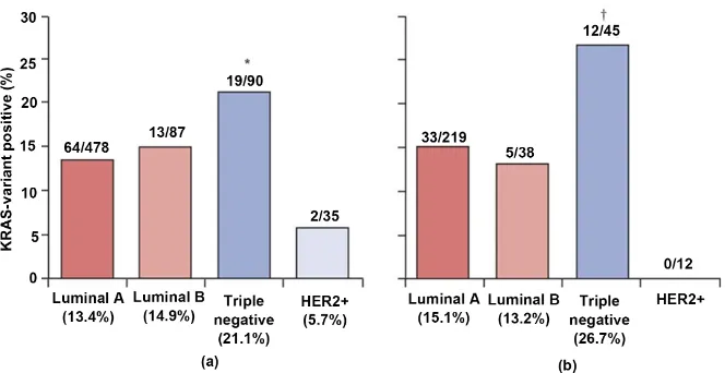
Taken together, these reports imply that a further study needs to analyze a larger patient sample to determine a more reliable analysis of the frequency of
these gene variants in breast cancer (Figure 2).
2.11. Consensus in Literature but Few Studies on SOS
In humans there are two Son of Sevenless (SOS) homologues, hSOS1 and hSOS2. SOS1 is a guanine nucleotide exchange factor that promotes RAS and
RAC activation downstream of EGFR and other growth factor receptors [74].
The guanine nucleotide exchange factor promotes Ras by catalyzing the
conver-sion of the inactive GDP-bound form of Ras to its active GTP-bound form [75].
SOS proteins comprise of multiple domains that are functionally unique. The REM and the CDC25H domains map next to each other between the helical linker (HL) and the proline-rich motif, while the N-terminal region contains the histone-like domain, the Dbl homology domain, the pleckstrin domain, and the
HL which is the most frequently mutated [76]. The REM domain is where SOS
binds to Ras-GTP, although this is only possible when SOS is localized to the cell membrane.
DOI: 10.4236/abcr.2018.72010 148 Advances in Breast Cancer Research
Figure 2. Distribution of the KRAS variant in breast-cancer subtypes in all women (a) and premenopausal (≤51 years) women (b) non premenopausal women 70.
membranes, lamellipodia protrusions), cell invasion and migration [77]. Some
speculate that higher expression of SOS1 may contribute to the more aggressive
phenotype and poor prognosis in African American women [78]. However,
since African women are up to three times more likely to have TNBC [79], this
correlation with SOS1 may be due to the aggressiveness of TNBC. DAG Involved in Cell Growth but Not Well Studied in Breast Cancer Diaclglycerol (DAG) is a lipid which functions as a second messenger and is involved in multiple signal transduction pathways. Significantly, it activates both the classical and novel PKC. Specifically, both classical (cPKC) and novel PKC (nPKC) respond to DAG generation, although atypical PKC (aPKC) activation is DAG-independent
[80]. aPKC cannot bind to DAG due to the absence of DAG-binding motifs.
Re-gardless, it can be concluded that DAG is a critical molecule in cellular biology and may have significant effects on cell growth. DAG is a product of the hydro-lysis of the phosphatidylinositol-4,5-bisphosphate (PIP2) by the enzyme phos-pholipase C (PLC) that also produces inositol 1,4,5-triphosphate (IP3) impli-cated in Ca2+ release from the endoplasmic reticulum [81].
Despite DAGs involvement in cell growth there has been very little current research on DAGs involvement in breast cancer. However, it has been deter-mined that inhibition of DAG synthesis with Triacsin C completely reversed
stearate-induced caspase 3 activity in two breast cancer cell lines [82]. Caspase 3
is known for its apoptotic functions, showing that DAG somehow is involved in cell survival during breast cancer.
2.12. CAMs Significant Interactions with Multiple Proteins
Calmodulin (CAM) is a small ~16 kDa protein that is ubiquitously expressed
throughout the human body and can bind to the secondary messenger Ca2+,
thereby regulating multiple cellular processes via changing the concentration of
DOI: 10.4236/abcr.2018.72010 149 Advances in Breast Cancer Research folding, metabolic homeostasis and numerous others. The highly conserved CAM is composed of two globular domains and one peptide chain with four Ca2+ binding sites [83]. While its regulation of free Ca2+ is a major factor in
leading to all of these cellular processes, another major factor is its ability to bind to a multitude of other proteins. Consequently, CAM is a difficult protein to target without severe consequences in drug therapy. Regardless, during cell cycle progression, the concentration of CAM progressively increases, reaches high le-vels at the G1/S transition, and remains high during the ensuing progression of
the cell cycle [84]. CAM regulates apoptotic processes both positively and
nega-tively mediating elevated intracellular Ca2+ mediating elevated intracellular Ca2+,
which can have both growth promoting and cell death-inducing consequences [85].
While there is literature assessing CAMs involvement in cancer, there is less research on its involvement in breast cancer. It has been shown that CAM can bind directly to death receptor 5 in a calcium-dependent manner in both a ER+
and a TNBC cell line [86]. By binding to the death 5 receptor this could prevent
the receptor from mediating apoptosis in these cancer cells. Another study shows that there is a strong relation between the k63-linked ubiquitination pro-tein and the CAM-like propro-tein 5 (CALML5) and the carcinogenesis of the breast
in young women [87]. Lastly, inhibition of CAM function or disruption of
CAM-HER2 interaction reduced HER2 phosphorylation and HER2-stimulated
cell growth [88]. Regardless of the protein bound to CAM, it appears that CAM
serves a critical role in preventing programmed cell death in breast cancer cells.
2.13. Not all Rafs Created Equal in Breast Cancer
Currently, there are 3 known isoforms of Raf, ARaf, Braf, and Craf, all of whom are serine/threonine kinases activated by recruiting to Ras and dimerizing. There
is high homology and similar domain organization between the isoforms [89].
While all of the Rafs share the same substrate, MEK1/2 kinase, their activity va-ries between them. The general Raf structure can be split into a regulatory N-terminal region, containing the Ras binding site, which is critical for activa-tion as well as inhibitory phosphorylaactiva-tion sites, and a catalytic C-terminal re-gion, which includes phosphorylation sites necessary for the kinase activation
[90]. Since these regions are conserved and found in all isoforms, it is not
sur-prising that all members of Ras can bind to all isoforms of Raf. Most research has been conducted on CRaf, however BRaf has recently caught the attention of
some studies, while there have been very limited studies regarding ARaf [91].
The CRaf isoform has been observed and associated with breast cancer before any other isoform. Although, some research has recently been aimed at BRaf in breast cancer, its functions and activity in breast cancer is not fully understood. Research on BRaf may be difficult since, BRaf is only mutated in about 3% of
breast cancer patients [92]. CRaf has been shown that its knockdown inhibited
breast cancer cell survival and promoted them to apoptosis cells [93]. CRaf has
aggres-DOI: 10.4236/abcr.2018.72010 150 Advances in Breast Cancer Research sive breast cancer subset, TNBC. It was found that CRaf was frequently upregu-lated in TNBC in comparison to 3 other main breast tumor subgroups, further highlighting the heterogeneity of the TNBC subgroup and the difficulty of find-ing a specific target [94].
Some mechanisms have been speculated for CRafs effect on proliferation in breast cancer. For instance, it is suggested that CRaf amplification induced by RAD51 downregulation may be a “necessary but not sufficient” effect of RAD51 on breast tumor initiating cells, and these compensatory mechanisms might help sustain stem/progenitor cell survival under DNA damage and genomic instabili-ty, which could further lead to deregulated signaling changes enabling breast
tumor initiating cells to outgrow and expand [95]. However, other more general
to Raf protein mechanisms have been proposed. Using a robust 3D culture mod-el that approximates formation of mammary acini, it was shown that hyperactive Raf/MEK/extracellular signal-regulated kinases (ERK/MAPK) pathway leads to
increased MMP9, which destroys tissue polarity and growth control [96]. In
terms of migration, it is suspected that EGF and SDF-1α mediated migration of
M13MDA435-1 hybrid cells was most likely attributed to an active Raf-Akt
crosstalk in these cells [97]. While these general mechanisms are useful for
con-structing a mutated ESR pathway of breast cancer, ARaf has not been taken into account and therefore should be examined to determine their role in the pathway.
2.14. MEK Synergy with EGFR
Mitogen activated protein kinase kinase (MEK) is a family of 7 proteins that are involved in the ESR pathway, often regulating apoptotic behavior. While the MEKs are similar to one another, they are involved in different protein activa-tions. MEKs serve vital roles among species as the regulated phosphosites are highly conserved in all species of human, mouse, Arbidopsis, Drosphila C.
ele-gans, and even yeast [98]. All Meks comprise of an N-terminal domain and a
C-terminal domain, as well as a catalytic domain. MEK1 and MEK2 are known to participate in the Ras/Raf/MEK/ERK signal, Mek3 and Mek6 phosphorylate residues in the p 38 MAPK, Mek4 and Mek7 activate JNK protein kinases, while
Mek5 interacts with MEKK2, MEKK3 or ERK5 [99]. Mek1 and Mek2 are the
most studied out of the Mek family. Mek1/2 are serine/threonine kinases that have dual-specificity and when activated can catalyze the phosphorylation of ERK1/2 [100].
Currently, it is known that MEK is associated with tumor growth and forma-tion, despite disappointing clinical trials. This most likely occurs either from cy-totoxicity or from overlapping functions of other proteins. Specifically, some TNBC cell lines have shown resistance to MEK inhibitors in pre-clinical studies and early clinical trials have not shown activity of these agents in different tumor types, thus underlining the need to better understand the mechanisms regulating
resistance to these drugs [101]. By combining inhibitors there has been some
DOI: 10.4236/abcr.2018.72010 151 Advances in Breast Cancer Research
tested [102]. However, this was a relatively small study and it would be beneficial
to further study the effects of EGFR and MEK inhibitors not only in TNBC but in other cancer cell lines to evaluate any differences and effects. One study ob-servers that inhibition of just MEK was capable of completely abolishing
an-chorage-independent growth, cell invasion, and cell migration in TNBC [103].
2.15. Tumorigenesis and Invasion via FKBP52
FK506 binding proteins (FKBPs) function as protein folding chaperones for a multitude of proteins. Specifically, FKBP51 and FKBP52 are Hsp90 co-chaperones
that modify steroid hormone receptor (SHR) activity [104]. However, it is
im-portant to note that FKBP51 and FKBP52 only interact and regulate some SHRs but not all. Structurally, FKBP52 consists of a C-terminal Hsp90-binding TPR domain, an N-terminal FK1 domain that contains a functional peptidyl/prolyl isomerase (PPIase) active site to which the immunosuppressive ligand FK506 binds, and a middle FK2 domain that is similar to FK1 but lacks PPIase activity
[105]. Altogether these regions make up a ~51 kDa protein which is a family
member of the immunophilins, functioning as endogenous cytosolic
pep-tidyl-prolyl isomerases [106]. Consequently, it is expected that FKBPs are
in-volved in the immune system and immune pathologies; however, it may not be so apparent that they are also involved in cancer.
It is interesting to note, that while these proteins have been associated with numerous cancers, whether it promotes or inhibits tumors is not predictable. For example, FKBP5 is found to be down regulated in pancreatic tumor tissue,
while it is overexpressed in melanoma [107]. Most research regarding breast
cancer however has shown that FKBPs are overexpressed and promote tumor development. One such study explains that because depletion of FKBP5 led to decreased CDK4 protein levels and CDK4 kinase activity in breast cancer, con-sidering that CDK4 is hyperactivated in tumorigenesis, it indicates that FKBP5
promotes oncogenesis in part by stabilizing CDK4 [108]. Additionally, the
in-creased expression of FKBP5 in breast cancer cell lines has also been associated
with invasion in breast cancer cells as well as tumorigenesis [109]. Other FKBPs
have also been associated to drugresistant cell lines which may explain the inva-siveness and responinva-siveness of these cell lines. Specifically, it was shown that FKBP4 was upregulated in the drug-resistant cell lines SK-BR-3 (ER negative) and MCF7 (ER+) in comparison to other cell lines, which indicates that FKBP4
expression correlates to drug responsiveness [110]. However, these studies do
not look into other FKBP proteins, and it would thus be useful to examine other FKBPs to have a comprehensive understanding of their effects in breast cancer.
2.16. The Vasodilator NO Participates in Angiogenesis
DOI: 10.4236/abcr.2018.72010 152 Advances in Breast Cancer Research cGMP-independent manner, typically through NO targeting the heme compo-nent of soluble guanylyl cyclase, which further undergoes coupling with c-GMP-dependent protein kinase G and phosphodiesterases as well as cyclic
nucleotide gated channels [111]. When produced the, relatively small free
radi-cal, water soluble, NO is capable of diffusing out and stimulating a diverse range of cellular events, such as vasodilation and inflammation, however, NO is unsta-ble and breaks down easily. While NO function is critical for healthy cells, its dysregulating has been implicated as a causal or contributing to pathophysiolog-ical conditions including cancer [112].
One of the key contributors of NOs involvement in cancer, including breast cancer, is its participation in vasodilation, as this process facilitates angiogenesis in tumors. Although this hasn’t been replicated in breast cancer is has been shown that NO induces angiogenesis by modulating the level of the angiogenesis
inhibitor thrombospondin 2 via EGFR signaling pathway, VEGF, and p53 [113].
It would be therefore interesting to examine the association of these proteins during breast cancer development. Furthermore, it is proposed that NO can modulate tumor aggression in breast carcinoma through the inhibition of
en-zymes linked to DNA repair machinery [114][115]. However, the latter source
suggests that while there were increased levels of NO in breast cancer patients, there was no correlation between metastatic and non-metastatic patients, indi-cating that NO is not involved in such events.
2.17. CREBs Involvement with MMPs Promotes Metastasis
As a ubiquitous transcription factor, cAMP response element binding protein (CREB) binds to DNA and subsequently regulates diverse biological processes and pathways. In fact, CREB targets genes that are involved in cell metabolism, survival, proliferation, differentiation, cell cycle, DNA repair, immortalization of
cells, inflammation, immune modulation and more [116]. Although there are
many downstream molecular targets of CREB some of these include brain-derived neurotrophic factor, B-cell lymphoma 2 protein, c-Fos transcription factor, and
corticotropin release factor [117]. Depending on the target gene, CREB can act
as a repressor or activator for transcription. Activation occurs from numerous extracellular signals including but not limited to hormones, growth and
neuro-trophic factors, neurotransmitters, and membrane depolarization [118]. In order
to be activated CREB must be phosphorylated at the Ser-133 residue which then enables it to bind to the cAMP-responsive element sequence “TGACGTCA”
[119]. The binding of DNA is facilitated by CREB’s leucine zipper domain by
dimerizing two DNA regions. Enzymes that catalyze the phosphorylation of CREB include protein kinase A (PKA), Akt, CAM-dependent protein kinase,
and MEK/ERK [120].
DOI: 10.4236/abcr.2018.72010 153 Advances in Breast Cancer Research critical roles in stress-induced angiogenesis through VEGF signaling, which is
vital for tumor growth and metastasis [121]. However, this study is not breast
cancer specific and would consequently need to be tested in breast cancer cell lines. Although recent studies have not focused on angiogenesis and CREB, stu-dies have largely focused on CREBs association with metastasis in breast cancer. One study shows that CREB signaling in breast cancer regulated the gene ex-pression of parathyroid hormone-related peptide, MMP2 and MMP9, and os-teoprotegerins, which are closely involved in cancer metastasis and bone
de-struction [122]. This is supported by another study which found that CREB2 was
not only over expressed in breast tumors but was associated with lymph node metastasis in infiltrating breast carcinoma [123].
One way CREB may be involved in metastasis by its ability to mediate cell
survival signaling [124]. Essentially, this would enable cells to be
anchoragein-dependent unlike healthy cells which undergo apoptosis when unanchored. Cell survival signaling would benefit cell cycle progression, despite DNA damage causing mutations. These effects are demonstrated by the downregulation of CREB in HER-2 transformed cells which causes reduced cell proliferation by cell-cycle arrest, cell migration, MMP expression, but with increased fibronectin
adherence [125]. Overall, this research provides a glimpse into the functions of
CREB, however other variations of CREB remain to be analyzed. Therefore, fu-ture research should observe other CREB variations in order to see if there are further correlations between CREB and breast cancer. Regardless, these studies have shown that CREB is specifically critical for breast cancer metastasis and cell survival, which should be considered during prognosis of patients.
3. Varied Correlations to Breast Cancer
This section includes genes where there are either inconsistencies in the litera-ture, or whether there is consensus on a gene being both a tumor suppressor and promoter depending on factors such as breast cancer subset and chemical mi-croenvironment.
3.1. Possible Resolution of E2 Discrepancies
17β-Estradiol (E2) is an integral component in the regulation of the female
re-productive cycles. E2 is biosynthesized from progesterone (arrived at in two
steps from cholesterol, via intermediate pregnenolone) [126] and is a ligand to
DOI: 10.4236/abcr.2018.72010 154 Advances in Breast Cancer Research In one report, it is shown that E2 is able to induce complete neoplastic trans-formation of human breast epithelial cells, as proven by the trans-formation of tumors
in severe combined immunodeficient mice [128]. Though these results facilitate
our understanding of the formation of breast cancer, it must be noted that this report basis its analysis off severe combined immunodeficient mice and conse-quently is less representative of results that may happen in humans. Moreover, it is now suggested, that while E2 has a role in tumor formation, the important is-sue for the decision of breast cancer cells to survive or die in response to estra-diol depends entirely on the cell populations present in an estrogenized
envi-ronment or following estrogen deprivation [129]. Based on the laboratory data
they assessed, the decision is survival or death, respectively. E2 may also be in-volved in breast cancer cell movement and invasion by the extra-nuclear
activa-tion of the actin-binding protein ezrin [130]. Furthermore, it has been proposed
that E2 induces proliferation via SDF-1/CXCR4-mediated activation of EGFR
that in turn activates both Akt and ERK1/2 cascade [131]. The study speculates
that besides a role in cell migration, Akt activation by E2 and SDF-1 is also in-volved in breast cancer cell proliferation. Although some literature seems to be in accordance with this research, it would be beneficial to further explore the in-terrelatedness of these genes. While this acts as a platform to begin constructing a model of the mechanisms and pathways of E2 in breast cancer, it is not yet a completely reliable and accurate representation of E2s role in tumor progression.
3.2. Each Variation of MMP Has Unique Function
The endopeptidases matrix metalloproteinase (MMPs) function was originally restricted to tissue remodeling and maintenance, as they are capable of degrad-ing extracellular matrix (ECM) proteins. However, recent studies have revealed that they participate in innate and adaptive immunity, inflammation,
angioge-nesis, bone remodeling, and neurite growth [132]. MMPs are capable of many
functions because unlike most proteins they are not specific to a particular li-gand, enabling them to bind to a multitude of ligands. They are multi-domain proteins and their activities are regulated by tissue inhibitors of
metalloprotei-nases [133]. So far, there are more than 24 gene variants of MMPs that have been
observed in humans. With so many known variants it is unsurprising that MMPs represent the most abundant ECM regulator within the tissue
microen-vironment [134]. All MMP variants except MMP-23 comprise a pro-domain
up-stream of the catalytic domain, and activation proceeds through its removal [135].
While MMPs are necessary in humans to prevent tissue disruption, numerous MMPs have been associated with breast cancer, especially during metastasis and invasion. One report observed that MMP-1, -9, -11, -15, -25, and -25 were upregulated in breast cancer tissues compared to normal breast tissue, whereas
MMP10 and MMP19 were downregulated [136]. Although it is agreed upon in
DOI: 10.4236/abcr.2018.72010 155 Advances in Breast Cancer Research MMPs vary from report to report. Even though this report takes into account 39 samples from breast cancer and 16 from healthy patients, the results differ from other reliable sources. For instance, another report suggests that MMP2 is
over-expressed and is more active in metastatic breast cancer [137]. It would seem
that particular MMPs are involved in specific stages of breast cancer and conse-quently further research needs to be done regarding MMPs and their roles and functions throughout tumor progression.
Since there are varied results for which MMPs are associated with breast can-cer, it is understandable that the exact mechanistic nature of MMPs during breast cancer is varied as well. However, with that being said, the general outline is that during malignant progression, MMP activity becomes deregulated, which contributes toward the disruption of normal tissue ECM, and also the abnormal
regulation of several signaling pathways [138]. It is believed that altogether
MMPs play a critical role in breast cancer initiation, growth, angiogenesis,
inva-sion, and metastasis [139]. Some MMPs have been more thoroughly investigated
than others, for example MMP1 has been observed to regulate the levels of
transforming growth factor α which in turn affects the activation of EGFR in
breast cancer cells [140]. EGFR is relevant to breast cancer as its ligand has been
linked to promoting heparanase function and topoisomerase I localization in
brain metastasizing breast cancer cells [141]. This information can then facilitate
our medical community in development of drugs and treatment, however, to improve it is vital that we pinpoint which MMPs the most critical in breast can-cer and how they function during tumor development.
3.3. GABA and OPRM1 Located in the Brain Yet Influence Breast
Cancer
Even though gamma-aminobutyric acid (GABA) is the primary inhibitory neu-rotransmitter in the mature human brain, it is known to be involved in a large
spectrum of functions throughout the body. Although the µ1-opioid receptor
(OPRM1) is not a neurotransmitter itself, it is a receptor for neurotransmitters and is predominately located in the brain with GABA. GABA can act as a troph-ic factor during nervous system development to influence cellular events in-cluding proliferation, migration, differentiation, synapse maturation, and cell
death [142]. In fact, GABA has been shown to influence the development of a
variety of tissues and organs, including but not limited to the pancreas [143],
liver [144], and even stem cells [145]. When bearing in mind that GABA is
in-volved and contributes to cell proliferation throughout the body, it becomes ap-parent to why GABA would be evident in multiple types of cancers, including breast cancer. On the other hand, it is not so apparent how OPRM1 is involved in breast cancer.
DOI: 10.4236/abcr.2018.72010 156 Advances in Breast Cancer Research as a tumor promoter or as a tumor repressor depending on the particular cancer. For instance, in liver cancer GABA suppressed the cancer cells from migration
and invasion [146], while in pancreatic cancer it has been speculated that GABA
promotes tumor progression [147]. It is suspected that GABA can significant
promote the invasive ability of prostate and renal cancer cells through the
pro-duction of MMPs [148]. Due to the varied response of GABA during cancer, it
would be especially relevant to explore its function in breast cancer. Whereas GABA has been studied in some cancers, due to OPRM1s association with morphine/opium, the receptor is less established in the study of cancer let alone breast cancer. However, recent studies have linked OPRM1 and gene variants to breast cancer. For instance, breast cancer-specific mortality was significantly
re-duced in patients with a genetic variant (A118G) in the µ-opioid receptor that
reduces opioid response [149]. Even though the same study admits that one
li-mitation of their study is that gene association studies cannot definitively show that particular outcomes are actually caused by a specific mutation/gene. Despite this limitation, other studies concur with these findings. Particularly, one study reports that carriers of the same polymorphism had more than three times in-creased breast cancer risk than both healthy female and the entire control group [150].
Although there is growing evidence for an effect of the family of OPRM1 in
mediating tumor metastasis, the reason for this effect remains unclear [151].
One process of thought is that opioids appear to suppress a number of aspects of immune system function, and some of these effects have been shown to be
me-diated by OPRM1 activation [152]). Other data suggests that OPRM increases
PTEN/p53 via PI 3 kinase Phosphatidylinositol-4,5-bisphosphate 3-kinase and protein kinase B (PI3K and Akt respectively) signaling pathway in some cell
lines, including MCF7, T47D, and MDA-MB231 [153]. Since the information on
OPRM1s involvement and function in breast cancer is so limited it would be ef-ficacious to determine whether any other variants are involved in breast cancer. Moreover, a molecular dynamics study would offer insight on some of the asso-ciations and mechanisms of OPRM1 with other genes in the ESR pathway.
3.4. Inhibitors of AC Cause Conflicting Effects
Cyclic adenosine monophosphate (cAMP) is a secondary messenger molecule and is typically produced by the cleavage of adenosine triphosphate (ATP) cata-lyzed by adenylate cyclase (AC). AC is bound to the cell membrane and its activ-ity is regulated by the concentrations of hormones and chemical signals. As a secondary messenger, cAMP is critical in multiple signal transduction cascades, and consequently AC is a vital molecule to analyze and investigate. In prostate cancer AC is observed to generate a rise in cAMP and subsequently proliferation from a cross-talk between cAMP signaling and androgen receptors via PKA ac-tivation [154].
DOI: 10.4236/abcr.2018.72010 157 Advances in Breast Cancer Research agent, Resveratrol, stimulates AC in human breast cancer cells through
cPLA2-dependant pathway [155]. Paradoxically, another report suggests that
antiproliferative effects in cancer therapy occur via the inhibition of AC [156].
However, there is little recent data on AC and its appearance in breast cancer particularly, so it would be efficacious to know its presence and regulation pat-terns in breast cancer through gene expression data analysis methods such as, Hierarchical Clustering, Self-Organized Map, and Consensus Clustering.
3.5. Localization of Estrogen Receptor Is Significant in Breast
Cancer
While membrane-bound Estrogen Receptors (mERs) are similar to nuclear ERs there are subtle differences in their functions and effects in the cellular environ-ment. Non-genomic pathway regulates more genes than just genomic action of
ER alone, such as proliferation, apoptosis, and survival [157]. However, it must
be noted that most ERs are not restricted to the membrane or cytosol. For
in-stance, ERα36 can be located in plasma membrane, nuclear, and cytosolic
frac-tions. Although, it is more common to detect ERα36 bound to the plasma
mem-brane. It is known that mERs rapidly signal as GPCRs to generate calcium flux, stimulate cAMP and cGMP production, and trigger phosphoinositide 3-kinase
(PI3K) and ERK pathway activation [158]. In fact, one mER exists as a G
pro-tein-coupled estrogen receptor (GPER). Although it was once referred to as the G protein-coupled receptor 30. It was later changed as it was determined that GPER can bind to estradiol and consequently is now considered an ER. The seven transmembrane-domain protein mediates the effects of estrogens in a wide number of cell types to produce rapid non-genomic biological responses [159].
Even though all ERs have been associated with breast cancer, mERs should be explored separately since their mechanisms by which they function are unique. This is illustrated in one report which observes that when a xenograft model of MCF-7 human breast cancer cells were injected into nude mice, engagement of only mERs by an estrogenic compound failed to stimulate proliferation of the
tumor [160]. The study later proposes that communication between extranuclear
and nuclear ERs is likely to be important to promote the growth of human breast tumors. In contrast to this, one study reports that specifically membrane
locali-zation of ERα36 in TNBC provides a survival benefit of 16 months, while no
correlation between intracellular ERα36 and prognosis [161]. Similarly GPER
was found to inhibit growth of ERα+ breast cancer, which may indicate better
prognosis [162]. However, the same study finds that GPER can induce
prolifera-tion in another breast cancer cell line. While this informaprolifera-tion is significant as it could be applied to determine prognosis factors for patients, it is specific to-wards a subset of breast cancer, and should be further explored in other subsets.
DOI: 10.4236/abcr.2018.72010 158 Advances in Breast Cancer Research
EGFR and ERα36 positively regulate each other in TNBC, however ERα36 may
dynamically change its partners during estrogen signaling in regards to EGFR
and Src/Shc [163]. A mechanism for GPER has also been speculated. Specifically,
upregulation, stabilization, and nuclear translocation of p53 by activation of
GPER is involved in G-1-induced growth arrest of ER− breast cancer cells [164]
[165]. However, this speculation is only for one subset of breast cancer, and
cannot be assumed for other subsets, especially because GPER has also been
as-sociated with promoting tumor development [166][167] [168]. Although one
mechanism for cell proliferation is proposed by Ignatov et al., which suggests
that activation of GPER results in stimulated proliferation via EGFR transactiva-tion. Since there are discrepancies in the literature, a study should aim to eluci-date the cause for the tumor suppressor and promoter activity of the ERs.
3.6. Model of Shc
Src-homology collagen (Shc) proteins can be recruited to cell surface growth factor RTKs. The resulting complex relays and amplifies an exquisitely fine-tuned regulation of multiple downstream signaling events, which depending on
cellu-lar context, mediate specific biological response [169]. However, RTKs more
typically recruit Grb2 than Shc, although others recruit both. Even so, Shc pro-teins interact with diverse signaling molecules in addition to Grb2, thereby
en-gage in Grb2-independent pathways and biological functions [170][171][172]
[173][174]. Within the Shc family there are four members, two of them only
detected in certain human tissues, one of them only found in mice, and lastly one of them, ShcA, is expressed ubiquitously in the human body. ShcA is the only family member that has been implicated in human breast cancer and has three isoforms: p46Shc, p52Shc, and p66Shc. However, Shc isoforms functions are complex and can seem contradictory, particularly in cancer. As shown in
colorectal cancer cells where they induced migration of cancer cells [175], while
in another was shown to suppress migration in lung cancer cells [176].
While typically Shc has been correlated to promoting metastasis and tumor progression in breast cancer, there are some studies which show otherwise. However, one report suggests that the association between high p66ShcA levels and good outcome is reflective of the fact that p66ShcA is enriched in luminal
breast cancers which have a better prognosis than other subsets [177]. This
sug-gests that p66Shc enhances signaling downstream of the Met RTK and that Mets activation is required for p66Shc to induce epithelia-mesenchymal transition in luminal breast cancer. While Shc is a member of the ESR pathway, it must be noted that there are other genes that aren’t found in the normal ESR pathway that it can interact with. One study associates p66Shc with ARF1 and ARF6 (as
illustrated in Figure 2) which then may lead to cell proliferation and tumor cell


![Figure 3. Model of ARF1 and ARF6 activation downstream of the EGFR in MDA MB-231 cells [178]](https://thumb-us.123doks.com/thumbv2/123dok_us/9293607.426180/27.595.236.521.72.312/figure-model-arf-arf-activation-downstream-egfr-cells.webp)
