Comment on “Quantitative Evaluation of Commercially
Available Test Kit for Ciguatera in Fish”
Joanne S. M. Ebesu, Cara E. Campora ToxiTec, Honolulu, USA.
Email: [email protected]
Received May 11th, 2012; revised August 10th, 2012; accepted August 17th, 2012
ABSTRACT
This letter is in regards to the paper, “Quantitative evaluation of commercially available test kit for ciguatera in fish” [1]. We were compelled to respond because the entire premise of this paper is flawed, thus invalidating its stated conclu-sions. The data presented in the paper is derived from the opinions of four independent readers who evaluated identical Cigua-Check® test sticks to screen fish samples for ciguatoxin (CTX), the results of which were then compared with
corresponding samples tested in a non-specific bioassay with questionable statistics (see Table 1 [1]). In addition to several factual errors presented in the paper, we have identified several issues with this study, such as insufficient detail and questionable data analyses, that make its interpretations unreliable.
Keywords: Ciguatera; Cigua-Check®; Ciguatoxin; Neuroblastoma Cell Assay
First, while the sodium channel-specific N2a neuroblas-toma cell bioassay used for comparative analysis in this study is a useful tool for detecting and measuring general cytotoxicity as a result of exposure to CTXs and related toxins, it is not specific for CTXs. Like the Cigua-Check® test, the N2a bioassay is a screening method, and the N2a bioassay does not provide “actual ciguateric status” as claimed by the authors in the Discussion. Instead, it is a non-specific sodium channel assay that requires an ana-lytical method in order to definitively identify specific neurotoxins in a given sample. This bioassay is not capa-ble of discriminating between different neurotoxins that act in a similar manner [2]. Indeed, the N2a bioassay has been used to detect and measure brevetoxins, saxitoxins, neosaxitoxin, gonyautoxin II plus III, decarbamoylsaxi-toxin, and palydecarbamoylsaxi-toxin, in addition to ciguatoxin [3-9], and requires confirmation of the identity of specific toxic compounds using analytical methods whenever possible [2]. Even the US Food and Drug Administration (FDA) employs a two-tiered protocol to test fish for CTXs, in-cluding an in vitro assay such as the N2a bioassay and an analytical chemistry technique (liquid chromatography- mass spectrometry, LC-MS) because the N2a screening procedure does not specifically identify the sodium chan-nel active agent present in fish samples [10]. Given that all comparative analyses in this paper were based solely on positive or negative determinations made using the N2a neuroblastoma cell bioassay, any comparisons must be approached with caution.
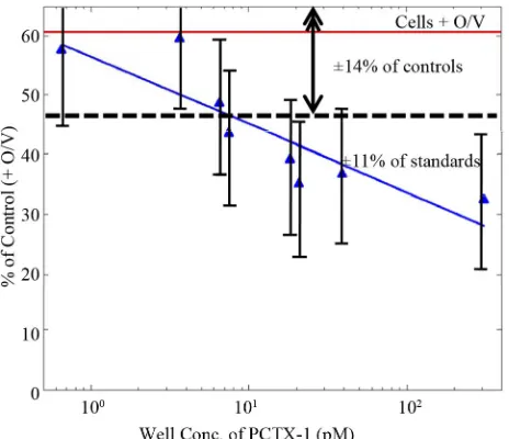
Figure 1. Dose-response curve for the N2a neuroblastoma bioassay to pacific ciguatoxin. Signal strengths in response to a range of PCTX-1 concentrations are given as % of con-trol wells (n = 20) following addition of ouabain and veratr- idine. Relative standard deviations about the means for stan-dards and controls averaged 11% and 14%, respectively [1].
[image:2.595.57.289.365.565.2]Note: This figure has been modified from the original to illustrate the aver-aged relative standard deviations about the means for the standards and controls given in the original reference.
Figure 2. Dose-response curve for the N2a neuroblastoma bioassay to pacific ciguatoxin. Signal strengths in response to a range of PCTX-1 concentrations are given as % of con-trol wells (n = 20) following addition of ouabain and veratr- idine. Relative standard deviations about the means for stan-dards and controls averaged 11% and 14%, respectively [1].
these reagents are too high and might in fact mask the true cytotoxicity of the toxin, thereby significantly influ-encing the results.
Third, the authors do not define the threshold values used in the N2a assay to determine the definitive P-CTX-1
content of the fish samples tested, nor do they provide information on their source or the purity of the P-CTX-1 standard used. They state that results were statistically analyzed using Students t-test to identify significant dif-ferences between various controls and fish sample means, but do not present any of these statistical analyses. To-gether with the dubious dose-response curve shown in Figure 2, using the given data it is impossible to com-pare the sensitivity of the N2a bioassay the authors used against the accuracy of the Cigua-Check® method.
More-over, because their standard curve does not deviate from the control group until the last data point (see Figure 2), it cannot be used to accurately determine P-CTX-1 con-centrations, nor can it be used as the basis for any of their comparisons or conclusions. Since commercial CTX stan-dards do not exist, clear information about the reference CTX used is relevant.
Fourth, the maximum concentration of tissue equiva-lent (TE) recommended for use in the N2a bioassay ranges from 2 mg TE/ml [13] to no more than 20 mg TE/ml because an excessive matrix quantity of fish sam-ples may in fact be cytotoxic (see [2]). In the Bienfang, et al. [1] paper, the authors briefly described a sample ex-traction process using 10 g of each fish, but they did not specify how much of this fish extract was used per sam-ple in the N2a assay. If they used 10 g of fish per samsam-ple, then the TE would be in the range of 50 g/ml, assuming the maximum volume of 200 µl per well in a 96-well plate, which may explain the extremely high rate of posi-tive samples (70% for roi and 57% for kole). The overall lack of detail in the Materials and Methods section of the Bienfang, et al. [1] paper prevents accurate replication of their experiments for verification.
Fifth, comparing the results of a test that uses the means of triplicate samples (n = 3) against the results of a dif-ferent test that uses single samples (n = 1) is not reason-able. In this paper, the authors used n = 1 for Cigua-Check®
samples and n = 3 for the N2a samples. Furthermore, the fish samples tested in the N2a assay were extracted using a more rigorous method, including lyophilization, extrac-tion with methylene chloride, sonicaextrac-tion, evaporaextrac-tion, and resuspension in methanol. Cigua-Check® uses only a sim-ple methanol extraction of the fish tissues, which is not explained in the Materials and Methods section of the paper. This difference in fish tissue extraction procedures alone would definitely result in varying toxin extraction efficiencies and subsequent quantification.
Sixth, Cigua-Check® test results will fade slightly over
time, even within 15 minutes. The paper does not make it clear whether the Cigua-Check® sticks were read by
the results, which would likely affect the interpretation between readers.
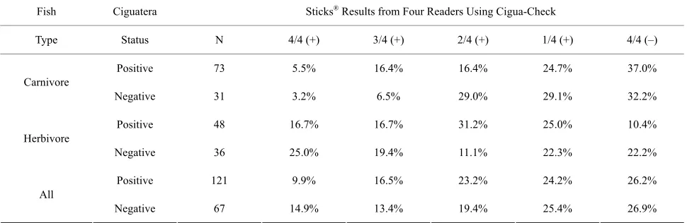
Given that the flawed standard curve in Figure 1 could not have been used to assess the accuracy of the Cigua- Check® test results, the data in Table 1 of the Bienfang,
et al. publication [1] are also flawed and inconclusive. This table depicts data distribution, but without being subjected to statistical analysis such as Fleiss’ kappa to assess the reliability of agreement between readers, and is not an analysis by itself. If the raw Cigua-Check® test
results are evaluated simply by calculating the percentage of agreement among the four independent readers, the majority of readers (3/4 and 4/4) agreed on the same re-sult for each stick in 78.2% (147/188) of the samples, while a minority of readers (2/2 and 1/2) disagreed on the same result for each stick in 21.8% (41/188) of the sam-ples. Home pregnancy tests (HPTs), which are commer-cially available antibody-based assays, have been shown to be misread by 1 in 4 women; this translates into a 25% disagreement/75% agreement rate among independent readers [14]. While most of these HPTs claim “over 99% accuracy” or a similar statement on their packaging or insert [15], their actual accuracy rates can range from 8.3% to 97% [14,16]. The high false negative rates for these tests may be caused by many factors, including the length of time after a missed menstrual period at the point the test is conducted, late implantation, an ectopic or het-erotopic pregnancy, heterophilic antibodies, spontaneous abortion, or non-compliance with the HPT instructions [17,18]. It would be expected that Cigua-Check® kits have
a similar rate of interpretation.
Additional factual errors in this paper include the state-ment that, “Polyclonal antibodies in an assay for detect-ing haptens via a luminescent probe was pursued over years [19-21], and subjected to considerable analytical controversy [22-26].” While polyclonal antibody to CTX
was used in the first two references [19,20], a mono-clonal antibody to CTX (MAb-CTX) was used in [21]. None of the assays utilized luminescent probes: the first assay used 125I in a radioimmunoassay [20]; the second
assay used horseradish peroxidase in an enzyme immu-noassay [19]; and the third assay employed dyed latex particles in a membrane immunobead assay [21]. The “analytical controversy” referred to in the five references cited was primarily due to mistaken identity and differ-ences or flaws in protocol. A similar immunobead test (Ciguatect) developed by a separate research group [22], which is often confused with the Cigua-Check® method,
showed high false positive and negative detection rates [23]. The evaluation of the Ciguatect test by Dickey, et al. [23] has often been incorrectly identified with the Cigua- Check® method, and this mistaken perception has been
continued by other researchers citing this study when referring to Cigua-Check® (including [24,25]. The
Cigu-atect test employed a different antibody that was not spe-cific for CTXs because the antigen (hapten) used to gen-erate this antibody was crude fish extract and not purified CTX, which was used to develop the MAb-CTX. A study of fish samples from Hong Kong utilizing the Cigua- Check® method also resulted in high false negative and
positive rates [26], but this was probably due to slight yet important differences in the proper protocol for the Cigua- Check® method, and for comparison against the mouse
bioassay, which is not specific nor as sensitive as the Cigua- Check® method for CTX [27].
[image:3.595.56.541.577.735.2]The authors also mention that “anecdotal information from researchers and fishermen who used these kits por-trayed shortcomings (i.e., false positives and false nega-tives) in their application,” without citing any factual sup-port. Additionally, in the last paragraph of the Bienfang et al. [1] paper, the authors mention that they used the
Table 1. Summary data from four independent readers evaluating identical Cigua-Check® test sticks. Ciguatera status was
determined by N2a neuroblastoma bioassays. N gives the number of fish specimens that were evaluated. 4/4 (+) indicates that all four readers agreed that the samples were positive, and 4/4 (−) indicates that all four readers agreed that the samples were negative, etc. [1].
Fish Ciguatera Sticks® Results from Four Readers Using Cigua-Check
Type Status N 4/4 (+) 3/4 (+) 2/4 (+) 1/4 (+) 4/4 (–)
Positive 73 5.5 %% 16.4 %16.4 %24.7 %37.0
Carnivore
Negative 31 3.2 %% 6.5 %29.0 %29.1 %32.2
Positive 48 16.7 %% 16.7 %31.2 %25.0 %10.4
Herbivore
Negative 36 25.0 %% 19.4 %11.1 %22.3 %22.2
Positive 121 9.9 %% 16.5 %23.2 %24.2 %26.2
All
Cigua-Check® kit to test orange “roughly” [sic], chicken,
and two types of cheeses, which all yielded positive test results. Due to the nature of antibodies and the lipid poly-ether structure of CTX, samples containing high amounts of lipid and/or similar structures can cross-react with the MAb-CTX. Thus, the Cigua-Check® kits should be used
only to test tropical reef fish, for which the method has been designed and has been extensively tested. Past re-search has proven that CTX screening with various im-munological methods utilizing the MAb-CTX, employed in the Cigua-Check® method, is effective in preventing
ciguatera poisoning and in detecting CTX in fish samples from clinically documented ciguatera cases [19,20,28-31]. Using a modified enzyme-linked immunosorbent assay (ELISA) technique, the MAb-CTX has been demonstrated to detect Pacific (P-CTX-1, P-CTX-2, P-CTX-3) and Caribbean (C-CTX-1) CTX congeners at concentrations ranging from 0 to 5.0 ppb, with statistically insignificant cross-reactivity at similar concentrations to okadaic acid, palytoxin, and domoic acid [32]. A study by Dierking and Campora [33] of 30 Cephalopholis argus fish samples showed strong and significant correlation (r = 0.64, P < 0.001) between results obtained using the MIA and N2a assays. Furthermore, over 50,000 Cigua-Check® tests for
CTX have been commercially sold or distributed since 1997, with no verified reported incidences of false negatives.
Due to missing information and incomplete analyses in the Bienfang et al. [1] paper, its conclusions are not reli-able. Because the N2a bioassay and the Cigua-Check®
immunobead assay are both considered screening meth-ods, a more reliable, robust analysis should include the ciguatoxic status of individual fish samples obtained us-ing an analytical method.
REFERENCES
[1] P. Bienfang, S. DeFelice and A. Dowling, “Quantitative Evaluation of Commercially Available Test Kit for Cigua- tera in Fish,” Food Nutrition Sciences, Vol. 2, No. 6, 2011, pp. 594-598.
[2] A. Caillaud, P. de la Iglesia, H. T. Darius, S. Pauillac, K. Aligizaki, S. Fraga, M. Chinain and J. Diogène, “Update on Methodologies Available for Ciguatoxin Determina- tion: Perspectives to Confront the Onset of Ciguatera Fish Poisoning in Europe,” Marine Drugs, Vol. 8, No. 6, 2010, pp. 1838-1907. doi:10.3390/md8061838
[3] R. L. Manger, L. S. Leja, S. Y. Lee, J. M. Hungerford, and M. M. Wekell, “Tetrazolium-Based Cell Bioassay for Neurotoxins Active on Voltage-Sensitive Sodium Chan-nels: Semiautomated Assay for Saxitoxins, Brevetoxins, and Ciguatoxins,” Analytical Biochemistry, Vol. 214, No. 1, 1993, pp. 190-194. doi:10.1006/abio.1993.1476 [4] R. L. Manger, L. S. Leja, S. Y. Lee, J. M. Hungerford, Y.
Hokama, R. W. Dickey, H. R. Granade, R. Lewis, T. Ya-sumoto and M. M. Wekell, “Detection of Sodium Chan-nel Toxins: Directed Cytotoxicity Assays of Purified Ci-
guatoxins, Brevetoxins, Saxitoxins, and Seafood Extracts,” Journal of AOAC International, Vol. 78, No. 2, 1995, pp. 521-527.
[5] K. Kogure, M. Tamplin, U. Simidu and R. Colwell, “A Tissue Culture Assay for Tetrodotoxin, Saxitoxin and Re- lated Toxins,” Toxicon, Vol. 26, No. 2, 1988, pp. 191- 197. doi:10.1016/0041-0101(88)90171-7
[6] J. Jellett, L. Marks, J. Stewart, M. Dorey, W. Watson- Wright and J. Lawrence, “Paralytic Shellfish Poison (Saxitoxin Family) Bioassays: Automated Endpoint De-termination and Standardization of the in Vitro Tissue Culture Bioassay, and Comparison with the Standard Mouse Bioassay,” Toxicon, Vol. 30, No. 10, 1992, pp. 1143-1156. doi:10.1016/0041-0101(92)90430-D
[7] J. Jellett, J. Stewart and M. Laycock, “Toxicological Evaluation of Saxitoxin, Neosaxitoxin, Gonyautoxin II, Gonyautoxin II Plus III and Decarbamoylsaxitoxin with the Mouse Neuroblastoma Cell Bioassay,” Toxicol in Vi-tro, Vol. 9, No. 1, 1995, pp. 57-65.
doi:10.1016/0887-2333(94)00194-Y
[8] W. A. Catterral and M. Gainer, “Interaction of Brevetoxin A with a New Receptor Site on the Sodium Channel,” Toxicon, Vol. 23, 1985, pp. 497-504.
doi:10.1016/0041-0101(85)90034-0
[9] R. Dickey, E. Jester, R. Granade, D. Mowdy, C. Mon-creiff, D. Rebarchik, M. Robl, S. Musser and M. Poli, “Monitoring Brevetoxins during a Gymnodinium breve Red Tide: Comparison of Sodium Channel Specific Cy-totoxicity Assay and Mouse Bioassay for Determination of Neurotoxic Shellfish Toxins in Shellfish Extracts,” Natural Toxins, Vol. 7, No. 4, 1999, pp. 157-165.
doi:10.1002/(SICI)1522-7189(199907/08)7:4<157::AID-NT52>3.0.CO;2-#
[10] M. A. Friedman, L. E. Fleming, M. Fernandez, P. Bien-fang, K. Schrank, R. Dickey, M.-Y. Bottein, L. Backer, R. Ayyar, R. Weisman, S. Watkins, R. Granade and A. Reich, “Ciguatera Fish Poisoning: Treatment, Prevention and Management,” Marine Drugs, Vol. 6, No. 3, 2008, pp. 456-479. doi:10.3390/md6030456
[11] E. Caňete and J. Diogène, “Comparative Study of the Use of Neuroblastoma Cells (Neuro-2a) and Neuroblastoma x Glioma Hybrid Cells (NG108-15) for the Toxic Effect Quantification of Marine Toxins,” Toxicon, Vol. 52, No. 4, 2008, pp. 541-550. doi:10.1016/j.toxicon.2008.06.028 [12] A. Caillaud, E. Caňete, P. de la Iglesia, G. Giménez and J.
Diogène, “Cell-Based Assay Coupled with Chromato- graphic Fractioning: A Strategy for Marine Toxins Detec-tion in Natural Samples,” Toxicol in Vitro, Vol. 23, No. 8, 2009, pp. 1591-1596. doi:10.1016/j.tiv.2009.08.013 [13] R. W. Dickey, “Ciguatera Toxins: Chemistry, Toxicology,
and Detection,” In: L. M. Botana, Ed., Seafood and Freshwater Toxins: Pharmacology, Physiology, and De-tection, 2nd Edition, CRC Press, Boca Raton, 2008, pp. 479-500. doi:10.1201/9781420007541.ch22
doi:10.1185/03007990802120572
[15] S. A. Butler, S. A. Khanlian and L. A. Cole, “Detection of Early Pregnancy Forms of Human Chorionic Gonadotro-pin by Home Pregnancy Test Devices,” Clinical Chemis-try, Vol. 47, 2001, pp. 2131-2136.
[16] L. A. Cole, “The Utility of Six Over-the-Counter (Home) Pregnancy Tests,” Clinical Chemistry and Laboratory Medicine, Vol. 49, No. 8, 2011, pp. 1317-1322.
doi:10.1515/cclm.2011.211
[17] S. Davies, F. Byrn and L. A. Cole, “Human Chorionic Gonadotropin Testing for Early Pregnancy Viability and Complications,” Clinics in Laboratory Medicine, Vol. 23, No. 2, 2003, 257-264.
doi:10.1016/S0272-2712(03)00026-X
[18] B. G. Valanis and C. S. Perlman, “Home Pregnancy Test-ing Kits: Prevalence of Use, False-Negative Rates, and Compliance with Instructions,” American Journal of Pub- lic Health, Vol. 72, No. 9, 1982, pp. 1034-1036.
doi:10.2105/AJPH.72.9.1034
[19] Y. Hokama, “A Rapid, Simplified Enzyme Immunoassay Stick Test for the Detection of Ciguatoxin and Related Polyethers from Fish Tissues,” Toxicon, Vol. 23, No. 6, 1985, pp. 939-946. doi:10.1016/0041-0101(85)90386-1 [20] Y. Hokama, A. H. Banner and D. B. Boylan, “A
Radio-immunoassay for the Detection of Ciguatoxin,” Toxicon, Vol. 15, No. 4, 1977, pp. 317-325.
doi:10.1016/0041-0101(77)90014-9
[21] Y. Hokama, W. E. Takenaka, K. L. Nishimura and J. S. M. Ebesu, “A Simple Membrane Immunobead Assay (MIA) for Detecting Ciguatoxin and Related Polyethers From Human Ciguatera Intoxication and Natural Reef Fishes,” Journal of AOAC International, Vol. 81, No. 4, 1998, pp. 727-735.
[22] D. L. Park, “Evolution of Methods for Assessing Cigua-tera Toxins in Fish,” Reviews of Environmental Conta- mination & Toxicology, Vol. 136, 1994, pp. 1-20. doi:10.1007/978-1-4612-2656-7_1
[23] R. W. Dickey, H. R. Granade and F. D. McClure, “Evalua- tion of a Solid-Phase Immunobead Assay for Detection of Ciguatera-Related Biotoxins in Caribbean Finfish,” Me- moirs of the Queensland Museum, Vol. 34, 1994, pp. 481- 488.
[24] A.-M. Legrand, T. Teai, P. Cruchet, M. Satake, K. Murata and T. Yasumoto, “Two Structural Types of Ciguatoxins Involved in Ciguatera Fish Poisoning in French Polyne-sia,” In: B. Reguera, J. Blanco, M. L. Fernandez and T. Wyatt, Eds., Harmful Algae, Xunta de Galicia and
Inter-Governmental Commission of UNESCO, 1998, pp. 473- 475.
[25] S. Pauillac, M. Sasaki, J. Naar, M. Inoue, P. Branaa, P. Cruchet, M. Chinain and A.-M. Legrand, “Immunochemi- cal Methods for Ciguatoxins Detection in Pacific Her-bivorous and Carnivorous Fish,” In: B. Seret and J.-Y. Sire, Eds., Society of French Ichthyologists, Paris, Pro-ceedings of the 5th Indo-Pacifique Fisheries Conference, Noumea, 1999, pp. 759-773.
[26] C. K. Wong, P. Hung, K. L. Lee and K. M. Kam, “Study of an Outbreak of Ciguatera Fish Poisoning in Hong Kong,” Toxicon, Vol. 46, 2005, pp. 563-571.
doi:10.1016/j.toxicon.2005.06.023
[27] J. S. Yoshikawa Ebesu and Y. Hokama, “Study of an Outbreak of Ciguatera Fish Poisoning in Hong Kong,” Toxicon, Vol. 48, No. 4, 2006, pp. 467-469.
doi:10.1016/j.toxicon.2006.06.006
[28] Y. Bentur and E. Spanier, “Ciguatoxin-Like Substances in Edible Fish on the Eastern Mediterranean,” Clinical Toxicology, Vol. 45, 2007, pp. 695-700.
doi:10.1080/15563650701502865
[29] Y. Hokama, “Simplified Solid-Phase Immunobead Assay for Detection of Ciguatoxin and Related Polyethers in Fish Tissue,” Journal of Clinical Laboratory Analysis, Vol. 4, No. 3, 1990, pp. 423-235.
doi:10.1002/jcla.1860040313
[30] J.-L. Pérez-Arellano, O. P. Luzardo, A. P. Brito, M. H. Cabrera, M. Zumbado, C. Carranza, A. Angel-Moreno, R. W. Dickey, and L. D. Boada, “Ciguatera Fish Poisoning, Canary Islands,” Emerging Infectious Diseases, Vol. 11, No. 12, 2005, pp. 1981-1982.
doi:10.3201/eid1112.050393
[31] H. D. Luo, Y. Y. Bai and N. Zhou, “Study of Three Ciguatera Fish Poisoning Cases in Xiamen City, in 2005,” Chinese Journal of Preventive Medicine, Vol. 45, No. 6, 2011, pp. 512-515.
[32] C. E. Campora, Y. Hokama and J. S. M. Ebesu, “Com- parative Analysis of Purified Pacific and Caribbean Ci- guatoxin Congeners and Related Marine Toxins Using a Modified ELISA Technique,” Journal of Clinical Labo-ratory Analysis, Vol. 20, No. 3, 2006, pp. 121-125. doi:10.1002/jcla.20113
[33] J. Dierking and C. E. Campora, “Ciguatera in the Intro-duced Fish Cephalopholis argus (Serranidae) in Hawaii and Implications for Fishery Management,” Pacific Sci-ence, Vol. 63, No. 2, 2009, pp. 193-204.