Original Article
CXCR4 promotes metastases to the lung
of osteosarcoma xenografts in mice
Shunyou Chen1,2*, Tao Zhang2*, Yuancheng Pan2, Ran Lin2, Dongdong Chen2, Yan Weng2, Bin Yu1, Yiyuan Zhang2
1Department of Orthopedics and Traumatology, Nanfang Hospital Affiliated to Southern Medical University,
Guangzhou, Guangdong, PR China; 2Department of Orthopedics, Fuzhou Second Hospital Affiliated to Xiamen
University, Fuzhou, Fujian, PR China. *Equal contributors.
Received August 21, 2017; Accepted June 9, 2018; Epub September 15, 2018; Published September 30, 2018
Abstract: Objectives: To investigate the effect of CXCR4 on the metastasis of osteosarcoma cells. Methods: The in vitro invasion and migration of osteosarcoma MG-63 cells was assessed by transwell assay. Bioluminescence was applied to evaluate the metastasis of osteosarcoma cells in a mouse xenograft model. Changes in the expression of the metastasis-related cytokines vascular endothelial growth factor (VEGF), matrix metallopeptidase 9 (MMP-9) and
tissue inhibitor of metalloproteinase-1 (TIMP-1) were assessed by western blotting. Results: A significantly (p<0.05) higher invasion ability was observed in MG-63 cells with CXCR4 overexpression by transwell assay. A similar size of orthotopic osteosarcoma but more pulmonary tumors were found in mice injected with CXCR4-overexpressing MG-63 cells than the control. Moreover, the expressions of VEGF and MMP-9 in the groups with CXCR4 overexpression
were significantly (p<0.05) higher than those in the control; however, the opposite results were found for TIMP-1 in cultured MG-63 cells and in mouse orthotopic osteosarcoma and pulmonary tumors. Conclusions: CXCR4 up-regu-lates the expression of VEGF and MMP-9 but down-reguup-regu-lates TIMP-1, thereby promoting lung metastases of human
osteosarcoma cells. Our findings suggest that CXCR4 is a potential target for the treatment of human osteosarcoma.
Keywords: CXCR4, osteosarcoma, pulmonary metastasis, VEGF, TIMP-1
Introduction
Osteosarcoma (OS) is a bone malignant tumor in children and teenagers. OS is the most com-mon pediatric bone tumor, accounting for 5% of all pediatric tumors. OS derives from stromal cells that evolve into tumor osteoid tissue and bone tissue in the cartilage phase. OS is char-acterized by aggressiveness, high malignancy, and early pulmonary metastasis [1, 2]. Due to
a lack of efficient methods for the early detec -tion of metastases and effective drugs target-ing micro-metastases, the prognosis of OS pa- tients is very poor with a 5-year survival rate of approximately 20% [3-5]. It is thus of high importance to elucidate the molecular mecha-nisms and identify biomarkers for the early dia- gnosis and targeted therapy of OS metastasis. Cancer cell metastasis is an extremely com- plex process including deregulated cell prolif-eration, as well as the initiation, promotion, progression and invasion of metastatic cells.
metasta-sis of OS cells both in cell culture and mouse xenograft models, as well as the underlying molecular mechanisms.
Materials and methods
Cell culture
Osteosarcoma MG-63 cells were purchased from the Global Bioresource Center (ATCC, USA)
and were grown in Dulbecco’s modified Eagle
medium (DMEM) and F-12K supplemented with 10% fetal bovine serum (FBS) (OriCell, Australia)
and 100 μg/mL penicillin/streptomycin in a hu-midified incubator under 5% CO2 at 37°C. Cell culture medium was replaced with fresh medi-um every two days.
Establishment of CXCR4-overexpressing OS cells
CXCR4 protein-encoding cDNA was obtained
from MG-63 cells by RT-PCR amplification
us-ing primers for CXCR4 (sense: 5’-ATGGAGGG- GATCAGTATATACA-3’ and anti-sense: 5’-TTAGC- TGGAGTGAAAACTTGA-3’). PCR products were digested with XhoI and BamHI restriction en- donucleases (Fermentas, Canada) and insert-ed into the corresponding sites of the vector pcDNA-3.1(+) digested with the same restric-tion enzymes. Constructed recombinant
plas-mid pcDNA-3.1(+)-CXCR4 was purified and veri
-fied by DNA sequencing (Sangon, Shanghai,
China). OS MG-63 cells were transfected with 3.1(+)-CXCR4 or empty vector pcDNA-3.1(+) with Lipofectamine 2000 (Invitrogen, USA) for 24 h, followed by the detection of pro-tein expression level by western blotting.
Transwell assay
Matrigel (BD, USA) was diluted using DMEM
medium with a ratio of 1:8, 100 μL of which
was used to cover the upper chamber of the Transwell plate, followed by incubation at 37°C for 30 min. OS cells were collected and sus-pended by serum-free DMEM medium contain-ing 20% bovine serum albumin (BSA) to 5×10⁵/
mL. Then, a 100-μL cell suspension was added into each upper well, while 600 μL medium with
20% FBS was added to the lower wells followed by incubation in a 5% CO2 incubator at 37°C for
24 h. Transwells were washed by PBS twice,
fix-ed with methanol for 30 min and stainfix-ed with crystal violet dye. Cells without invasion on me-
mbranes were cleared with cotton swabs and washed by PBS three times. Five visions were chosen randomly under 400× microscopy to count the cell number of the well surface. The mean value was used to represent the invas- ion ability of OS cells.
Real-time RT-PCR
Total RNA from OS cells was extracted using Trizol reagent (Life Technologies, USA) accord-ing to the manufacturer’s instructions. cDNA was obtained by reverse transcription using the PrimeScript 1st Strand cDNA Synthesis Kit (Takara, Japan) according to the manufactur-er’s protocol. The expression of CXCR4 in OS cells was detected by Real-Time PCR using the SYBR Green method on a Corbett Rotor-gene 3000 real-time thermal cycler (Corbett Rese- arch, Concorde, Australia). The PCR reaction conditions were 95°C for 2 min followed by 40 cycles of 95°C for 1 min, 55°C for 1 min and 72°C for 1 min, followed by 72°C for 7 min. The relative expression levels are presented as 2-ΔCt. The PCR primers were as follows:
β-actin (sense: 5’-TGGGCATGGGTCAGAAGGA-3’ and anti-sense: 5’-AAGCATTTGCGGTGGACGA- 3’); CXCR4 (sense: 5’-CCCCATCCTCTATGCT-3’ and anti-sense: 5’-ATGTCCACCTCGCTTT-3’).
Establishment of a mouse tumor xenograft model
BALB/c nude mice (n=12) of 5 weeks old with a weight of 21 ± 3 g were obtained from labora-tory animal center of Fujian Medical University. The mice were anesthetized with 1% sodium
pentobarbital and fixed in a mouse holder to
expose the anus. Ten-microliter cell suspen-sions (5×10⁵ MG-63 cells) were injected into the medullar cavity of the left tibia of individ-
ual mice. The mice were sacrificed after one
Western blotting
Cells were lysed in RIPA lysis buffer (Invitrogen, USA) on ice for 30 min, followed by
centrifuga-tion and quantificacentrifuga-tion. An equal amount of pro -teins was separated by SDS-PAGE (10%) gel electrophoresis and transferred onto a PVDF
membrane. The membrane was subsequently
incubated with 5% non-fat milk intris buffered
saline(TBS) for 1 h to block nonspecific binding
sites, a primary antibody (diluted 1:500) over-night at 4°C and then an appropriate peroxi-dase-conjugated secondary antibody (diluted
1:4,000) (Abcam, UK) for 2 h. After the final
washing, signals were developed by an ECL
detection system and the images were quanti -tated and assessed by E-Gel Imager (Thermo Fisher, USA). The used antibodies were VEGF, MMP-9, tissue inhibitor of TIMP-1 and CXCR4; all these materials were purchased from Ab- cam (UK).
In vivo bioluminescence assay
Mice were intraperitoneally injected with 200
μL of 15 mg/mL luciferin for 8 min before being anesthetized with isoflurane. Imaging was per -formed using a MIIS imaging system (Molecular
Devices, USA). After 5 seconds of exposure, the
total flux of the region of interest was recorded
as photons/second for each mouse.
Statistical analysis
Data are expressed as the mean ± SD
(stan-dard deviation) of triplicates. Statistical signifi -cances of differences between two groups we-
re assessed using t test; Statistical significanc -es of differenc-es between multiple groups were assessed using a one-way ANOVA test. All data were analyzed using SPSS 19.0 software. A p
value <0.05 was considered to be statistically
significant.
Results
Overexpression of CXCR4 promotes the inva-sion of MG-63 cells
To explore CXCR4 as a therapy target for osteo-sarcoma, we assessed the ectopic overexpres-sion of CXCR4 in the invaoverexpres-sion and metastasis of OS MG-63 cells. Real-time RT-PCR showed that transfection of MG-63 cells with plasmid
pcD-NA-3.1(+)-CXCR4 led to significant
upregulat-ion of the mRNA level of CXCR4 compared to that transfected with empty vector pcDNA-3.1(+) (Figure 1A). Similarly, western blot ana- lysis demonstrated that the expression of CX- CR4 in MG-63 cells transfected with CXCR4 was higher than that in control (Figure 1B). To assess CXCR4 overexpression in the invasion of MG-63 cells, we performed a Transwell assay with these cells transfected with either pcDNA-3.1(+)-CXCR4 or pcDNA-3.1(+). We found that MG-63 cells with CXCR4 overexpression had higher invasion ability than control (p=0.02,
Figure 2).
CXCR4 overexpression increases the metasta-sis of osteosarcoma cells in mouse xenografts
To assess the role of CXCR4 overexpression in OS metastasis in mice, an in vivo biolumines-cence assay was performed to monitor the metastatic status of mice bearing MG-63 tumor xenografts. Bioluminescence was observed on the area near the left leg and upper region of the mice (Figure 3A). There was no significant
[image:3.612.92.286.73.281.2]difference between the number of photons on the left leg near the location of injection (ortho-topic xenografts) in mice injected with MG-63 cells transfected with empty vector pcDNA-3.1(+) (control group) and those injected with Figure 1. CXCR4 overexpression in OS MG-63 cells.
A. MG-63 cells were transfected with plasmid pcD-NA-3.1(+)-CXCR4 (overexpression) or empty vector pcDNA-3.1(+) (control) for 48 hrs. Total RNAs were extracted for real time RT-PCR analysis of the mRNA
levels of CXCR4 with β-Actin as an internal control.
*p<0.05 vs control. B. Protein expression of CXCR4 in MG-63 cells was detected by western blotting with
CXCR4 overexpressing MG-63 cells (overex-pression group) (p>0.05). However, the number of photons on the lung in the CXCR4-overex-
pressing group was significantly higher than
that in the control group (p<0.01, Figure 3C). After tumor removal, we found obvious tumors on both the left legs and lung in the control
group and CXCR4-overexpression group (Fig- ure 3B). Consistent with the bioluminescence
assay, there were no significant differences
between the size of OS in the left legs in the control and CXCR4 overexpression group (p<
0.05); however, significantly larger tumors were
[image:4.612.89.520.75.192.2]found in the lungs of the CXCR4-overexpressing Figure 2. Overexpression of CXCR4 resulted in enhanced invasion of MG-63 cells. (A) MG-63 cells were transfect-ed with plasmid pcDNA-3.1(+)-CXCR4 (overexpression) or empty vector pcDNA-3.1(+) (control) for 24 hrs, followtransfect-ed
by assessment of invasion by transwell assay (magnification ×200). (B) Quantification of the invaded cells of (A).
*P<0.05.
[image:4.612.91.517.271.563.2]group compared to the control group (p<0.05,
Figure 3C).
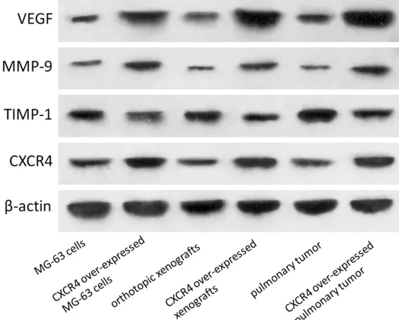
CXCR4 overexpression promotes the expres-sion of VEGF and MMP-9 but reduces TIMP-1 expression
To explore the underlying mechanisms of the enhancement of OS metastasis by CXCR4, we analyzed the protein expression of VEGF, MMP-9, TIMP-1 and CXCR4 by western blotting in cultured MG-63 cells, mouse orthotopic xeno-grafts of osteosarcomas and pulmonary meta-static tumors, respectively. The expression of VEGF and MMP-9 in MG-63 cells with CXCR4 overexpression were higher than those in the control. However, TIMP-1 in MG-63 cells with
CXCR4 overexpression was significantly lower
than that in the control. Similar, VEGF, MMP-9 and TIMP-1 protein expression patterns were found in orthotopic xenografts and pulmonary tumors (Figure 4).
Discussion
Patients with OS usually have a low 5-year sur-vival rate predominantly due to metastasis. Metastasis is an extremely complicated multi-step process, and its mechanism has not yet
for the CXCR4-CXCR12 signaling axis in the metastasis of OS and CXCR4-CXCR12 as a novel target for OS treatment.
The present study investigated the effect of CXCR4 overexpression on the metastasis of MG-63 cells and osteosarcoma xenografts in a mouse model. CXCR4 overexpression enhan- ced the invasion of MG-63 cells. The size and luciferase intensity of orthotopic xenografts with CXCR4 overexpression were larger and higher than normal orthotopic xenografts but
were not significantly different. Consistent with the results from a previous study [3], signifi -cantly higher luciferase intensity and more pul-monary tumors were observed in orthotopic xenografts with CXCR4 overexpression, sug-gesting that CXCR4 preferentially promotes orthotopic OS metastasis to the lungs.
VEGF is a vascular endothelial cell specific
growth factor and plays a crucial role in the induction of new blood vessels in the body [8, 21]. MMP-9 is a member of MMPs that degrade a variety of proteins in the extracellular matrix and destroy the histologic barrier in the pro-cess of tumor cell invasion and metastasis [22, 23]. TIMP-1, an inhibitor of
[image:5.612.92.376.72.300.2]metalloprotein-ase, specifically inhibits the activity of MMP-9,
Figure 4. CXCR4 overexpression increased VEGF and MMP-9 but reduced TIMP-1 expression. Group MG-63 cells and CXCR4 over-expressed MG-63 cells were in vitro experiments; group orthotopic xenografts, CXCR4 over-ex-pressed orthotopic xenografts, pulmonary tumor and CXCR4 over-exover-ex-pressed pulmonary tumor were in vitvo experiments.
thereby preventing tumor invasion and metas-tasis [24, 25]. We analyzed the changes in the expression of those metastasis-related cyto-kines after CXCR4 overexpression both in vitro
and in vivo. Significantly higher expressions of
VEGF and MMP-9 and a lower expression of TIMP-1 were observed in MG-63 cells with CXCR4 overexpression. The same expression pattern was found in orthotopic xenografts and pulmonary tumors. These results suggest that CXCR4 may promote OS tumor metastasis at least in part by enhancing the expression of VEGF and MMP-9 while suppressing TIMP-1 expression. However, the molecular mecha-nism by which the CXCR4-CXCR12 signaling axis regulates these cytokines warrants further investigation. In conclusion, the present results demonstrated that CXCR4 promotes metasta-ses to the lung of human osteosarcoma xeno-grafts in mice, likely through regulating the ex- pression of metastasis-related cytokines such
as VEGF, MMP-9 and TIMP-1. Our findings sug -gest that the development of CXCR4-blocking reagents is a promising approach for the treat-ment of metastasizing OS in combination with current treatment modes.
Acknowledgements
This study is funded by Key Clinical Specialty Discipline Construction Program of Fuzhou, Fujian province, China; 2015 Fuzhou Science and technology project (No. 2015-S-141-10) and 2016 Fuzhou Science and Technology Project (No. 2016-S-123-13).
Disclosure of conflict of interest
None.
Address correspondence to: Bin Yu, Department of Orthopedics and Traumatology, Nanfang Hospital
Affiliated to Southern Medical University, 1813
No-rth Guangzhou Road, Guangzhou 510515, Guang- dong, PR China. Tel: +86138-0254-3387; E-mail: yubin_nfykdx@126.com; Yiyuan Zhang, Department of Orthopedics, Fuzhou Second Hospital, 47 Shang- teng Road, Fuzhou 350007, Fujian, PR China. Tel: +8613905022511; E-mail: zhangyiyuan_fj@126. com
References
[1] Lettieri CK, Appel N, Labban N, Lussier DM, Blattman JN and Hingorani P. Progress and
opportunities for immune therapeutics in os-teosarcoma. Immunotherapy 2016; 8: 1233-1244.
[2] Li C, Cong Y, Liu X, Zhou X, Zhou G, Lu M, Shi X and Wu S. The progress of molecular diagnos-tics of osteosarcoma. Front Biosci (Landmark Ed) 2016; 21: 20-30.
[3] Brennecke P, Arlt MJ, Campanile C, Husmann K, Gvozdenovic A, Apuzzo T, Thelen M, Born W and Fuchs B. CXCR4 antibody treatment sup-presses metastatic spread to the lung of in-tratibial human osteosarcoma xenografts in mice. Clin Exp Metastasis 2014; 31: 339-349. [4] Gorlick R, Anderson P, Andrulis I, Arndt C,
Beardsley GP, Bernstein M, Bridge J, Cheung NK, Dome JS, Ebb D, Gardner T, Gebhardt M, Grier H, Hansen M, Healey J, Helman L, Hock J, Houghton J, Houghton P, Huvos A, Khanna C, Kieran M, Kleinerman E, Ladanyi M, Lau C, Malkin D, Marina N, Meltzer P, Meyers P,
Scho-field D, Schwartz C, Smith MA, Toretsky J, Tso -kos M, Wexler L, Wigginton J, Withrow S, Scho- enfeldt M and Anderson B. Biology of child-hood osteogenic sarcoma and potential tar-gets for therapeutic development: meeting summary. Clin Cancer Res 2003; 9: 5442-5453.
[5] Gorlick R and Khanna C. Osteosarcoma. J Bone Miner Res 2010; 25: 683-691.
[6] Chang PY, Hsieh MJ, Hsieh YS, Chen PN, Yang JS, Lo FC, Yang SF and Lu KH. Tricetin inhibits human osteosarcoma cells metastasis by tran-scriptionally repressing MMP-9 via p38 and Akt pathways. Environ Toxicol 2017; 32: 2032-2040.
[7] Rojiani MV, Ghoshal-Gupta S, Kutiyanawalla A, Mathur S and Rojiani AM. TIMP-1 overexpres-sion in lung carcinoma enhances tumor kinet-ics and angiogenesis in brain metastasis. J Neuropathol Exp Neurol 2015; 74: 293-304. [8] Zhang P, Dong L, Yan K, Long H, Yang TT,
Dong MQ, Zhou Y, Fan QY and Ma BA.
CXCR4-mediated osteosarcoma growth and pulmo-nary metastasis is promoted by mesenchymal stem cells through VEGF. Oncol Rep 2013; 30: 1753-1761.
[9] Balkwill F. Chemokine biology in cancer. Semin Immunol 2003; 15: 49-55.
[10] Nguyen DX, Bos PD and Massague J.
Metasta-sis: from dissemination to organ-specific colo -nization. Nat Rev Cancer 2009; 9: 274-284. [11] Markiewicz K, Zeman K, Kozar A and
Golebio-wska-Wawrzyniak M. [Evaluation of selected cytokines in children and adolescents with osteosarcoma at diagnosis-preliminary report. Med Wieku Rozwoj 2011; 15: 25-31.
[12] Sun B, Chen L, Fu H, Guo L, Guo H and Zhang N. Upregulation of RICTOR gene transcription
NF-kappaB pathway contributes to the metastasis of renal cell carcinoma. Tumour Biol 2016; 37: 4457-4466.
[13] Tu B, Peng ZX, Fan QM, Du L, Yan W and Tang
TT. Osteosarcoma cells promote the produc-tion of pro-tumor cytokines in mesenchymal stem cells by inhibiting their osteogenic differ-entiation through the TGF-beta/Smad2/3 pa- thway. Exp Cell Res 2014; 320: 164-173. [14] Muller A, Homey B, Soto H, Ge N, Catron D,
Bu-chanan ME, McClanahan T, Murphy E, Yuan W, Wagner SN, Barrera JL, Mohar A, Verastegui E and Zlotnik A. Involvement of chemokine re-ceptors in breast cancer metastasis. Nature 2001; 410: 50-56.
[15] Cespedes MV, Unzueta U, Alamo P, Gallardo A, Sala R, Casanova I, Pavon MA, Mangues MA,
Trias M, Lopez-Pousa A, Villaverde A, Vazquez E and Mangues R. Cancer-specific uptake of a li -ganded protein nanocarrier targeting aggres-sive CXCR4+ colorectal cancer models. Nano-medicine 2016; 12: 1987-1996.
[16] Chen Y, Wang S, Bu S, Xu M and Lai D. Low-dose cisplatin-induced CXCR4 expression pro-motes proliferation of ovarian cancer stem-like cells. Acta Biochim Biophys Sin (Shanghai) 2016; 48: 282-289.
[17] Kuribayashi N, Uchida D, Kinouchi M, Takama-ru N, Tamatani T, Nagai H and Miyamoto Y. The role of metabotropic glutamate receptor 5 on the stromal cell-derived factor-1/CXCR4 sys-tem in oral cancer. PLoS One 2013; 8: e80773. [18] Liu WT, Jing YY, Yan F, Han ZP, Lai FB, Zeng JX,
Yu GF, Fan QM, Li R, Zhao QD, Wu MC and Wei
LX. LPS-induced CXCR4-dependent migratory properties and a mesenchymal-like phenotype of colorectal cancer cells. Cell Adh Migr 2017; 11: 13-23.
[19] Sarchio SN, Scolyer RA, Beaugie C, McDonald
D, Marsh-Wakefield F, Halliday GM and Byrne
SN. Pharmacologically antagonizing the CX-CR4-CXCL12 chemokine pathway with AMD- 3100 inhibits sunlight-induced skin cancer. J Invest Dermatol 2014; 134: 1091-1100. [20] Sobolik T, Su YJ, Wells S, Ayers GD, Cook RS
and Richmond A. CXCR4 drives the metastatic phenotype in breast cancer through induction of CXCR2 and activation of MEK and PI3K pathways. Mol Biol Cell 2014; 25: 566-582. [21] Lin F, Zheng SE, Shen Z, Tang LN, Chen P, Sun
YJ, Zhao H and Yao Y. Relationships between levels of CXCR4 and VEGF and blood-borne metastasis and survival in patients with os- teosarcoma. Med Oncol 2011; 28: 649-653. [22] Han J, Yong B, Luo C, Tan P, Peng T and Shen J.
High serum alkaline phosphatase cooperating with MMP-9 predicts metastasis and poor pro- gnosis in patients with primary osteosarcoma in Southern China. World J Surg Oncol 2012; 10: 37.
[23] Peng TS, Qiu JS, Wu HX, Liang HZ and Luo CQ. [Expressions of CD44s, MMP-9, and Ki-67:
possible association with invasion, metastasis, and recurrence of osteosarcoma]. Ai Zheng 2002; 21: 745-750.
[24] Su Y, Wan D and Song W. Dryofragin inhibits the migration and invasion of human osteosar-coma U2OS cells by suppressing MMP-2/9 and elevating TIMP-1/2 through PI3K/AKT and p38 MAPK signaling pathways. Anticancer Drugs 2016; 27: 660-668.