Review Article
Comparison of short-segment versus long-segment
fixation for the treatment of thoracolumbar
burst fracture: a meta-analysis
Jun Li, Lei Liu
Department of Orthopaedics, West China Hospital, Sichuan University, 37# Wainan Guoxue Road, Chengdu 610041, People’s Republic of China
Received August 7, 2016; Accepted November 25, 2016; Epub February 15, 2017; Published February 28, 2017
Abstract:Background: Thoracolumbar burst fracture is one of the most common spinal traumas. It was still indeter-minate whether short-segment (SS) fixation was more effective and preferable than long-segment (LS) fixation. We therefore conducted this meta-analysis to assess the comparative safety and efficacy of SS and LS fixation for the treatment of thoracolumbar burst fracture. Methods: A literature search was conducted between January 1990 and September 2014. The sources of electronic searching included EMBASE, PubMed, Google Scholar, Ovid, Cochrane Library and Springer. Study eligibility was based on predefined criteria. The data were extracted by two authors independently and were managed using the Review Manager (RevMan) 5.2. A meta-analysis was conducted under the suggestion of PRISMA guideline standards. Results: Finally only 8 eligible articles were included in the current study, including a total of 455 thoracolumbar burst fracture patients who underwent the treatment with SS fixation (n=239) and LS fixation (n=216). The outcomes of this study indicated that final outcome in regard to implant fail-ure, anterior body height loss (ABHL), cobb angle (CA) and restoration ratio of compromised canal is better in the LS fixation group than those of SS fixation group. However, SS group significantly reduced surgical time when compared with LS group. There was no significant difference regarding blood loss, complication rate, Sagittal Index (SI) and kyphotic angle (KA). Conclusions: The main results of radiographic indexes and implant failure are better in the LS fixation group than those of SS fixation group. Nevertheless, LS fixation prolonged the duration of operation. Clinical outcomes suggested that there was no difference between the SS and LS fixation. The choice of the appropriate method has to be made cautiously and on an individualized basis.
Keywords: Thoracolumbar burst fracture, short-segment, long-segment, meta-analysis
Introduction
Thoracolumbar burst fractures, which count for 10%-20% of spinal fractures, are common spi-nal traumas that comprise serious instability of vertebral column and spinal canal compres-sion, resulting in increased hazards of
neuro-logical deficits, kyphosis and osteoporosis of
the spine [1]. A number of studies have proved good therapeutic effect of thoracolumbar burst fractures [2-4]. A consensus has been reached by the majority of experts that surgical therapy is necessary for symptomatic, unstable burst fractures [5, 6], nevertheless, it is still contro-versial with respect to the best surgical approach for thoracolumbar burst fractures [7-12].
The objectives of surgical treatment for thora-columbar burst fractures are to reestablish sta-bility of the vertebral column as well as to decompress the spinal canal, which is the foun-dation of early mobilization [7-12]. Among all the surgical modalities, posterior
transpedicu-lar screw fixation has been more preferable
when dealing with thoracolumbar burst frac-tures [15, 16].
Pedicle screws were introduced for the treat-ment of thoracolumbar fractures by Roy-Camille et al [17]. In 1963 and developed by Dick et al
[18] in 1985, since then, transpedicular SS fixa
-tion has become increasingly prevalent [18].
This modality involves pedicle screw fixation at
that SS fixation is in relation to unacceptable
implant failure rate, inadequate stability and postoperative loss of kyphosis correction [7, 8, 20]. Additional transpedicular procedures such as vertebroplasty and bone graft to maintain the anterior column has been provided as one of the choices to avoid this failure [21-24]. An alternative is to use longer segmental instru-mentation to reduce the load on each screw.
Hence, performing posterior fixation with two or
more segments above and below the fractured vertebra appears to be associated with less
rate of failure, yet significant increased verte
-bral immobility, dorsalgia and implant failure
was also detected in LS fixation [11, 13, 19,
25]. Therefore, it was still indeterminate
wheth-er SS fixation was more effective and prefwheth-era
-ble than LS fixation. So we conduct this
meta-analysis, trying to systematically assess the
comparative safety and efficacy of SS fixation
and LS fixation for the treatment of thoracolum
-bar burst fracture with the best available evidence.
Methods
Study selection criteria
Study eligibility was based on predefined crite
-ria. Allowing for the fact that only a small num-ber of randomized controlled trials were avail-able in the literature, non-randomized compar-ative studies (retrospective and prospective) were also included. The eligibility criteria were as follows: 1) the study included a comparative
design of SS fixation versus LS fixation in
patients with thoracolumbar burst fracture; 2)
patients without confirmed pathological thora
-columbar burst fractures according to imaging results; and 3) at least one of the following out-comes should be reported: perioperative results (operative time, blood loss, or hospital stay), kyphotic angle, sagittal index, anterior body height loss (ABHL), cobb angle, implant failure or complications. Studies were excluded if they 1) included patients who had thoraco-lumbar surgery in the previous 12 months; 2)
reported insufficient data, e.g. did not report
measures of variability. Lead authors were con-tacted in order to achieve additional data as required.
Literature search and data sources
A literature search was conducted between January 1990 and September 2014. The sourc-es of electronic searching included EMBASE,
PubMed, Google Scholar, Ovid, Cochrane Library and Springer. The initial key words used were “thoracolumbar burst fracture”, with the
search limited to “short-segment fixation ver
-sus long-segment fixation”. Other key words
that were used (without any search limitations)
were “fixation of thoracolumbar burst frac
-tures”, “treatment of thoracolumbar burst frac-tures with various segment stabilization” and “different instrumentation strategies for frac-tured thoracic and lumbar spine”. Additionally, conference full text articles were also reviewed to identify more articles. The results of unpub-lished studies were not sought.
Data extraction
The data were extracted by two authors inde-pendently based on the following categories: 1) Study year, country and study design; 2) Basic study characteristics including patients’ inclu-sion/exclusion criteria, enrolled number, age and sex proportion; 3) Baseline comparison information of confounding factors, such as diagnosis, surgical level and concomitant dis-eases; 4) Surgical information, including detailed spinal level and numbers, instrumen-tation and bone graft; 5) Perioperative out-comes such as operative time, blood loss, and hospital stay; 6) Imaging outcome improvement at last follow-up, including kyphotic angle, sag-ittal index, anterior body height loss (ABHL), cobb angle and canal compromise; 7) Fusion assessment method, fusion success criteria, and fusion rate at last follow-up; 8) Complication types and complication rates. Any differences were determined by consensus at the conclu-sion of full text review.
Data synthesis and statistical analysis
Data were managed using the Review Mana- ger 5.2, a software provided by Cochrane Centre. Dichotomous outcomes (e.g. presence/ absence of pain) were reported as proportions and were directly compared (difference in
pro-portions). We used these proportions to calcu
-late risk ratios (RRs) with 95% confidence inter
-vals (CIs). For continuous data (e.g. kyphotic angle, sagittal Index, anterior body height loss (ABHL)), results are presented as weighted
mean differences (WMD). The heterogeneity
was explored using the chi-squared test with
significance set at a P-value less than 0.05,
and I2 was used to estimate total variation
adopted if there was no statistical evidence of heterogeneity, and a random-effect model was
adopted if significant heterogeneity was evi -dent. In addition, the review was conducted under the suggestion of PRISMA guideline stan-dards [26]. The possibility of publishing bias was not evaluated because of the small num-ber of studies assessed.
Search results
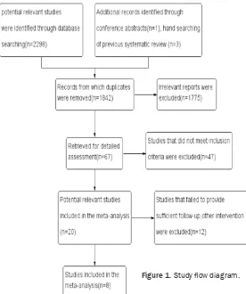
Firstly, a total of 2298 potentially relevant
reports were identified through database
searching (MEDLINE, n=186; PubMed, n=1692; Google Scholar, n=139; Ovid, n=110; Cochrane Library, n=46; Springer, n=125). 4 additional
literatures identified through conference
abstracts (n=1), hand searching of previous systematic review (n=3). After the duplicates were removed, 1842 potentially relevant stud-ies were reserved, 1775 of which were exclud-ed because they were not relatexclud-ed to the thora-columbar burst fracture. The full texts of the left 67 studies were assessed and 47 were excluded (24 only reported thoracolumbar
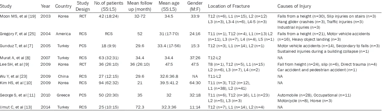
The study characteristics of the 8 trials includ-ed in the meta-analysis were summarizinclud-ed in Table 1, of which five are RCSs (retrospective
comparative studies) [8, 10, 13, 25], two are PCSs (prospec-tive comparative studies) [7, 11] and 2 are RCTs (randomized controlled trials) [9, 19]. The male to female ratio is 1.65:1. The mean age was 40.2 years. The mean follow-up duration ranged from 13.73 to 49.75 months. 93.6% of the fractures were located at T11 to L2 level. The cause of injury were reported in
five studies, among which falls from the height
and motor vehicles accidents were the top two inducements. Preoperative spinal Canal en- croachment were observed in two studies [8, 13] and anterior body height loss were assessed in six studies [7, 9-11, 19, 25]. Four papers provided information about preopera-tive kyphotic angle [8, 10, 19, 25]. The main complications reported in the included studies were implant failure and donor site pain.
Operative time
Four studies investigated significant reduced
surgical time in the SS group (Figure 2). All burst fracture data while not for comparison of short and
long segment fixation; 23 due
to no available data). After a thorough review, 12 studies were eliminated as they failed
to provide sufficient follow-up
or received other intervention. Finally, only 8 eligible articles were included in current study.
The flow chart of the study
selection is shown in Figure 1. The eligible studies were pub-lished between 2003 and 2014 including a total of 455 thoracolumbar burst fracture patients who underwent the treatment with short segment (SS, n=239) and long segment (LS, n=216) instrumentation. The age of the patients ranged from 13 to 70 years old. The smallest sample size was 18 and largest was 94. The fol-low-up time of the included studies was from 3 to 104 months.
[image:3.612.94.370.71.401.2]Table 1. The study characteristics of the trials that included in the meta-analysis
Study Year Country Study Design No.of patients (SS:LS) Mean follow up (month) Mean age (SS:LS) Gender (M:F) Location of Fracture Causes of Injury
Moon MS, et al [19] 2003 Korea RCT 42 (18:24) 32-72 34.5 33:9 T12 (n=6), L1 (n=15), L2 (n=12) L3 (n=3), L3-4 (n=6), L4-5 (n=3)
Falls from a height (n=30), Slip injuries on stairs (n=3)
Hang glider crashes (n=3), Traffic injuries (n=3)
Industrial injuries (n=3) Gregory F, et al [25] 2004 America RCS RCS 52 31 (17-70) 24:16 T11 (n=1), T12 (n=4), L1 (n=13) L2
(n=11), L3 (n=7), L4 (n=4), L5 (n=1)
Falls from a height (n=21), Motor vehicle accidents (n=16), Heavy object landing (n=3)
Gunduz T, et al [7] 2005 Turkey PCS 18 (9:9) 29.6 33.4 (17-56) 15:3 T12 (n=3), L1 (n=14), L2 (n=1) Motor vehicle accidents (n=14), Secondary to falls (n=3) Sustained injuries during a building collapse (n=1) Murat A, et al [8] 2007 Turkey RCS 63 (32:31) 34.4 34.4 37:26 T12-L2 NA
Lee SH, et al [9] 2009 Korea RCT 36 (26:10) 36 (26:10) 47.5 47.5 T8 (n=1), T12 (n=5), L1 (n=15)
L2 (n=6), L3 (n=7), L4 (n=2) Fall from height (n=24), slip (n=6), Direct trauma (n=4) Car accident and pedestrian accident (n=1)
Wu Y, et al [23] 2009 China RCS 27 (12:15) 29.6 32.6:36.8 NA T11-L2 NA Kim HS, et al [10] 2009 Korea RCS 94 (62:32) 21 39.5:41.2 64:30 T11 (n=3), T12 (n=12),
L1 (n=38), L2 (n=41)
NA George S, et al [11] 2010 Greece PCS 50 (20:30) 35 32 32:18 T11 (n=6), T12 (n=16), L1 (n=23)
L2 (n=5), L3 (n=3)
Automobile (n=28), Occupational (n=11) Motorcycle (n=8), Horse (n=3)
Umut C, et al [13] 2014 Turkey RCS 25 (10:15) 72.3 32.3:36 11:14 T12 (n=7), L1 (n=14), L2 (n=4) NA
studies showed SS group significantly reduced
[image:5.612.95.519.76.181.2]surgical time when compared with LS group. Overall, the WMD was -24.49 (95% CI: -42.07 to -6.91, P=0.006) in favor of the SS group. There
[image:5.612.97.521.341.654.2]Figure 2. Forest plot of comparison: operative time.
Figure 3. Forest plot of comparison: blood loss.
was obvious evidence for statistically signifi -cant heterogeneity (I2=81%; P=0.001).
Blood loss
Three trials reported perioperative blood loss (Figure 3) [7, 11, 25]. Pooling of relevant data
showed no statistically significant difference between the two groups, the WMD was -76.71
(95% CI: -202.94 to 49.53, P=0.23). There was
obvious evidence for statistically significant
heterogeneity (I2=79%; P=0.008).
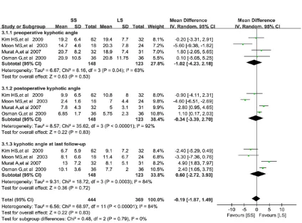
Kyphotic angle (KA)
Information of kyphotic angle was extracted from four trials (Figure 4) [8, 10, 19, 25]. In
each trial, the difference was not significant
between the two groups with regard to
preop-erative kyphotic angle (WMD=-1.02, 95% CI:
-4.23 to 2.18, P=0.53), postoperative kyphotic
angle (WMD=-0.34, 95% CI: -3.39 to 2.70,
P=0.83) and kyphotic angle at last follow-up
(WMD=0.60, 95% CI: -2.72 to 3.93, P=0.72).
[image:6.612.93.523.74.374.2]There were obvious evidence for statistically
Figure 5. Forest plot of comparison: anterior body height loss.
[image:6.612.90.523.409.528.2]significant heterogeneity in preoperative, post -operative and last follow-up ABHL results (I2=63%; P=0.04, I2=92%; P<0.00001 and I2=84%; P=0.0003 separately).
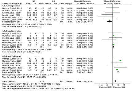
Anterior body height loss (ABHL)
Anterior body height loss were assessed in seven eligible studies (Figure 5) [7, 9-11, 19,
25]. Pooled estimates also revealed no signifi
-cant difference in preoperative ABHL between
the two groups (WMD=-2.40, 95% CI: -4.96 to
0.15, P=0.07), whereas the ABHL at postopera-tive and last follow-up in LS group were less than those of SS group, the statistical
differ-ence were significant (WMD=0.94, 95% CI:
0.37 to 1.52, P=0.001 and WMD=5.06, 95%
CI: 4.51 to 5.62, P<0.00001 respectively). There was no obvious evidence for statistically
significant heterogeneity in preoperative and
postoperative ABHL (I2=0%; P=0.66 and I2=0%; P=0.83 separately), while moderate heteroge-neity existed among the last follow-up results (I2=84%, P<0.00001).
Implant failure
Data regarding implant failure were
document-ed in five studies (Figure 6) [7, 9-11, 19]. The
participants in LS group were at a statistically
significant lower risk to undergo implant failure
than that of SS groups (RR=2.72, 95% CI: 1.27 to 5.83, P=0.01). There was moderate obvious
evidence for significant heterogeneity (I2=42%;
P=0.14).
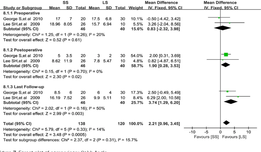
[image:7.612.95.518.75.322.2]Figure 7. Forest plot of comparison: Cobb Angle.
Cobb angle (CA)
Two of the included studies provided data on cobb angle (Figure 7) [9, 11]. The difference
was not significant between the two groups in preoperative results (WMD=0.83, 95% CI:
-2.32 to 3.98, P=0.61), nevertheless the Cobb Angles at postoperative and last follow-up in LS group were smaller than those of SS group, the
statistical difference were significant (WMD=
1.90, 95% CI: 0.28 to 3.53, P=0.02 and
WMD=3.74, 95% CI: 1.29 to 6.20, P=0.003
respectively). There was no obvious evidence
for statistically significant heterogeneity in pre -operative and post-operative results (I2=20%; P=0.26 and I2=0%; P=0.70 separately), while moderate heterogeneity existed among the last follow-up results (I2=50%, P=0.16).
Complications
Seven studies [9, 11] reported data on the total number of complications (including implant fail-ure, nerve root injuries, deep wound infections and reoperation etc.) (Figure 8). Total
complica-tions rates did not differ significantly between
SS and LS group (17.8% and 16.9%, respective-ly; P=0.40, RR=1.27, 95% CI: 0.73 to 2.21), and
heterogeneity between studies was not signifi
-cant (I2=16%, P= 0.31).
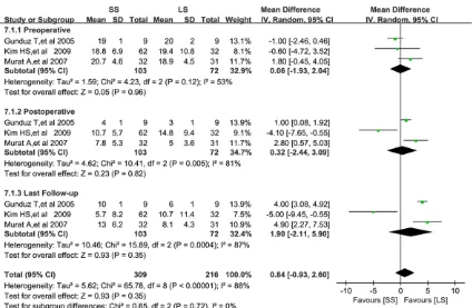
Sagittal index (SI)
Data on SI were available from three studies (Figure 9) [7, 8, 10]. No significant differences
was detected between the two groups with
regard to preoperative SI (WMD=0.06, 95% CI:
-1.93 to 2.04, P=0.96), postoperative SI
(WMD=0.32, 95% CI: -2.44 to 3.09, P=0.82) and SI at last follow-up (WMD=1.90, 95% CI:
-2.11 to 5.90, P=0.72). There were obvious
evi-dence for statistically significant heterogeneity
in preoperative, postoperative and last follow-up SI results (I2=53%; P=0.12, I2=81%; P=0.005 and I2=88%; P<0.00001 separately).
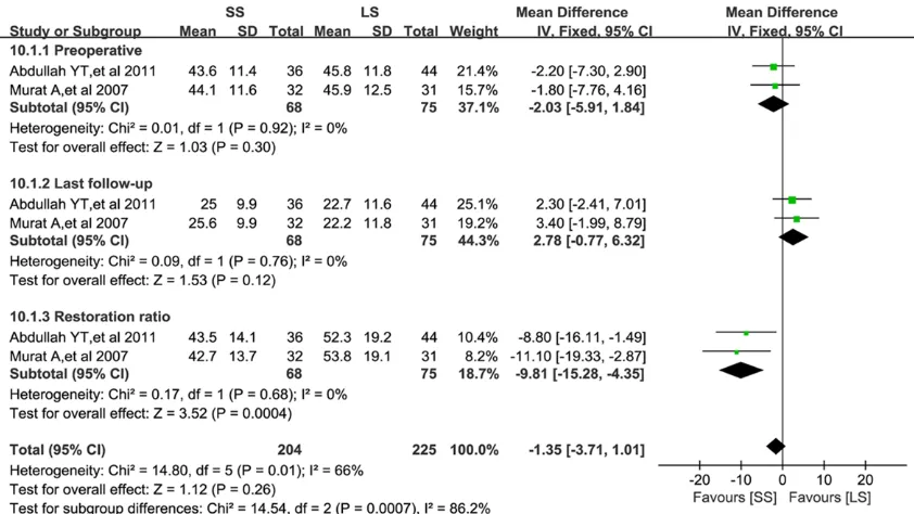
Canal compromise
Data regarding canal compromise were docu-mented in two studies (Figure 10) [8, 13]. No
significant differences was detected between
the two groups in preoperative and last
follow-up results (WMD=-2.03, 95% CI: -5.91 to 1.84, P=0.03 and WMD=2.78, 95% CI: -0.77 to 6.32,
[image:8.612.95.519.70.347.2]P=0.12 respectively) while the restoration ratios in LS group were higher than those of SS
group, the statistical difference were significant (WMD=-9.81, 95% CI: -15.28 to -4.35,
P=0.0004). No obvious heterogeneity was detected in each subgroup results (I2=0%, P=0.92; I2=0%, P=0.76 and I2=0%, P=0.68 separately).
Discussion
There are numerous matters to be taken into account in the disposal of patients with thora-columbar burst fractures. Prime disposal issues comprise medical stabilization, immobi-lization and acquisition of spinal alignment. Decisive therapeutic strategies are based on treatment expenses, spinal stabilization at the injured area, and the requirement for nerve decompression, followed by proper rehabilita-tion to optimize the patient’s funcrehabilita-tional recov-ery. It was demonstrated that the indirect expenses of conservative management out-number the direct expenses up to now, make up 95.4% of the total expenses, which is 71.6%
in surgical therapy [27]. Allowing for cost effi
-ciency, the surgical therapy of thoracolumbar burst fractures is more preferable. Never- theless, the optimal surgical approaches for the treatment of thoracolumbar burst fractures remains controversial.
Short segment (SS) fixation has been exten
-sively used by surgeons throughout the world in
previous decades [28, 29]. and has been fre-quently regarded as prefered surgical option because it provides advantages such as fusing fewer motion segments, reducing operative time, decreasing perioperative bleeding as well as avoiding loss of lumbar lordosis that is
asso-ciated with flat back syndrome [30, 31]. However, it was reported that SS fixation tend
to bring about fairly high rates of implant fail-ure, ranging from 9% to 54% [7, 8, 17, 32-36]. Lee SH et al [9]. Declared that LS pedicle instru-mentation can reduce implant failure rate, but
it also sacrifices additional motion segments
and ultimately reduces the range of motion [8, 9, 37]. Our meta-analysis showed that the par-ticipants in LS groups was at a statistically
sig-nificant lower risk to undergo implant failure
than that of SS group.
In general, the most significant objective of the
surgical intervention of thoracolumbar burst fractures is to switched back the patients’ nor-mal lives. Our review indicated that despite the
SS fixation was associated with less inco rpo-rated motion segments, it was not associated with a greater enhancement in Mean Dennis
Work Scores or a higher incidence of return to
[image:9.612.95.516.74.311.2]work. Among all the nine documented studies, only Gunduz T et al [7] reported Low Back Outcome Score(LBOS), which was devised by Fraser and Greenough [38], the results showed
that there was no significant difference in pain
reduction between SS and LS group. Murat Altay et al [8] demonstrated that restoration of canal compromise (CC) was well maintained in both SS and LS group, but the results were bet-ter in LS group and correction loss in LS group was much less than in SS Group. George S et al [11]. compared the back index in SS and LS
instrumentation, but no statistically significant
difference was detected between their preop-erative and postoppreop-erative values.
In this meta-analysis, we found significant dif
-ferences in duration of operation between the two groups. The LS group was associated with longer operative times, while use of the SS instrumentation could reduce the risks associ-ated with longer duration of operation. Pooling
of relevant data showed no statistically signifi
-cant difference between the two groups regard-ing perioperative blood loss, in which the
sur-geon’s proficiency, the size of the screws and
internal anatomy would play a part [25]. Lee et al [39] concluded that easy cage insertion and conveniently designed expandable cage would enable to reduce the duration of operation and the amount of hemorrhage during operation. Our meta-analysis also suggested that the main outcomes of the radiographic indexes
studied are better in the LS fixation group than
that of SS fixation group. The ABHL at postop
-erative and last follow-up in LS group were less than that of SS group, the statistical difference
were significant. Several study also indicated
that LS fixation can provide more secure fixa
-tion and better correc-tion than SS fixa-tion in
unstable thoracolumbar burst fractures, there-fore avoiding correction loss [40]. Some stud-ies demonstrated that anterior column recon-struction via cement augmentation techniques in combination with short segment pedicle screw constructs was a useful modality for tho-racolumbar burst fractures [41-43]. The
differ-ence was not significant between the two
groups with regard to kyphotic angle, yet LS group showed better results in cobb angle and sagittal index at postoperative and last
follow-up, the statistical difference were significant.
For complication rate, no significant differenc
-es were detected between the two groups. The inducements for complication were similar
among the studies, including superficial infec
-tion, pedicle screw dislodgement or implant
breakage and epidural hematoma. Lee SH et al [9]. reported two cases of implant removal in
LS fixation group, as there was a risk of skin
breakdown due to the irritation by the rods, and
one case of screw breakage in SS fixation
group.
We acknowledge that some limitations exists in
our meta-analysis. Firstly, despite several rele-vant studies have been reported, the majority were small and of poor quality, all datas and subsequent analysis were on the basis of only
395 patients. Secondly, statistically significant
heterogeneity was detected in the literatures especially when we incorporated the continu-ous outcomes. The heterogeneity, which lead-ed to high I2 values for our incorporated results for operative time, kyphotic angle, ABHL, SI and cobb angle, might be interpreted by the research design, inadequate baseline compari-sons, and the distinct result measurements. Furthermore, the pooled literatures were most-ly released in English and this may result in a cultural, language, and/or publication bias. Therefore, more randomized controlled trials with high quality are still required in the future. Conclusion
The main results of this review indicated that
the final outcomes of radiographic indexes in
regard to ABHL, cobb angle and SI were better
in the LS fixation group than those of SS fixa
-tion group. The implant failure is less in the LS
fixation group than that of SS fixation group. However, LS fixation prolonged the duration of operation significantly. Furthermore, clinical
outcomes suggested that there was no
differ-ence between the SS and LS fixation. Hdiffer-ence it is difficult to recommend one modality over the
other, the choice of the appropriate method have to be made cautiously and on a
individual-ized basis. It is significant that more high-quali -ty, randomized controlled trials be designed to direct the selection of the surgical modality in patients with thoracolumbar burst fractures.
Acknowledgements
The study was financially supported by the
Disclosure of conflict of interest None.
Address correspondence to: Dr. Lei Liu, Department of Orthopaedics, West China Hospital, Sichuan University, 37# Wainan Guoxue Road, Chengdu 610041, People’s Republic of China. E-mail: mror-thopedics@sina.com
References
[1] Suzuki T, Abe E, Miyakoshi N, Murai H, Kobayashi T, Abe T, Kikuchi K and Shimada Y. Anterior decompression and shortening recon-struction with a titanium mesh cage through a posterior approach alone for the treatment of lumbar burst fractures. Asian Spine J 2012; 6: 123-130.
[2] Dai LY, Jiang LS and Jiang SD. Conservative treatment of thoracolumbar burst fractures: a long-term follow-up results with special refer-ence to the load sharing classification. Spine (Phila Pa 1976) 2008; 33: 2536-2544. [3] Post RB, van der Sluis CK, Leferink VJ and Ten
DH. Long-term functional outcome after type A3 spinal fractures: operative versus non-oper-ative treatment. Acta Orthop Belg 2009; 75: 389-395.
[4] Thomas KC, Bailey CS, Dvorak MF, Kwon B and Fisher C. Comparison of operative and nonop-erative treatment for thoracolumbar burst frac-tures in patients without neurological deficit: a systematic review. J Neurosurg Spine 2006; 4: 351-358.
[5] Heary RF, Salas S, Bono CM and Kumar S. Complication avoidance: thoracolumbar and lumbar burst fractures. Neurosurg Clin N Am 2006; 17: 377-388.
[6] La Rosa G, Conti A, Cardali S, Cacciola F and Tomasello F. Does early decompression im-prove neurological outcome of spinal cord in-jured patients? Appraisal of the literature us-ing a meta-analytical approach. Spinal Cord 2004; 42: 503-512.
[7] Tezeren G and Kuru I. Posterior fixation of tho-racolumbar burst fracture: short-segment ped-icle fixation versus long-segment instrumenta-tion. J Spinal Disord Tech 2005; 18: 485-488. [8] Altay M, Ozkurt B, Aktekin CN, Ozturk AM,
Dogan O and Tabak AY. Treatment of unstable thoracolumbar junction burst fractures with short- or long-segment posterior fixation in magerl type a fractures. Eur Spine J 2007; 16: 1145-1155.
[9] Lee S, Pandher D, Yoon K, Lee S and Oh KJ. The effect of postoperative immobilization on short-segment fixation without bone grafting
for unstable fractures of thoracolumbar spine. Indian J Orthop 2009; 43: 197-204.
[10] Kim HS, Lee SY, Nanda A, Kim JY, Park JO, Moon SH, Lee HM, Kim HJ, Wei H and Moon ES. Comparison of surgical outcomes in thora-columbar fractures operated with posterior constructs having varying fixation length with selective anterior fusion. Yonsei Med J 2009; 50: 546-554.
[11] Sapkas G, Kateros K, Papadakis SA, Brilakis E, Macheras G and Katonis P. Treatment of un-stable thoracolumbar burst fractures by indi-rect reduction and posterior stabilization: short-segment versus long-segment stabiliza-tion. Open Orthop J 2010; 4: 7-13.
[12] Moon MS, Choi WT, Sun DH, Chae JW, Ryu JS, Chang H and Lin JF. Instrumented ligamento-taxis and stabilization of compression and burst fractures of dorsolumbar and mid-lum-bar spines. Indian J Orthop 2007; 41: 346-353.
[13] Tabak AY, Gunay MC, Altay M and Turker HB. [Effects of short- and long-segment posterior instrumentation on spinal canal remodeling in thoracolumbar vertebra burst fractures]. Ulus Travma Acil Cerrahi Derg 2011; 17: 141-148. [14] McLain RF. The biomechanics of long versus
short fixation for thoracolumbar spine frac-tures. Spine (Phila Pa 1976) 2006; 31: S70-S79, S104.
[15] Akbarnia BA, Crandall DG, Burkus K and Matthews T. Use of long rods and a short ar-throdesis for burst fractures of the thoracolum-bar spine. A long-term follow-up study. J Bone Joint Surg Am 1994; 76: 1629-1635.
[16] Bastian L, Lange U, Knop C, Tusch G and Blauth M. Evaluation of the mobility of adja-cent segments after posterior thoracolumbar fixation: a biomechanical study. Eur Spine J 2001; 10: 295-300.
[17] Koller H, Acosta F, Hempfing A, Rohrmuller D, Tauber M, Lederer S, Resch H, Zenner J, Klampfer H, Schwaiger R, Bogner R and Hitzl W. Long-term investigation of nonsurgical treatment for thoracolumbar and lumbar burst fractures: an outcome analysis in sight of spi-nopelvic balance. Eur Spine J 2008; 17: 1073-1095.
[18] Dick W, Kluger P, Magerl F, Woersdorfer O and Zach G. A new device for internal fixation of thoracolumbar and lumbar spine fractures: the ‘fixateur interne’. Paraplegia 1985; 23: 225-232.
[20] Akbarnia BA, Crandall DG, Burkus K and Matthews T. Use of long rods and a short ar-throdesis for burst fractures of the thoracolum-bar spine. A long-term follow-up study. J Bone Joint Surg Am 1994; 76: 1629-1635.
[21] Schroeder GD, Kepler CK, Koerner JD, Oner FC, Fehlings MG, Aarabi B, Schnake KJ, Rajasekaran S, Kandziora F, Vialle LR and Vaccaro AR. Can a thoracolumbar injury sever-ity score be uniformly applied from T1 to L5 or are modifications necessary? Global Spine J 2015; 5: 339-345.
[22] Dai J, Lin H, Niu S, Wu X, Wu Y and Zhang H. Correlation of bone fragments reposition and related parameters in thoracolumbar burst fractures patients. Int J Clin Exp Med 2015; 8: 11125-11131.
[23] Verlaan JJ, Diekerhof CH, Buskens E, van der Tweel I, Verbout AJ, Dhert WJ and Oner FC. Surgical treatment of traumatic fractures of the thoracic and lumbar spine: a systematic review of the literature on techniques, compli-cations, and outcome. Spine (Phila Pa 1976) 2004; 29: 803-814.
[24] Panchal RR, Matheis EA, Gudipally M, Hussain MM, Kim KD and Bucklen BS. Is lateral stabili-zation enough in thoracolumbar burst fracture reconstruction? A biomechanical investiga-tion. Spine J 2015; 15: 2247-2253.
[25] Guven O, Kocaoglu B, Bezer M, Aydin N and Nalbantoglu U. The use of screw at the fracture level in the treatment of thoracolumbar burst fractures. J Spinal Disord Tech 2009; 22: 417-421.
[26] Stahl S, Vida D, Meisner C, Lotter O, Roth- enberger J, Schaller H and Stahl AS. Systematic review and meta-analysis on the work-related etiology of de Quervainʼs tenosynovitis: a criti-cal appraisal of its recognition as an occupa-tional disease. Plast Reconstr Surg 2013; 132: 1479-91.
[27] Dimar JN, Wilde PH, Glassman SD, Puno RM and Johnson JR. Thoracolumbar burst frac-tures treated with combined anterior and pos-terior surgery. Am J Orthop (Belle Mead NJ) 1996; 25: 159-165.
[28] Wang L, Li J, Wang H, Yang Q, Lv D, Zhang W, Tang K, Shang L, Jiang C, Wu C, Ma K, Wang B, Liu Y, Zhang R, Shang X, Kou D, Jia X, Yang X, Tang Y, Zhang M, Wang P, Xu Y and Wang S. Posterior short segment pedicle screw fixation and TLIF for the treatment of unstable thoraco-lumbar/lumbar fracture. BMC Musculoskelet Disord 2014; 15: 40.
[29] Parker JW, Lane JR, Karaikovic EE and Gaines RW. Successful short-segment instrumenta-tion and fusion for thoracolumbar spine
frac-tures: a consecutive 41/2-year series. Spine (Phila Pa 1976) 2000; 25: 1157-1170. [30] Khare S and Sharma V. Surgical outcome of
posterior short segment trans-pedicle screw fixation for thoracolumbar fractures. J Orthop 2013; 10: 162-167.
[31] Audige L, Cornelius CP, Di Ieva A and Prein J. The first AO classification system for fractures of the craniomaxillofacial skeleton: rationale, methodological background, developmental process, and objectives. Craniomaxillofac Trauma Reconstr 2014; 7: S6-S14.
[32] Daniaux H, Seykora P, Genelin A, Lang T and Kathrein A. Application of posterior plating and modifications in thoracolumbar spine injuries. Indication, techniques, and results. Spine (Phila Pa 1976) 1991; 16: S125-S133.
[33] Knop C, Fabian HF, Bastian L, Rosenthal H, Lange U, Zdichavsky M and Blauth M. Fate of the transpedicular intervertebral bone graft af-ter posaf-terior stabilisation of thoracolumbar fractures. Eur Spine J 2002; 11: 251-257. [34] Leferink V, Zimmerman K, Veldhuis E, Ten
Vergert E and Ten Duis H. Thoracolumbar spi-nal fractures: radiological results of transpe-dicular fixation combined with transpetranspe-dicular cancellous bone graft and posterior fusion in 183 patients. Eur Spine J 2001; 10: 517-523. [35] Hegde A, Babu R and Shetty A. Management of
unstable thoraco-lumbar fractures with pedic-ular screw instrumentation: a series of 30 cases. J Clin Diagn Res 2013; 7: 2563-2566. [36] Heini PF. Comment on “Transpedicle body
aug-menter in painful osteoporotic compression fractures” (Kung-Chia Li, Anna F.-Y. Li, Ching-Hsiang Hsieh, Ching-Hsiang-Ho Chen). Eur Spine J 2007; 16: 599-600.
[37] Aebli N, Timm K, Patrick M and Krebs J. Short-segment posterior instrumentation combined with anterior spondylodesis using an autolo-gous rib graft in thoracolumbar burst fractures. Acta Orthop 2014; 85: 84-90.
[38] Hegde A. Management of unstable thoraco- lumbar fractures with pedicular screw instru-mentation: a series of 30 cases. J Clin Diagn Res 2013; 7: 2563-6.
[39] Lee G, Lee J, Hur H, Jang J, Kim T and Kim S. Comparison of clinical and radiologic results between expandable cages and titanium mesh cages for thoracolumbar burst fracture. J Korean Neurosurg S 2014; 55: 142.
[41] Hartensuer R, Gehweiler D, Schulze M, Matuszewski L, Raschke MJ and Vordemvenne T. Biomechanical evaluation of combined short segment fixation and augmentation of incomplete osteoporotic burst fractures. BMC Musculoskelet Disord 2013; 14: 360.
[42] Oner FC, Dhert WJ and Verlaan JJ. Less inva-sive anterior column reconstruction in thoraco-lumbar fractures. Injury 2005; 36 Suppl 2: B82-B89.