A molecular phylogeny of nuclear and mitochondrial
sequences in
Hymenolepis nana
(Cestoda) supports
the existence of a cryptic species
M. G. MACNISH1, U. M. MORGAN-RYAN1, P. T. MONIS2, J. M. BEHNKE3
andR. C. A. THOMPSON1*
1
WHO Collaborating Centre for the Molecular Epidemiology of Parasitic Infections and Division of Veterinary and Biomedical Sciences,Murdoch University,Murdoch,Western Australia,Australia 6150
2
Microbiology Unit,Australian Water Quality Centre,South Australian Water Corporation,Hodgson Road,
Bolivar 5110,Australia
3School of Life and Environmental Sciences,University of Nottingham,University Park,Nottingham NG7 2RD,UK
(Received 18 March 2002; revised 11 July 2002; accepted 15 July 2002)
S U M M A R Y
Since isolates ofHymenolepis nanainfecting humans and rodents are morphologically indistinguishable, the only way they can be reliably identified is by comparing the parasite in each host using molecular tools. In the current study, isolates of
H. nanafrom rodent and human hosts from a broad geographical range were sequenced at the ribosomal first internal transcribed spacer (ITS1), the mitochondrial cytochromecoxidase subunit 1 (C01) gene and the nuclear paramyosin gene loci.#Twenty-three isolates ofH. nanawere sequenced at the ITS1 locus and this confirmed the existence of spacers
which, although similar in length (approximately 646 bp), differed in their primary sequences which led to the separation of the isolates into 2 clusters when analysed phylogenetically. This sequence variation was not, however, related to the host of origin of the isolate, thus was not a marker of genetic distinction betweenH. nana from rodents and humans. Sequencing of a 444 bp fragment of the mitochondrial cytochromecoxidase 1 gene (C01) in 9 isolates ofH. nanafrom rodents and 6 from humans identified a phylogenetically supported genetic divergence of approximately 5 % between some mouse and human isolates. This suggests thatH. nanais a species complex, or ‘ cryptic ’ species (=morphologically identical yet genetically distinct). A small segment of the nuclear gene, paramyosin, (625 bp or 840 bp) was sequenced in 4 mouse and 3 human isolates ofH. nana. However, this gene did not provide the level of heterogeneity required to distinguish between isolates from rodent and human hosts. From the results obtained from faster evolving genes, and the epidemiological evidence, we believe that the life-cycle ofH. nanathat exists in the north-west of Western Australia is likely to involve mainly ‘ human to human ’ transmission.
Key words :Hymenolepis nana, cryptic species, ribosomal ITS1, mitochondrial C01, paramyosin.
I N T R O D U C T I O N
The tapewormHymenolepis nanawas first described asTaenia nanaby Von Siebold in 1852 as a parasite found in humans. In 1906 Stiles described a mor-phologically identical parasite from a rodent host and named it Hymenolepis nana var. fraterna (see Joyeux, 1920 and Skrabin & Matevosan, 1945 in Baer & Tenora, 1970). Controversy over their status as a single or dual species and host specificity has existed ever since (Baer & Tenora, 1970 ; Schantz, 1996). It is not entirely clear whether the speciesHymenolepis
nanaandHymenolepis fraternaare 2 distinct species, each highly host specific ; whether they are 2 distinct species but capable of infecting both human and rodent hosts or whether they are simply the same species found in either host (see Brumpt, 1949 and Yamaguti, 1959 in Baer & Tenora, 1970 ; Ferretti, Gabriele & Palmas, 1981).
Further nomenclature difficulties are encountered with the re-classification of Hymenolepidids with armed rostella (hooks present) as Rodentolepis
(Spasskii, 1954). Thus,H. nana(von Siebold, 1852) and H. fraterna (Stiles, 1906) are now classified as
Rodentolepis nana and R. fraterna respectively by some taxonomists. Despite this revised nomencla-ture, the original confusion of speciation and host specificity remains to be solved. In a recent study, the oral inoculation of 51 samples ofH. nanaof human origin into specific pathogen-free hamsters, 4 mouse strains and 2 rat strains failed to establish infections (Macnishet al. 2002). Furthermore, inoculation of the same samples into thymus deficient- and cortisone
* Corresponding author : Murdoch University, South Street, Murdoch, WA 6150. Tel :+61 8 9360 2466. Fax :
+61 8 9310 4144. E-mail : andrew_t@central.murdoch. edu.au
# Nucleotide sequence data published here have been
submitted to GenBankTMand are available under accession numbers AF461124 and AF461125 (18S–28S) ; AY121842 and AY121843 (cytochrome c oxidase 1) ; AY1844 and AY121845 (paramyosin).
acetate treated-mice, rats and hamsters also failed to establish infections, providing supportive evidence for the hypothesis that the species in mice may be non-infective to humans, and thus, represent a host-specific ‘ strain ’ (Macnish et al. 2002).
The form/‘ strain ’ ofH. nanapresent in Australia has never been identified with certainty. Further-more, it is not well understood which form of transmission commonly occurs in Australian com-munities, whether the ‘ strain/species ’ present in the north-west of Western Australia is infective to human and rodent hosts, or whether humans harbour their own ‘ strain/species ’ ofHymenolepis. Since isolates of
H. nanainfecting humans and rodents are morpho-logically identical, the only way they can be reliably distinguished is by comparing the parasite in each host using molecular techniques.
To date, no comprehensive study of the molecular characteristics of H. nana isolates from humans or rodents has been carried out. In one study, sequences of the internal transcribed spacer 2 (ITS2) region of ribosomal DNA and partial sequences of the mito-chondrial cytochrome c oxidase subunit 1 (C01) gene were compared between an isolate ofH. nana, collected from a laboratory mouse (Mus musculus) from Japan, and a laboratory golden hamster ( Meso-cricetus auratus) from Uruguay. No sequence dif-ferences were found in the ITS2 between both isolates and only 2 base differences were detected in the C01 locus (Okamotoet al. 1997).
Understanding the status of the 2 putative species
H. nanaandH. fraterna, and their host predilection, is of biological, epidemiological and taxonomic im-portance. Molecular characterization of these para-sites, collected from both human and rodent hosts from a wide geographical distribution, will provide further evidence towards their identity. Understand-ing the genotypic characteristics ofHymenolepis iso-lates in different hosts will also help in determining host specificity and transmission patterns and thus
allow a more appropriate approach to the control of infections in endemic communities. The aim of this study was, therefore, to sequence the ribosomal ITS1, mitochondrial C01 and part of the paramyo-sin gene of numerousHymenolepisisolates collected from humans and mice from several geographically separated regions to ascertain whether any signifi-cant genetic differences existed between H. nana
isolates from the two host types.
M A T E R I A L S A N D M E T H O D S
Collection of parasite material
Sources of all parasites used in this study are listed in Table 1. A reference isolate ofHymenolepis nanawas obtained from Dr Akira Ito, Gifu University, Japan. Approximately 2000 H. nana eggs were inoculated into 5-week-old male BALB/c mice. Adult worms were dissected from the small intestine approxi-mately 14 days post-inoculation then washed re-peatedly in phosphate-buffered saline (PBS) and stored atx80xC until DNA extraction.H. diminuta adult worms were obtained by dissection of infected 6-week-old male Wistar rats maintained by the Murdoch University Parasitology teaching resource. Adult worms ofH. citelli (hamsters) and H. micro-stoma (mice) preserved in dimethyl sulphoxide-saturated NaCl were supplied by Dr Jerzy Behnke.
Purification of DNA from adult worms and cysticercoids
DNA was purified from H. nana, H. diminuta,
[image:2.595.71.543.113.242.2]H. microstomaandH. citelliusing the QIAmp tissue purification kit (Qiagen, Hilden, Germany) with some minor modifications. Briefly, 10ml of glass milk matrix (Bio-Rad, California, USA) was substituted from the QIAmp spin columns as suggested by Morganet al. (1998). DNA was eluted in 300ml of Table 1. Source and geographical location of parasite material used in this study
(AI, Dr Akira Ito, Gifu University, Japan ; MC, Dr Margherita Conchedda, Universita` degli Studi di Cagliari, Italy ; MUPTR, Murdoch University Parasitology Teaching Resource ; JB, Dr Jerzy Behnke, University of Nottingham, UK ; GS, Dr Grant Singleton, CSIRO, NSW, Australia ; MUPS, Murdoch University Parasite Survey.)
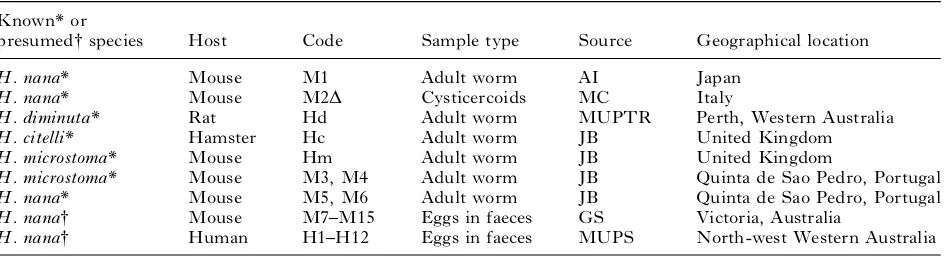
Known* or
presumed#species Host Code Sample type Source Geographical location
H. nana* Mouse M1 Adult worm AI Japan
H. nana* Mouse M2D Cysticercoids MC Italy
H. diminuta* Rat Hd Adult worm MUPTR Perth, Western Australia
H. citelli* Hamster Hc Adult worm JB United Kingdom
H. microstoma* Mouse Hm Adult worm JB United Kingdom
H. microstoma* Mouse M3, M4 Adult worm JB Quinta de Sao Pedro, Portugal
H. nana* Mouse M5, M6 Adult worm JB Quinta de Sao Pedro, Portugal
H. nana# Mouse M7–M15 Eggs in faeces GS Victoria, Australia
H. nana# Human H1–H12 Eggs in faeces MUPS North-west Western Australia
* Morphologically identified adult worm (except M2D=cysticercoids).
Tris–EDTA (TE) and 1ml of the diluted extract was added to the polymerase chain reaction (PCR) mix.
Purification of DNA from human and mouse faeces
DNA was purified from mouse faecal samples as previously described (Morganet al. 1998). DNA was purified from human faecal samples using a method first described by Walsh, Metzger & Higuchi (1991) and modified by Paxinoset al. (1997). Some further modifications were used in our laboratory. Briefly, a small plug of faecal material was suspended in 250ml of 10 % ChelexÒ100 (Bio-Rad, California, USA) in TE buffer, boiled for 7 min and vortexed vigorously. Samples were boiled again for 7 min then centri-fuged at full speed for 5 min. The supernatants were de-proteinized usingProCipitateTM(LigoChem
Inc, USA), which is a non-hazardous alternative to phenol/chloroform. Briefly, the supernatant was mixed with an equal volume of ProCipitateTM and
mixed gently for 5 min at room temperature. The samples were centrifuged at full speed for 5 min. The supernatant was further concentrated using standard sodium acetate/ethanol precipitation then eluted in 50ml of TE. Usually 2.5ml of template DNA was used for subsequent PCR reactions.
Primer design for amplification of ITS1
DNA sequences of H. nana and H. diminuta
spanning the 3kend of the 18S rRNA gene, internal transcribed spacer 1 (ITS1), 5.8S, ITS2 and the 5k end of the 28S rRNA gene (GenBankTM accession
numbers AF461124 and AF461125 respectively) were used to identify regions conserved between the 2 species of interest. Primers from these regions, designated F3 (5k GCGGAAGGATCATTACAC-GTTC 3k) and R3 (5k GCTCGACTCTTCATC-GATCCACG 3k) were designed using the software package Amplify 2.1 (Bill Engels, University of Wisconsin) to allow the amplification of the ITS1 regions ofH. nana andH. diminuta.
PCR amplification and sequencing of ITS1
DNA was amplified in 67 mM Tris–HCl (pH 8.8), 16.6 mM (NH4)2SO4, 2 mM MgCl2, 0.5 unit of Tth plus (Fisher Biotech, Perth, Australia), 200mM of each dNTP and 12.5 pmoles of each primer. Reac-tions were performed on a PE 2400 (Perkin Elmer, Foster City, California) thermal cycler. Samples were heated to 94xC for 2 min, 63xC for 2 min, 72xC for 1 min, followed by 50 cycles of 94xC for 20 sec, 63xC for 20 sec, 72xC for 45 sec and a final cycle of 72xC for 7 min. Usually 0.5 unit ofTaqExtender (Strategene, USA) was added to the PCR mix to improve ampli-fication efficiency.
Amplification products were purified using the QIAquick-spin PCR purification kit (Qiagen,
Germany) and sequenced in both directions with F3 and R3 primers using an ABI PrismTM Dye
Terminator Cycle Sequencing Ready Reaction kit (Applied Biosystems, Foster City, California) accord-ing to manufacturer’s instructions with some modi-fications. Briefly, the reagent volumes were halved and the annealing temperature was raised to 60xC. In some instances, 2ml of HalfTERM (Genpak Inc, Stony Brook, New York) was substituted for 2ml of dye terminator mix as this reduced the cost of the reaction without compromising the quality of the sequence. The sequences were analysed using SeqEd v1.0.3 (Applied Biosystems).
When direct sequencing of the PCR product yielded poor results PCR products were cloned into a pCRÒ2.1 T-vector (Invitrogen, USA) and trans-formants were screened by PCR. Plasmid DNA was purified from overnight cultures using the Flexi-PrepÒkit (Pharmacia Biotech Inc, USA). At least 3 positive clones were sequenced in both directions using universal M13 primers.
PCR amplification and sequencing of mitochondrial C01
A segment of the mitochondrial cytochrome c oxi-dase subunit 1 (C01) was amplified using primers and conditions described by Okamoto et al. (1997) with a single modification which was to increase the annealing temperature from 42xC to 55xC. This modification was required to prevent non-specific amplification because problems were encountered with non-specific primer binding from DNA ex-tracted from faecal samples.
When direct sequencing of the C01 fragment yielded poor results the PCR product was cloned in the manner described for the ITS1 fragments and sequenced using universal M13 forward and reverse primers.
Primer design, PCR amplification and sequencing of nuclear paramyosin(pmy)
A degenerate forward primer, designated Pmy-F (5k
AAYCAYYTVAGTCCGAGATGGAAC 3k) and
located approximately 1550 bp downstream of the 5k end of pmywas designed using available sequences of the closely related speciesEchinococcus granulosus
(GenBankTM accession number Z21787), Taenia
of primers, Ext-F (5k AGAAAGAGCACCACTCG-CAC 3k) were located just 3k of the Pmy-F primer. A conserved new external reverse primer, Ext-R (5k GACAGTAATCTCACGGATCTC 3k) was located just 3kof the Pmy-R primer. The external set of pri-mers, Ext-F and Ext-R amplified a 700 bp product. The internal set of primers, Int-F (5k
ATTTCTGA-GATGGAGGTCAGATTTAAG 3k) and Int R
(5kTTTGCGAAGAGTTTCAGCACGCTTG 3k), amplified a 625 bp product. DNA was amplified in 25ml vol. reactions as for the ITS1 and C01 loci, except that 25 pmol of each primer was used and the MgCl2concentration was increased to 3 mM.
For the primary PCR reaction samples were heated to 94xC for 3 min followed by 50 cycles of 94xC for 30 sec, 58xC for 20 sec, 72xC for 45 sec and a final cycle of 72xC for 7 min. Oneml of the primary PCR reaction was used as a template for the secondary nested PCR reaction. Samples were heated to 94xC for 1 min, followed by 50 cycles of 94xC for 3 sec, 70xC for 20 sec, 72xC for 45 sec and a final cycle of 72xC for 7 min.
Phylogenetic analyses
Nucleotide sequences were aligned using Clustal X (Thompson et al. 1997). Distance-based and parsi-mony analyses were performed using PAUP* (Swof-ford, D. L. 1999. PAUP*. Phylogenetic Analysis Using Parsimony (*and Other Methods). Version 4.0b2 Sinauer Associates, Sunderland, Massachu-setts). Maximum Likelihood analyses were per-formed using PUZZLE (version 4.1, (Strimmer & von Haeseler, 1996)). Distance-based analyses were conducted using Tamura-Nei distance estimates and trees were constructed using the Neighbour-Joining algorithm. Parsimony analyses were conducted using either the branch and bound or heuristic search methods. Bootstrap analyses were conducted using 1000 replicates. Trees were drawn using the Tree-View program (Page, 1996).
R E S U L T S
Sequence analysis of ITS1
The ITS1 region was sequenced from 23 isolates of
H. nana(11 human, 12 mouse). Amplified ITS1 was cloned for 11 isolates and between 2 and 6 clones were sequenced for each isolate resulting in a total of 37 clones being analysed. Amplicons from the re-maining isolates (5 human, 7 mouse) were sequenced directly. Sequence analysis determined that the PCR products fromH. nanaincluded 22 bp of the 3kend of the 18S, 571 bp of the ITS1 and 53 bases of the 5k end of the 5.8S. Although 23 isolates were analysed, only 6 distinct sequence types were identified. Of these 6 sequences, 13 isolates (H7, M7, M1, M12, H10, M2, H8, M9c1, H11c3, H11c1, M11c3, H2c3)
possessed one sequence and 5 (M5, M6, H4c2, H4c1, H6c2) possessed another. The 2 predominant sequences were obtained from both cloned and directly sequenced amplified DNA’s.
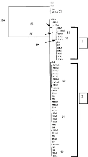
Phylogenetic analysis of ITS1
Analysis of ITS1 nucleotide sequences was con-ducted usingH. microstomaas an outgroup. Due to the uncertainties regarding the basis for the high levels of variability among cloned sequences from individual isolates of H. nana, each clone was in-cluded in the final phylogenetic analysis (Fig. 1). Analysis of ITS1 sequences provided confirmation that isolates M3, M4 and M15c4 areH. microstoma. Distance-based and Maximum Likelihood (ML) analyses identified 2 main clusters of isolates (Clusters 1 and 2, Fig. 1). Cluster 1, containing M5, M6, H3c1-c4, H4c1-c2, H6c1-c3, was supported by bootstrap analysis (89 %) (Fig. 1). The topology of the tree for the remaining isolates ofH. nanareceived poor bootstrap support. Parsimony analysis of these data was not possible due to the large number of trees with the same length generated. The most substantial sequence variation was seen between H. nana se-quences in Cluster 1 versus those in Cluster 2. Two directly sequenced isolates from Portugal (M5, M6) shared identical sequences with cloned isolates within Cluster 1. Some variation was also seen between the isolates within Cluster 2 itself ; however, this was usually low (98.8–99.4 %).
Sequence analysis of C01
Sequence analysis determined that the PCR product obtained by amplification with primers pr-a and pr-b was 444 bp for H. nana (PCR results not shown). Direct sequences were obtained for H. diminuta
(411 bp), H. microstoma (429 bp) and H. citelli
(425 bp). At the genus level,H. nana was 85, 81.3 and 81.7 % genetically similar to the 3 other Hymeno-lepidids H. microstoma, H. diminuta and H. citelli
respectively (data not shown). Intra-specific variation was not detected between the human isolates of
H. nanaand was very low (99.5–100 %) between the mouse isolates from Australia (M9, M11, M12, M13, M14), Japan (M1) and Italy (M2). However, exten-sive intra-specific variation was found between the 2 Portugese mouse isolates, M5 and M6, and the re-maining mouse isolates (M1, M2, M9, M11, M12, M13, M14) ranging from 95.0 to 96.0 %. Similarly, high levels of intra-specific variation between the human isolates and the 2 rodents isolates, M5, M6, were observed (96.1 %). Variation within an individual isolate, ascertained by sequencing 3 clones, was only observed for the H. nana isolate M6 and was low (98.8 %). The remaining isolates ofH. nanaand other
polymorphisms were found in the region sequenced for any species.
Phylogenetic analysis of mitochondrial C01
Analysis of C01 nucleotide sequences was conducted using H. diminuta and H. citelli as outgroups. Parsimony, distance-based and ML analyses pro-duced trees with similar topology (Fig. 2). The rodent
[image:5.595.168.469.66.600.2]isolates M3, M4 and M15 were identified as H. microstoma. Isolate M15 was placed into the same clade as theH. microstomareference sequence but this was poorly supported by bootstrap analysis. The isolates of H. nana were divided into 2 clades, one containing the mouse isolates M5 and M6 and the other containing the remaining human- and mouse-derived isolates ofH. nana. The topology within the latter clade suggests a division correlating with host
origin, with isolates from the same host species clus-tering with each other. However, this topology was not supported by bootstrap analysis.
Sequence analysis of paramyosin
A PCR product of approximately 840 bp was ob-tained from theH. nanaisolates M1, M2, M5, M6,
H. microstoma, M3,H. diminutaandH. citelliusing
the primers Pmy-F and Pmy-R (results not shown). Direct sequencing of approximately 840 bp PCR product, using the primers Pmy-F and Pmy-R, was achieved with the H. nana isolates M1, M2, M5, M6, the reference isolate ofH. microstoma, and the field isolate M3, H. diminuta and H. citelli. Unambiguous sequence of 782 bp, 775 bp, 796 bp, 788 bp was obtained for H. nana, H. microstoma,
[image:6.595.68.497.70.591.2]H. diminuta and H. citelli respectively. Direct
Fig. 2. Phylogram of distance-based analyses generated from the sequences of the mitochondrial cytochromec
oxidase subunit 1 (C01) gene region from human (H) and mouse (M) isolates ofHymenolepis nanaand fromH. diminuta
sequencing of the PCR product obtained using the nested PCR primers of H. nana isolates H7, H13, H14 andH. diminutaisolate H15 yielded poor results therefore the PCR products were cloned prior to sequencing. Sequence analysis of this PCR product confirmed the size of the PCR product was 625 bp which corresponded with the predicted frag-ment size using the secondary primers, Int-F and Int-R. Intra- and inter-specific variation between and within isolates ofH. nana was not detected.
Phylogenetic analysis of paramyosin
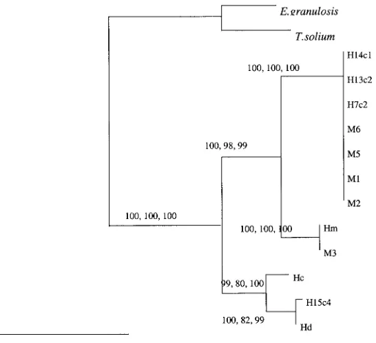
Parsimony, distance and maximum likelihood (heu-ristic, quartet puzzling) analyses produced trees with the same topology (Fig. 3). Isolates ofH. nana
(human and mouse) possessed identical paramyosin nucleotide sequences and were placed into a single clade.H. microstomawas placed as the closest rela-tive ofH. nana.Hymenolepis diminutaandH. citelli
were placed into the same clade and formed a sister group toH. nana/H. microstoma. The human isolate H15c4 was identified as H. diminuta based on se-quence similarity and phylogeny. All of the nodes of the tree were very highly supported by bootstrap analysis using the distance-based and ML methods (99–100 %). Bootstrap analysis using parsimony found high support (98–100 %) for the grouping of the H. nana and H. microstoma but lower support (80–82 %) for the grouping of H. diminuta with H. citelli. The monophyly of Hymenolepis was highly
supported (100 %) with respect to the outgroups used in the study.
D I S C U S S I O N
A total of 23 isolates (human and mouse) ofH. nana, representing a wide geographical distribution (Aus-tralia, Japan, Italy, Portugal) were characterized, by sequencing, at the ribosomal ITS1 locus. Of these, 14 isolates were also characterized at the mitochon-drial C01 locus and 7 at the paramyosin locus. More isolates were unable to be sequenced at the C01 and paramyosin loci due to insufficient material. Phylo-genetic analysis of the ITS1 region of these isolates identified 2 clusters whose composition did not cor-relate with host (mouse or human) or geographical origin of the isolates. Variation was found both between and within isolates ofH. nana. The basis of the variation within a single isolate was not deter-mined but could be due to either variation between eggs within a sample or variation between ITS re-peats within an individual egg. Detailed insight into ‘ strain resolution ’ between the mouse and human isolates was not possible using this locus due to the high levels of polymorphism (Vogler & DeSalle, 1994 ; Sorensen, Curtis & Mindhella, 1998 ; Jobst, King & Hemleben, 1998).
Sequencing of the mitochondrial cytochrome c
oxidase 1 gene (C01) in a number of isolates of
[image:7.595.56.329.68.317.2]H. nanafrom rodents and humans identified a phylo-genetically supported genetic divergence between
Fig. 3. Phylogram of distance-based analyses generated from the sequences of the paramyosin gene region from human (H) and mouse (M) isolates ofHymenolepis nanaand fromH. diminuta(Hd),H. microstoma(Hm),H. citelli
some mouse isolates in comparison with isolates of
H. nanafrom humans. This provided evidence that the mitochondrial C01 gene was useful for identify-ing genetic divergences in H. nana that were not resolvable using nuclear loci. The difficulties in amplifying mitochondrial genes from egg DNA meant that important information was lost for some isolates, such as H2, H3, H4 and H6, at the C01 locus. Genetic characterization of these particular isolates, at the C01 locus, would be invaluable for a direct comparison to be made between all the isolates characterized in this study.
In the current study, the placement of the 2 Portugese isolates (M5, M6) into a separate clade, as a result of 5.0 % genetic divergence at the C01 locus that is well supported by bootstrap analysis, is highly suggestive of the existence of ‘ cryptic species ’ of
H. nana (=genetically distinct yet morphologically identical). Given the geographical isolation of Portugal from Australia it is possible that distinct genotypes would evolve over time in this region. In addition, the separation of all the human isolates into a group within Cluster 2, whilst not supported by high bootstrap values, is well supported by biologi-cal data obtained in a previous study (Macnishet al. 2002) suggesting that a barrier to gene flow may be occurring in the Australian populations ofH. nana.
This may be due to environmental and/or ecological pressures caused by the documented absence of dom-estic mice species in close proximity to human dwell-ings, combined with the susceptibility or resistance of the host (genetic factors, host immunity, host diet). In addition, selection pressure by the host and/ or the parasite may contribute to the co-evolution of particular host–parasite relationships.
The region of thePmygene characterized in this study yielded phylogenetically informative data for the resolution of the relationships betweenH. nana,
H. microstoma,H. diminutaandH. citellithat corre-sponded with the relationships found using 2 other genetic loci ; the nuclear ribosomal ITS1 and the mitochondrial C01. However, the gene was too con-served to allow differentiation of H. nana isolates from different hosts.
Currently, the ‘ yardstick ’ for delineating ‘ species/ strains ’ on the basis of genetic differences is un-resolved in the literature and remains contentious (Morgan & Blair, 1995 ; Haaget al.1997 ; Sorensen
et al. 1998 ; Blouinet al. 1998). Although some have proposed that if ‘ within-species ’ variation at particu-lar genetic loci is low this supports the existence of a species (e.g. see Hunget al. 1999). Others suggest that caution is required in interpreting the genetic data in the absence of any supportive biological data (Thompson & Lymbery, 1990 ; Blouin et al. 1998 ; Thompson, Constantine & Morgan, 1998 ; Sorensen
et al. 1998 ; Tibayrenc, 1998). The inference, based on sequence data from the CO1 locus, thatH. nanais a species complex/cryptic species that differs in its
host range is consistent with biological data reported earlier for these isolates (Macnishet al. 2002). From an epidemiological viewpoint this provides highly useful information that helps identify whether trans-mission is likely to be occurring between rodent and human hosts.
In light of the data presented here, and the epi-demiological evidence (Macnishet al. 2002) we be-lieve that the life-cycle ofH. nanathat exists in the north-west of Western Australia is likely to involve mainly ‘ human to human ’ transmission. In other communities worldwide, where poor hygiene prac-tices help to promote the direct transmission of parasite species such asHymenolepis, it is not clear whether mice act as reservoirs of strains ofH. nana
that are transmissible to humans. From the results of this study it would appear that, where rodent hosts are minimal or absent, the potential exists for the route of transmission to become mainly direct (‘ human to human ’) in those communities also.
It is now recommended to sequence faster evolving loci to interpret the relationship between human and mouse isolates with more clarity. Recently, the nico-tinamide adenine dinucleotide dehydrogenase sub-unit 2 and 4 (ND2 and ND4 respectively) genes were characterized in 8 cestode species, includingH. nana
(Nakao et al. 2000). Further characterization of the ND4 mitochondrial gene in human and rodent iso-lates ofH. nanais facilitated by this recent research and is recommended for future characterization of this parasite.
The authors would again like to thank Dr Akira Ito, Gifu University, Japan, and Dr Margherita Conchedda, Cag-liari, Italy for their generous gifts of H. nana isolates. Funding provided by the Australian Federation of Uni-versity Women (WA) Inc. and the National Health and Medical Research Centre (NHMRC) is also gratefully acknowledged.
R E F E R E N C E S
BAER,J.G.& TENORA,F. (1970). Some species of
Hymenolepis(Cestoidea) from rodents and from primates.Acta Scientiarum Naturalium Academiae Scientiarum Bohemoslovacae Brno4, 1–32.
BLOUIN,M.S.,YOWELL,C.A.,COURTNEY,C.H.& DAME,J.B.
(1998). Substitution bias, rapid saturation, and the use of mtDNA for nematode systematics.Molecular Biology and Evolution15, 1719–1727.
FERRETTI,G.,GABRIELE,F.& PALMAS,C. (1981).
Development of human and mouse strain of
Hymenolepis nanain mice.International Journal for Parasitology11, 425–430.
HAAG,K.L.,ZAHA,A.,ARAUJO,A.M.& GOTTSTEIN,B. (1997). Reduced genetic variability within coding and
non-coding regions of theEchinococcus multilocularis
genome.Parasitology115, 521–529.
HUNG,G.C.,CHILTON,N.B.,BEVERIDGE,I.,ZHU,X.Q., LICHTENFELS,J.R.& GASSER,R.B. (1999). Molecular
minutus(Nematoda : Strongylidae).International Journal for Parasitology29, 285–291.
JOBST,J.,KING,K.& HEMLEBEN,V. (1998). Molecular
evolution of the internal transcribed spacers (ITS1 and ITS2) and phylogenetic relationships among species of the family Cucurbitaceae.Molecular Phylogenetics and Evolution9, 204–219.
LACLETTE,J.P.,LANDA,A.,ARCOS,L.,WILLMS,K.,DAVIS, A.E.& SHOEMAKER,C.B. (1991). Paramyosin is the
Schistosoma mansoni(Trematoda) homologue of antigen B fromTaenia solium(Cestoda).Molecular and Biochemical Parasitology44, 287–296.
MACNISH,M.G.,MORGAN,U.M.,BEHNKE,J.M.& THOMPSON, R.C.A. (2002). Failure to infect laboratory rodents with
humans isolates ofRodentolepis(=Hymenolepis)nana.
Journal of Helminthology76, 37–43.
MORGAN,J.A.T.& BLAIR,D. (1995). Nuclear rDNA
ITS sequence variation in the trematode genus
Echinostoma: an aid to establishing relationships within the 37-collar-spine group.Parasitology111, 609–615.
MORGAN,U.M.,PALLANT,L.,DWYER,B.W.,FORBES,D.A., RICH,G.& THOMPSON,R.C.A. (1998). Comparison of PCR and microscopy for detection ofCryptosporidium parvumin human fecal specimens : Clinical trial.Journal of Clinical Microbiology36, 995–998.
NAKAO,M.,SAKO,Y.,YOKOYAMA,N.,FUKUNAGA,M.& ITO,A. (2000). Mitochondrial genetic code in
cestodes.Molecular and Biochemical Parasitology111, 415–424.
OKAMOTO,M.,AGATSUMA,T.,KUROSAWA,T.& ITO,A.
(1997). Phylogenetic relationships of three
hymenolepidid species inferred from nuclear ribosomal and mitochondrial DNA sequences.Parasitology115, 661–666.
PAGE,R.D.M. (1996). TREEVIEW : an application to
display phylogenetic trees on personal computers.
Computer Applications in the Biosciences : CABIOS
12, 357–358.
PAXINOS,E.,MCINTOSH,C.,RALLS,K.& FLEISCHER,R.
(1997). A noninvasive method for distinguishing among canid species : amplification and enzyme restriction of DNA from dung.Molecular Ecology6, 483–486.
SCHANTZ,P.M. (1996). Tapeworms (Cestodiasis).
Gastroenterology Clinics of North America3, 637–653.
SORENSEN,R.E.,CURTIS,J.& MINDHELLA,D.J. (1998).
Intraspecific variation in the rDNA ITS loci of 37-collar-spined Echinostomes from North America : Implications for sequence-based diagnoses and phylogenetics.Journal of Parasitology84, 992–997.
STRIMMER,K.& VON HAESELER,A. (1996). Quartet puzzling :
a quartet maximum likelihood method for
reconstructing tree topologies.Molecular Biology and Evolution13, 964–969.
THOMPSON,J.D.,GIBSON,T.J.,PLEWNIAK,F.,JEANMOUGIN, F.& HIGGINS,D.G. (1997). The Clustal X windows
interface : flexible strategies for multiple sequence alignment aided by quality analysis tools.Nucleic Acids Research25, 4876–4882.
THOMPSON,R.C.A.,CONSTANTINE,C.C.& MORGAN,U.M.
(1998). Overview and significance of molecular methods : what role for molecular epidemiology ?
Parasitology117, S161–S175.
THOMPSON,R.C.A.& LYMBERY,A.J. (1990). Intraspecific
variation in parasites – What is a strain ?Parasitology Today6, 345–348.
TIBAYRENC,M. (1998). Genetic epidemiology of parasitic
protozoa and other infectious agents : the need for an integrated approach.International Journal for Parasitology28, 85–104.
VOGLER,A.P.& DESALLE,R. (1994). Evolution and
phylogenetic information content of the ITS-1 region in the tiger beetleCicindela dorsalis.Molecular Biology and Evolution11, 393–405.
WALSH,P.S.,METZGER,D.A.& HIGUCHI,R. (1991). Chelex 100 as a medium for simple extraction of DNA for PCR-bases typing from forensic material.BioTechniques