Amethod for
sequencing
restriction fragments withdideoxynucleoside
triphosphates
JanMaat* and Andrew J.H.Smith+
*Department
of Physiological Chemistry, Leiden University, Leiden, TheNetherlands,
and +MRCLaboratory
ofMolecular Biology, Hills Road,Cambridge
CB22QH,UK
Received 26 October 1978
ABSTRACT
A rapid enzymatic approach is described for the sequence analysis of a 5' terminally labelled restriction. fragmen.t. It involves limited nicking of the strands of the molecule throughout the sequence by pancreatic DNAase I. The 3' hydroxyl groups exposed by each nick are then. used to prime chain. exten.sion. by DNA polymerase I in. four separate reaction.s. Each reaction. uses on.e of the four chain terminating dideoxynucleoside triphosphates
(ddNTPSs),
together with the four deoxynucleoside triphosphates (dNTPs). In a single reaction. all the 3' ends are terminated in. positions of the same base, which is different for each of the four reaction.s. When. the products of these reactions are resolved by gel electrophoresis accordin.g to size, a sequence can be deduced from the pattern. of radioactive bands. Sequences can bedetermined onwards from 10-20 residues from the 5' labelled end. The length of sequence which can. be determined is only limited by the resolution. of the gel.
INTRODUCTION
A number of rapid methods have been. developed for DNA sequence analysis. In this paper a n.ew method is described which, like .the chemical degradation. procedure of Maxam and Gilbert
[1],
can. be used to sequence 5' termin.ally labelled restriction. fragments. This is an. enzymatic approach, which relies on. the ability of DNA polymerase I to use the chain. terminating 2',3'-dideoxynucleoside triphosphates (ddNTPs) as substrates in a nick-tran.slation. reaction.[2]
of the labelled fragment.Sanger et al.
[3]
have described amethod of rapid DNA sequencing. It involves the synthesis of a radioactive complementary copy of asingle
stranded target sequence with DNA polymerase, using the directlyadjacent
ann.ealed strand of a restriction, fragment as a primer. In. fourseparate
reactions, which each contain a differen.t ddNTPand,
in addition., the four deoxynucleoside triphosphates(dNTPs) (one
or more of which area-32
P-labelled),
there is apartial incorporation
of the terminator onto the radio-active elongation. products. In a single reaction a size range of labelledoligonucleotides is produced, all with a common 5' end, but with the 3? end terminating in. positions of the same base throughout the sequence. Parallel fractionation by denaturing polyacrylamide gel electrophoresis of the -products of the four separate reactions, which each con.tain. in turn. one of the four ddNTPs, resolves the oligonucleotides of different sizes. These reveal in. order the positions of each base, allowing a sequence to be determined.
The drawback of this method is that it requires a pure
single
stranded template. In. the approach described here, use is made of the ddNTPs to sequence double stran.ded DNA in the form of restriction. fragments, labelled at only one 5' en.d.In. the protocol a 5'
32
P-labelled fragment is incubated in. the presence of DNA polymerase I and pancreatic DNAase I in. four separate reactions. Each reaction. contains all four dNTPs and, in. turn., one different ddNTP. The pancreatic DNAase I introduces nicks,- the 3' hydroxyls of which serve as priming points for DNA polymerase I. Chain extension then. proceeds from the3' end of every nick, leading to a base specific chain termination. in. each reaction., as in. the method of Sanger et al.
[3].
The products of the reactions are fractionated on. denaturing polyacrylamide slab gels, resolving the oligonucleotides with the common. labelled 5? end.Although the nicking and subsequent chain. exten.sion. occurs on. both
strands, since only one is labelled a pattern. of radioactive ban.ds is
produced for only that strand, allowing its sequence to be deduced. Nicking of the fragment by DNAase I does not occur at every residue along its length but the fact that both deoxy- and dideoxy- NTPs are present in each reaction ensures that chain. extension con.tinues through several residues of the same base from the site of the nick. Every residue in. the sequence should there-fore give rise to a ban.d.
EXPERIMENTAL PROCEDURES Materials
Bovine pancreatic DNAase I, grade I, Boehringer Manrnheim (FRG); solution. of 1 mg/ml in. distilled water, stored at -20°C.
DNA polymerase I, grade I, Boehringer Mannheim (FRG); 5 units/,l. ddNTPs and dNTPs, P.-L. Biochemicals
(Milwaukee,
Wis.,USA);
dissolved to a concentration. of 10 mM in TE buffer(1
mMTris.HCl_pH
7.6, 0.05 M EDTA) and stored at-20°C.
T4
polynucleotide
kinase was made according to amodification. [4] of the 4538procedure of Richardson. [5].
y-32P-ATP, 3000 Ci/mmole, the Radiochemical Centre (Amersham, UK). DNA molecules, 5' terminally labelled, were prepared from adenovirus type 5 DNA which was a gift of B.M.M. Dekker.
All other materials used are as described in references 3, 4 an.d 6. 5' terminal labelling of restriction fragments
2-5 pmoles of restriction. fragments are labelled with T4 polynucleotide kinase and -y-32P-ATP as described in reference 4. In order to obtain DNA molecules with only one 3 P-labelled 5' terminus, the fragments are further digested with a restriction enzyme of different specificity, which cleaves at
one or more sites within the fragment. The resulting mixture of labelled
fragments is then fractionated by electrophoresis on, polyacrylamide slab gels (400 x 180 x 1.5 cm) in, 90 mliM Tris-borate pH 8.3, 1 mMv EDTA. The radioactive
fragments are recovered from the gel with the elution procedure described by Maxam and Gilbert [1] and precipitated with ethanol without carrier tRNA.
The precipitate is taken up in. 10 pl water.
Sequencing procedure
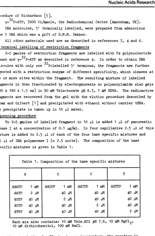
To 2-5 pnoles of labelled fragment in. 10 p1l is added 1 pl of pan.creatic DNAase I at a con.centration. of 0.1 pg/ml. In four capillaries 2.5 pl of this mixture is added to 2.5 il of each of the four base specific mixtures and 0.5 pl of DNA polymerase I (= 2.5 units). The composition. of the base specific mixtures is given in Table 1.
Table 1. Composition of the base specific mixtures
Each mix also contains: 10 mMi Tris.HCl pH 7.6, 10 mM
XgC12,
10 miM dithiothreitol, 100 mM NaCl.
After incubation for 30 min at room temperature, the reaction is
terminated by adding 15 p1 of formamide, containing 10 mM EDTA, 0.05%
XCPF
and 0.05% BPB. A G C T ddATP 1 mM ddGTP 1 mM ddCTP 1 mi ddTTP 1 mM dATP 2 pM
40
M40
p40
M dGTP 40 P 2pM
40 dIM40
P4 dCTP 40 p 40AV 2 p40
PM dTTP 40 M40
D40 ,V
2 MGel electrophoresis
5 pi of each sample in. formamide dye is then. heat denatured at 100°C for 3 min. and loaded in. a 5 mm wide well of an. 8% polyacrylamide slab gel (400 x
180 x 0.35 mm) containing 7 M urea. Gel electrophoresis is carried out according to Sanger and Coulson. [6]. En.ough material is left to allow several loadings on, the gel at differen.t times after the electrophoresis has been, started. In this way expan.sion. of the ban.d pattern. in differen.t regions of the sequence is obtained. On an. 8% gel, for example, up to 300 nucleo-tides from the 5' end can. be resolved.
The gels are wrapped in. Saranwrap and autoradiographed at -20°C on. pre-flashed Sakura QH n.o-screen. medical X-ray film. In. some cases enhanced auto-radiographic detection. is obtained with Ilford-fast tungstate 30 x 40 cm inten.sifying screen.s; exposure is at -70°C.
RESULTS
Figures 1 an.d 3 show examples of sequen.ces from part of the HindIII fragmen.t E of adenovirus type 5 DNA obtain.ed using the procedure described in. this paper. The HindIII fragmen.t E (between. map position.s 8.0 an.d 17.0) was
5' labelled and cleaved with HinfI to yield labelled fragments of
630
an.d 440 nucleotides long. The sequence deduced in. Figure 1 using the 440 nucleotide long fragmen.t starts at position. 2816, 10 nucleotides away from the 5'labelled end, an.d reads through to position. 3035 (coun.ting from the left terminus of adenovirus type 5 DNA in the 1-strand). Figure 2 shows the result of the Maxam and Gilbert procedure applied to the same fragmen.t. The sequen.ces obtained from Figures 1 and 2 are in agreemen.t. Figure 3 shows a part of the sequen.ce of the r-strand of the
630
nucleotide long fragmen.t. The label resides at the HindIII site at map position. 17.0. The sequence can. be deduced 17 nucleotides from the 5' end to a poin.t 200 nucleotides away. It also has been, shown to be in. full agreemen.t with the sequence deduced employing the Maxam and Gilbert method on. the same fragments(results
n.otshown).
DISCUSSION
The n.ick-tran.slation. sequencing procedure described here has been.. shown to be reliable, rapid and simple. The reaction. makes use of the in.tact DNA polymerase I enzyme which is necessary for carrying out
nick-tran.slation.;
the 5'-35'
exonuclease activity of the enzymehydrolyses
the5'
en.d of the nick asthe 3' end is simultaneously extended by the
5'-3' polymerase activity.
Experience has shown. that there is n.osign.ifican.t
loss of labelled materialrC0 wkFF3jF0_.,_
0:1
.3 U ;) t )< F-- <C) }C))01ss -O <r<fr-G0CO < O HO C . C-) I.D 4 E- F- <, 0 < u 11 F- <l i- k-C <0 4l O 0 0 0D ,".-C - 4 15:*nv6~St
s*S
65 S *0 S S e*sew
i X t - f 19 t)D|*! a1' ' --u *4Z"i4 W 94* a U l U a 91 a *] 5* h E l J C I) 0 0 I 0iC)
I u Q) 0 < 0 0 s-SQz 0 H-0D 0D C. 0D 9)5-I 0. Cd o 0 04)) F~02-4P P4 4 0 02 0~ H- 00 H- 4 a) P4Cd02 -PM Cd 0 A4 .t, 0 000 o 0 P., -1a 2Cd' 02 e H- 0 02 'H ) 'h 4)C 00 o 4- 0 O PQ q 12 4d)H0RCHC
'H O, G FIC\J 0 4)H 4A r 0 0 -Po h C) rd 4) 'Cd 'CdH Cd 4.) c 0 H 41) 0 P hP x H44) 0r a) to fi > o t- 4-1' H CH-'H* co020-P 4) 0 c- hCd 02 4) i) Cd 'b PR ZLI)4O 4-i c C c 0 -4F CDNucleic
Acids Research
IV
Ill
11
I
CPyAG
CPyAG*CPyA
G
CPyAG
q~~~~~~~~~~~~~~~~~~~~~~~~~~~~~~~~~~~~
*3as
.: '-'Ire a~~~~~~~~~ Sma
.*. A.W.a
A aw S4 a. dmk -a _ *a -. a 2530 -M" q A*^
'016 t. .4, .....
le t,. *-. K ._ --.,& wFigure 2. Result of the procedure of Maxam and Gilbert [1] applied to the same fragment as in Figure 1. Samples from each base specific chemical degradation were loaded in formamide and electrophoresed as described in the legend to Figure 1. A:
tw
.;... II
C C3 2, {3 0 < F-4 1- 0 L)
~0
0 < 0 00-)<
0 0<<40
0 1-0)
tj < 0 u < 0< 4 0< < 0 L) C) 0 00 4 Lo0 j c' D C_. :.51L 't o 0 <0 0< <0 < -7,D H-0 t) 0C-3
E: 0 (CI C) *7, H- t- V-0._ H- kC (C V GC 7, weW4 ~I* s% ~ ~ U 2)/|* * 4g5 *1 1W_ine*fhe.ieg
S S SJJ%t J1J *'www *. MDC tee[email protected]
::
IJ1 J*
9 c ; $'_-i # >OF w *,** A C-i. I----) eC . H.- F-F J% N A 4, Afi Itn0
e: rD <,e <-1.
L r Z-;D 3 = -(C ._ F-H--C 0 0 -- (C 0 Od Ce H a4) -d Cd H ) *H 02 -942 woR H H h o -H H Oq* d O U)r-@q
a)d P'C\ d "a w cd Cd o bH H *d a) -P hd (0i ., Cd ~I~
O H' w, H 4riH"r'dH
c) 42h la) 0 la)) R) d U) Ha) g ww (0 C. (0') 4)_ R o a) 0 CH - c c 0O L-.due to hydrolysis of the labelled 5' end by the 5'-3' exonuclease
activity. Preliminary experiments indicate that the Klen.ow subfragment of DNA polymerase I may also be suitable for this procedure. It may somehow be capable of displacing the 5' end of the nick as polymerisation. proceeds from the 3' end.
The sequencing approach can be applied to DYT.A fragments of any length. The sequence that can. be deduced is only limited by the resolving power of the gel electrophoresis
(which
is at present 300-400 nucleotides long). The conditions described in. the protocol are, on. the whole, suitable for applica-tion. to fragments of 100-500 nucleotides long. For longer fragments the DNA-ase I concentration may need to be increased in. order to obtain. most of the labelled DNA within. a size range of oligonucleotides below 300-400 nucleo-tides long.It is apparent from the results obtained using the nick-translation procedure that there are a number of peculiarities of the method, some of which are evident in Figures 1 and 3.
(a) Every residue gives rise to a band apart from the first 10-20 residues from the 5' labelled en.d. In some cases a sequence can. be deduced directly after the position of the strong band which ruans across all four tracks and appears to have a size
corres-ponding to an oligonucleotide of chain. length of 8-10 residues. No bands are seen. corresponding to oligonucleotides of smaller chain length than this. Generally bands corresponding to the first few residues of the sequence are very weak in intensity. In other cases not even. weak bands are seen. directly after the position. of this strong artefact band and sometimes sequences cannot be deduced until some 10 nucleotides from this position..
We do not completely understand the reasons for these phenomena.
(b)
Throughout the sequence band intensity is very variable. Thisparticularly happens in. a run of the same residue, where it decreases in, a 5' to 3' direction.. This is thought to be due in part to the fact that nicking of the fragment does not occur
randomly throughout the sequence and is
especially
infrequent in runs of the same residue. Chain extension through a run in most cases therefore takes place from the 3' end of a nick lying adjacent 5' to the run.. Partial incorporation of the chain. terninators at each residue in. a run, results in. an. exponential fall-off(in
a 5' to 3'direction.)
of the numbers ofnucleotides terminated at those residues.
(c) Although not seen. in. Figures 1 and
3,
DNA polymerase I sometimes has difficulties in. copying certain. sequences of the template stran.d. This leads to pile-ups of bands lying across all tracks of the gel. There seems to be no consistent feature ofthe sequences at these points.
In spite of the disadvantages mentioned above and the obvious limitation. of the method - that it can. only be applied to 5' termin.ally labelled
frag-ments (as opposed to 5' or 3' end labelled fragments, as is the case with the "Maxam-Gilbert" procedure) - we feel that this procedure may have use as a
general method of rapid DNA sequencing and have some advantages over existing methods. It has the advantage over the "classical" chain. terminator method
that it can. be applied directly to double stranded DNA. In. comparison with the chemical cleavage method of Maxam and Gilbert, the manipulations involved
are less laborious an.d more rapid, and the bands produced in. thin gels are sharper, enabling longer sequences to be read.
ACKNOWIEDGEGENTS
The authors thank Drs. F. Sanger, A. de Waard and H. van. Ormondt for advice an.d help in the course of their work.
The research reported in. this manuscript was supported in part by the Medical Research Council and in. part by the Netherlands Foundation. for Chemical Research (S.O.N.) with financial aid from the Netherlands Organ.isa-tion for Advan.cement of Pure Research (Z.W.O.)
A.J.H. Smith was supported by a Research Studentship from the Medical Research Council. J. Maat was supported by the Kon.ingin. Wilhemmina Fonds.
REFERENCES
1. Maxam, A.M. and Gilbert, W. (1977) Proc. Nat. Acad. Sci. USA 74, 560-564.
2. Kelly, R.B., Cozzarelli, N.R., Deutscher, M.P., Lehman., I.R. and
Kornberg, A. (1970) J. Biol. Chem 245, 39-45..
3. Sanger, F., Nicklen., S. and Coulson, A.R. (1977) Proc. Nat. Acad. Sci. USA 74,
5463-5467-4. van. Ormondt, H., Maat, J., de Waaxrd, A. and van, der Eb, A.J. (1978) Gene, in. press.
5. Richardson., C.C. (1972) Proc. Nucleic Acid Res. 2, 815-828. 6. Sanger, F. an.d Coulson, A.R. (1978) FEBS Letters 87, 107-110.
![Figure 2. Result of the procedure of Maxam and Gilbert [1] applied to the same fragment as in Figure 1](https://thumb-us.123doks.com/thumbv2/123dok_us/1650599.2725858/6.684.126.563.101.777/figure-result-procedure-maxam-gilbert-applied-fragment-figure.webp)