Ali Kemal KADİROGLU1
Kenan TURGUTALP2
Zulfikar YILMAZ2
Dede ŞIT1
İsmail Hamdi KARA3
Hasan KAYABAŞI2
Mehmet Emin YILMAZ1
1 Department of Nephrology,
Medicine Faculty in Dicle University,
2 Department of Internal
Medicine, in Dicle University, Diyarbakır,
3 Department of Family
Practice, Medicine Faculty in Düzce University, Düzce.
Corresponding Address /Yazışma Adresi:
Assoc. Prof. Dr. Ali Kemal Kadiroglu
Department of Nephrology, Medicine Faculty of Dicle University, 21280 Diyarbakır / TURKEY Phone: +90412 248 80 01 GSM: +90532 664 36 30 Fax No: +90412 248 81 71 akkadiroglu@hotmail.com ABSTRACT
Aim: Osteoporosis is a skeletal disorder that is characterized by low bone mass, micro-structural degeneration of bone and high risk of fracture. In this study our aim was to detect the frequency of osteoporosis in hemodialysis (HD) and continuous ambulatory peritoneal dialysis (CAPD) patients according to Parathyroid Hormone (PTH) after vitamin D therapy during the two years period.
Materials and method:18 HD patients (12 male, 6 female) and 12 CAPD patients (8 male, 4 female) undergoing to dialysis therapy in dialysis center of Medicine Faculty Hospital in Dicle University were enrolled to the study. The patients were evaluated with bone mineral density which was measured by left heel quantitative ultrasound before and after the active vitamin D therapy. The blood samples were collected for biochemical analysis in the morning after 12 hours fasting period before and after the active vitamin D therapy.
Results: After the therapy T and Z scores bone mineral density and ALP values were increased in the group that PTH values were between 120-250 pg/ml and more than 250 pg/ml. But these parameters were decreased in the group that PTH were lower than 120 pg/ml. Osteoporosis percentage were 23 % in PTH value <120 pg/ml, %20 in PTH value between 120-250 pg/ml and 20 % in PTH >250 pg/ml in the initial measurement. After the therapy these were 30 %, 0 % and 20 % relatively.
Conclusion:Before the treatment there was not a statistical difference between T score of 3 groups. After the treatment there was a statistically significant difference. Especially T score was better after the therapy in the second group that PTH values were between 120-250 pg/ml. Key Words:PTH, osteoporosis, dialysis, bone mineral density (BMD).
ÖZET
Amaç:Osteoporoz; düşük kemik kitlesi, kemikte mikroyapısal dejenerasyon ve yüksek fraktür riskiyle karakterize bir iskelet hastalığıdır. Bu çalışmada amacımız iki yıllık periodda vitamin D tedavisinden sonra Paratiroid hormone düzeyine göre hemodiyaliz ve devamlı ayaktan periton diyaliz tedavisi gören hastalarda osteoporoz sıklığını tespit etmektir.
Materyal ve metod:Dicle Üniversitesi Tıp Fakültesi Hastanesi Diyaliz Merkezinde diyaliz tedavisi gören 18 HD hastası (12 erkek, 6 kadın) ve 12 SAPD hastası (8 erkek, 4, kadın) çalışmaya alındı. Hastalar aktif vitamin D tedavisi öncesi ve sonrası sol topuk kantitatif ultrasonografi ile ölçülen kemik mineral dansitesiyle değerlendirildi. Biyokimyasal analizler için kan örnekleri aktif vitamin D tedavisi öncesi ve sonrasında 12 saatlik açlık periodundan sonra alındı.
Bulgular: Tedavi sonunda kemik mineral dansitesinin T ve Z skorları ile ALP, PTH değeri 120 -250 pg/ml olan grupla birlikte PTH değeri 250 pg/ml’den fazla olanlarda da yüksek bulundu. Bu değerler PTH değeri 120 pg/ml’nin altında olanlarda düşük bulundu. Başlangıç Osteoporoz oranı; PTH değeri < 120 pg/ml olanlarda % 23, 120 -250 pg/ml olanlarda % 20 ve 250 pg/ml’den büyük olanlarda ise % 20 olarak saptandı. Tedavi sonrasında sırasıyla % 30, % 0 ve % 20 olduğu görüldü.
Sonuç: Tedavi öncesi T skor değerinde her üç grup arasında istatistiksel farklılık yoktu. Tedaviden sonra, anlamlı istatistiksel farklılık saptandı. Özellikle tedavi sonrası PTH değeri 120 – 250 pg/ml olan ikinci grupta T skoru daha iyi bulundu.
Anahtar Kelimeler:PTH, osteoporoz, diyaliz, kemik mineral dansitesi.
The Frequency of Osteoporosis in Hemodialysis
and Continuous Ambulatory Peritoneal Dialysis
Patients According to PTH Levels after Active
Vitamin D Therapy during the Two Years Period
İki Yıllık Periodda Aktif Vitamin D Tedavisinden
Sonra Paratiroid Hormone Düzeyine Göre
Hemodiyaliz ve Devamlı Ayaktan Periton Diyaliz
Tedavisi Gören Hastalarda Osteoporoz Sıklığı
Submitted/Başvuru tarihi: 11. 06. 2009 Accepted/Kabul tarihi: 16. 12. 2009 Registration/Kayıt no: 09 06 44
©2010 Düzce Medical Journal e-ISSN 1307- 671X www.tipdergi.duzce.edu.tr duzcetipdergisi@duzce.edu.tr
DÜZCE MEDICAL JOURNAL
INTRODUCTION
Osteoporosis is a skeletal disorder that is characterized by low bone mass, microstructural degeneration of bone and high risk of fracture (1). Both matrix and mineral portion of bone are decreased but bone is in normal composition (2). World Health Organization (WHO) describes osteoporosis as a disorder that is characterized by high fracture risk according to low bone strength (3). Phosphate and PTH values increase when GFR decreases under the levels of 60 ml/minute and 1,25 (OH)2D3 level decreases (4,5). By increasing PTH levels, calcium is tried to be protected in normal levels by intact nephrons. Abnormal bone histology occurs in 50 % of patients when renal functions deteriorate in this level. When GFR decreases to 20-40 ml/minute, levels of 1,25 (OH)2D3 decreases under the normal level and calcium-phosphate balance can not be achieved. So, abnormal bone histology is occurred in patients with chronic kidney disease (5). Renal osteodystrophy is a term that is used to describe skeletal complication of end stage renal disease (4,6). Secondary hyperparathyroidism is still the most frequent cause of renal osteodystrophy (4,7). As the occurrence of renal osteodystrophy, osteoporosis which is characterized by low bone mass develops and bone fracture risk increases (5).
Hyperphosphatemia in dialysis patients is an important factor in the development of secondary hyperparathyroidism. High levels of calcium-phosphate product cause extra skeletal calcifications and high mortality risk (8).
In this study, bone densitometry of chronic hemodialysis (HD) and continuous ambulatory peritoneal dialysis (CAPD) patients were measured by heel quantitative ultrasound before and after D vitamin therapy and it is aimed to screen them for osteoporosis.
MATERIALS AND METHODS Subjects
12 men and 6 women totally 18 HD patients and 8 men and 4 women totally 12 CAPD patients that were undergoing to dialysis therapy at Dialysis Center of Medical Faculty in Dicle University were evaluated for their bone mineral densitometry by left heel quantitative ultrasound. At the same time blood samples were collected. 2 years later bone densitometry was measured by same method and also blood samples were collected. HD and CAPD patients for at least 6 months and with dialysis efficiency >1.2 for HD patients and > 1.7 for CAPD patients
measured by KT/V was included into the study. The investigation was conducted in accordance with the Declaration of Helsinki IV and the Guidelines of Good Clinical Practice.
None of the studied patients had suffered fractures of long bones, and none of the patients had undergone partial parathyroidectomy for severe secondary hyperparathyroidism. Gastric acid suppression therapy (either H2 receptor antagonists or proton pump inhibitors) was being taken by three HD patients, and warfarin therapy was being taken by only one CAPD patient. None of the post-menopausal females was receiving hormone therapy (HT). All patients were receiving daily <500 mg dietary calcium and rhuEPO therapy (weekly requiring erythropoietin dose was 2000±2030 IU in HD patients and 2000±2335 IU in CAPD patients subcutaneously). The studied patients had not iron or Vitamin B12 and folat deficiencies. The patients diet consist of 800 mg phosphorus and Vitamin D therapy was arranged according to the PTH levels.
Hemodialysis:The patients received 5 hour and three times per week HD with a low-flux polysulfone hollow fiber disposable dialyser (Fresenius Medical Care, Germany) and dialysers were never reused. HD was carried out using Braun-Dialog and Fresenius-4008S (Germany) dialysis machines and bicarbonate as dialysate. All patients were receiving heparin [low molecule weight heparin (LMWH)]. Machines were heat disinfected between treatments and chemically every month. There were no major changes in dialysis dose and efficiency during either study period. Continuous ambulatory peritoneal dialysis: Most CAPD patients were used four 2-liter exchanges daily. A minority was treated with four 1.5-liter exchanges daily if they couldn't tolerate 2 liters in the peritoneum. All patients received peritoneal dialysis via a Tenckhoff coil catheter. CAPD patients used a Baxter's Ultra Bag system (Baxter Healthcare Corp., USA) or Fresenius' Freedom Y-set system (Fresenius Medical Care, Germany).
Methods
Laboratory methods:Blood samples were collected after an overnight fast and before breakfast in CAPD patients and controls, and immediately before dialysis session in HD patients. Serum intact parathyroid hormone (1-84) (iPTH) (normal range: 10-55 pg/ml) was measured by RIA (Immulite 2000, DPC, Los Angeles, USA). Serum calcium (Ca+2) (normal values range from 8.5 to 10.9 mg/dl), phosphate (P) (normal range: 2.4 to 4.1 mg/dl) and alkaline phosphatase (ALP) (normal range: 44 to 147 IU/L)
were analysed using standard laboratory methods. Standard medications such as calcium carbonate (CaCO3), or calcium acetate (PhosEX®), were prescribed with meals and snacks to bind phosphorus in the bowel. 1,25 (OH)2D3 (calcitriol) or 1 alpha (OH)D3 (alpha calcidol) was administered orally at a low dose (0.25 mcg/day), or at a higher dose (0.5 to 1.0 mcg/day) (n=15, HD Group and n=6, CAPD Group).
Definition of Osteoporosis:The WHO definition was not suitable for use with speed of sound (SOS) measurements; therefore definition of osteoporosis was stated according to study of Knapp et al (13). Revised T-score thresholds for the diagnosis of osteoporosis of -2.6 and for osteopenia of -1.4 were used.
Bone mineral density:Quantitative ultrasound of the left heel examination was performed by measurement of broadband ultrasound attenuation (BUA, dB/MHz), speed of sound (SOS, m/s), and QUI [QUS index defined as (0.67 BUA) + (0.28 SOS)] using the Sahara Clinical Bone Sonometer (Hologic Inc, Bedford, MA, USA) by a single operator. There is no cut-off level for Osteoporosis criterion specific for men and women.
The reported coefficients of variance (CV) for estimated BMD, QUI, SOS, and BUA are 3, 2.6, 0.22, and 3.7%, respectively. One measurement of the left foot was obtained on all participants. A second measurement with repositioning of the foot was obtained if the first measurement was technically inadequate.
Some limitations of this study deserve comment. Firstly, QUS measurements may lack precision especially if the room temperature varies. To avoid this, the sonometer was calibrated with a standardized phantom daily and showed an in vitro precision error of 0.85% for BUA and 0.50% for SOS during the study period. We examined the subjects after at least 30 min rest in the test room, where the temperature was maintained at 25°C.
System components: The key components of the Sahara advanced clinical bone sonometer system include the ultrasound unit (including positioning aid), power supply, power cord, QC phantom, sahara ultrasound coupling gel, and an external desktop or laptop Windows-based PC.
Body Mass Index: The WHO (1997) classification of BMI was used for weight classification, i.e. underweight (BMI <18.5), normal weight (BMI 18.5-24.99), and overweight as moderate overweight (BMI 25.0-29.99) and obese subjects (BMI ≥30). Weight is
measured before and after dialysis in all patients. The weight used in this study was the average of three post-dialysis weights recorded in the week prior to entry.
Statistical methods: Analyses were done by SPSS (Statistical Package for Social Sciences) 11.5 PC program. Results were expressed as mean ± SD. The one-way ANOVA was used to compare independent parametric samples of different groups and the Pearson’s correlation test was used to determine the correlations. Differences between the means of multiple subgroups were assessed with a Kruskal-Wallis test and the Spearman correlation tests were used to determine the non-parametric correlations. Logistic regression analyses were performed with adjustment for clinical (dialysis type, PTH and smoking status), anthropometric (weight, age, gender and BMI) and QUS variables. A p<0.05 was accepted as statistically significant
RESULTS
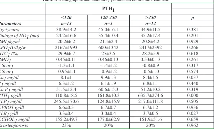
18 HD patients (12 men, 6 women) and 12 CAPD patients (8 men, 4 women) were included to study. 8 of CAPD patients were applying automated peritoneal dialysis. The parameters of patients before therapy is seen in table 1.
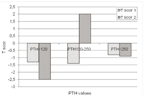
T and Z scores, bone mineral density and ALP levels of patients that PTH levels between 120-250 pg/ml and more than 250 pg/ml increased as it is seen in table 2 and these parameters were decreased in group that PTH levels were lower than 120 pg/ml.
Osteoporosis percentages were 23 %, 20 % and 20 % in PTH<120 pg/ml, PTH 120-250 pg/ml and PTH>250 pg/ml respectively. After the therapy these were 30 %, 0% and 20 % respectively. T scores of 3 groups before and after the therapy is showed in Figure 1. Especially T scores of group 2 that PTH levels between 120-250 pg/ml were better.
DISCUSSION
Chronic renal failure is a disease that causes deterioration in bone mineral metabolism (9). Renal osteodystrophy is related to morbidity and mortality. And this cause osteoporosis and fracture risk in dialysis patients (10). Renal osteodystrophy causes low bone mineral density in chronic renal failure. Dual Energy X-Ray Absorbsiometry (DEXA) is a noninvasive standard method to measure bone mineral density and it is essential in the diagnosis of the osteoporosis. Heel quantitative ultrasound is mobile, easing, has no radiation risk and is an useful tool for screening osteo-porosis among the risky groups such as chronic renal failure. It can also give information about fracture as enough as DEXA (2).
Table 2. Demographic and laboratory parameters after the treatment. PTH2 <120 120-250 >250 p Parameters n=3 n=7 n=20 Age (years) 38.9±14.2 45.0±16.1 34.9±11.5 0.381 Vintage of HD2 (mo) 43.6±13.7 53.4±10.2 54.2±14.7 0.141 BMI2 kg/m2 21.4±7.2 21.6±2.2 21.7±4.3 0.995 EPO2 IU/kg/w 2385±1557 1500±1414 4083±2968 0.065 HTC2 (%) 31±4.2 31±2.9 32.4±3.2 0.579 BMD2 0.41±0.13 0.47±0.12 0.54±0.15 0.122 T Scor2 -2.5±1.1- 1.96±1.43 -0.83±1.2 0.027 Z Scor2 -1.26±1.4 -0.8±1 -0.3±1 0.064 Ca2 mg/dl 9.7±1.1 9.6±0.86 8.4±0.7 0.066 P2 mg/dl 6±1.4 6±0.7 6.9±2 0.291 Ca.P2 mg/dl 54.2±13.6 56.9±7.5 62.1±17.9 0.424 PTH2 pg/dl 104.2±44.6 187.8±76.1 702.0±571.0 0.090 ALP2 mg/dl 260.6±154.1 114.2±86.4 236.0±98.0 0.668 T.PROT2 g/dl 7.1±0.4 7.1±0.4 7.1±0.6 0.997 ALB2 g/dl 3.6±0.2 3.1±0.3 3.5±0.5 0.019 T.CHOL2 mg/dl 183.1±38.4 178.4±60.9 141±27.5 0.030 % osteoporosis 30% 0% 20% 0.001
HD: Hemodialysis, BMI: Body mass index, EPO: erythropoietin, HCT: hematocrit, BMD: Bone mineral density, PTH: Parathormon, ALP: Alkalen phosphatase, ALB: Albumin, T.CHOL: Total Cholesterol.
Table 1. Demographic and laboratory parameters before the treatment.
PTH1 <120 120-250 >250 p Parameters n=13 n=5 n=12 Age(years) 38.9±14.2 45.0±16.1 34.9±11.5 0.381 Vintage of HD1 (mo) 24.2±16.6 35.4±10.4 35.2±17.4 0.201 BMI1kg/m2 20.2±6.2 21.1±2.4 20.8±4.2 0.929 EPO1IU/kg/w 2167±1993 600±1342 2417±2392 0.266 HTC1 (%) 29.9±6.7 27±3.5 28.2±5.9 0.618 BMD1 0.45±0.11 0.46±0.13 0.53±0.13 0.261 T Scor1 -1.3±1.1 -1.4±1.2 -0.8±0.9 0.317 Z Scor1 -0.95±1.1 -0.9±1.2 -0.5±1.0 0.574 Ca1 mg/dl 8.1±1 9.9±1.3 8.4±1.5 0.037 P1 mg/dl 6.3±1.2 6.1±1.9 6.8±1.1 0.440 Ca.P1 mg/dl 51.5±12.4 60.6±15.3 51.2±10.2 0.319 PTH1 pg/dl 110.8±18.5 161.8±10.3 635.7±274.6 0.000 ALP1 mg/dl 245.5±170.6 124.8±15.9 217.0±111.8 0.505 T.PROT1g/dl 6.6±0.3 6.7±0.7 6.7±1.2 0.936 ALB1 g/dl 3.3±0.4 3.0±0.4 3.7±0.5 0.027 T.CHOL1 mg/dl 155.2±49.7 177.0±62.9 151.9±51.6 0.659 % osteoporosis 23% 20% 20% 0.962
HD: Hemodialysis, BMI: Body mass index, EPO: erythropoietine, HCT: hematocrit, BMD: Bone mineral density, PTH: Parathormon, ALP: Alkalen phosphatase, ALB: Albumin, T.CHOL: Total Cholesterol.
Taal et al. (11) pointed that there is a correlation between the parameters of DEXA and heel quantitative ultrasound and bone mineral density in chronic HD patients. Speed of Sound (SOS) and Broad band Ultrasound (BUA) has 76% and 71 % sensitivity and 80% and 69% specificity relatively in the diagnosis of osteoporosis. The positive predictivity of BUA and SOS is 48% and 35% and negative predictivity is 93% and 91%, relatively. High negative predictivity emphasized that heel quantitative ultrasound can be used to screen BMD of non-osteoporotic patients. The diagnosis of osteoporotic patients that had low positive predictive value by heel quantitative ultrasound must be confirmed by DEXA.
Gallieni et al. (8) carried out a study that 239 patients were included to study. The mean age was 61,2 years, mean dialysis duration was 72,2 months and mean PTH value was 318±413 pg/ml. 43% of these patients had a PTH value of <100 pg/ml, 25.4% of them had PTH> 400 pg/ml and 19.5% had PTH 100-250 pg/ml. It is difficult to maintain PTH between 100 and 250 pg/ml in hemodialysis patients and hyperphosphatemia must be serially treated (8). Low phosphate dietary and strict phosphate control are the basis of preventing hyperphosphatemia related hyperparatiroidic bone disease. If PTH>450 pg/ml, it may reflect high turnover (hyperparathyroidism) bone disease. If PTH<120 pg/ml it may be low turnover bone disease and if PTH between 120-250 pg/ml mix type (hyperparathyroidism and low turnover) must be thought (12).
We measured T, Z scores and bone mineral density by heel quantitative ultrasound before and after D
vitamin therapy according to PTH values. Osteoporosis percent has increased after two years in the group that PTH<120 pg/ml. We established high turnover bone disease in 18 (60%) patients of PTH higher than 450 pg/ml in 2 years period.
As a result, there was a statistically meaningful difference between T score of 3 groups that D vitamin therapy and strict phosphate control was done. Especially in the second group that PTH values were between 120-250 pg/ml. T score improved and the frequency of osteoporosis decreased. In the group that PTH<120 pg/ml, osteoporosis frequency increased. The diagnosis of low turnover bone disease (adynamic bone disease, aluminum related bone disease, osteomalasia, osteitis fibrosa) in the group that PTH lower than 250 pg/ml must be confirmed by bone biopsy.
In conclusion, it is acceptable to maintain the level of PTH between 120 – 250 pg/ml for preventing osteoporosis risk in patients with chronic renal failure, undergoing dialysis therapy.
REFERENCES
1. Elaine D, Cyrus C : Osteoporosis. In Endocrinology and Metabolism. Pinchera A, Bertagna X, Fisher J. 14th New York. McGraw-Hill. pp: 271-282, 2001.
2. Robert L, Felicia C: Osteoporosis. In Harrison’s Principles of Internal Medicine. Braunwald E, Fauci AS, Kasper DL, Hauser SL, Longo DL, Jameson JL. 15th. New York. McGraw-Hill. pp: 2226- 2236, 2001.
3. Chesnut CH 3rd, Rosen CJ: Bone Quality Discussion Group. Reconsidering the effects of antiresorptive therapies in reducing osteoporotic fracture. J Bone Miner Res. 16(12):2163-2173, 2001.
4. Hruska K: New Concept in Renal Osteodystrophy. Nephrol Dial Transplant. 13:2755-2760, 1998.
5. Elder G: Pathophysiology and Recent Advances in the Management of Renal Osteodystrophy. J Bone Miner Res. 17:2094-2105, 2002.
6. Parfitt AM: A Structural Approach to Renal Bone Disease. J Bone Miner Res. 13(8):1213-1220, 1998.
7. Taal MW, Masud T, Gren D, Cassidy MJD: Risk Factors for Reduced Bone Density in Heamodialysis Patients. Nephrol Dial Transplant. 14:1922-1928, 1999.
8. Gallieni M,Cucciniello E, D’Amaro E: Calcium, Phosphate and PTH Levels in the Hemodialysis Population: A Multicenter Study. J Nephrol. 15(2):165-170, 2002.
9. Zayour D, Daouk M, Medawar W, Salamoun M: Predictors of Bone Mineral Density in Patients on Hemodialysis, Transplantation Proceedings. 36:1297-1301, 2004.
10. Hampson G, Vasa S, Evans C: Comparison of the Humoral Markers of Bone Turnover and Bone Mineral Density in Patients on Heamodialysis and Continuous Ambulatory Peritoneal Dialysis. Nephron. 91:94-102, 2002.
11. Taal MW, Cassidy MJ, Pearson D, Green D, Masud T: Usefulness of Quantitative Heel Ultrasound Compared with Dual-Energy X-ray Absorptiometry in Determining Bone Mineral Density in Chronic Haemodialysis Patients. Nephrol Dial Transplant. 14(8): 1917-1921, 1999.
12 Torres A, Lorenzo V, Hernandez D: Bone Disease in Predialysis, Hemodialysis and CAPD Patients: Evidence of a Better Bone Response to PTH. Kidney International. 47:1434-1442, 1995.