AIUM Practice Parameter for the Performance of
Ultrasound of the
Female Pelvis
© 2014 by the American Institute of Ultrasound in Medicine
Parameter developed in collaboration with the American College of Radiology (ACR), the American College of Obstetricians and Gynecologists (ACOG), the Society for Pediatric
The American Institute of Ultrasound in Medicine (AIUM) is a multidis-ciplinary association dedicated to advancing the safe and effective use of ultrasound in medicine through professional and public education, research, development of parameters, and accreditation. To promote this mission, the AIUM is pleased to publish, in conjunction with the American College of Radiology (ACR), the American College of Obstetricians and Gynecologists (ACOG), the Society for Pediatric Radiology (SPR), and the Society of Radiologists in Ultrasound (SRU), this AIUM Practice Parameter for the Performance of Ultrasound of the Female Pelvis. We are indebted to the many volunteers who contributed their time, knowledge, and energy to bringing this document to completion.
The AIUM represents the entire range of clinical and basic science interests in medical diagnostic ultrasound, and, with hundreds of volun-teers, the AIUM has promoted the safe and effective use of ultrasound in clinical medicine for more than 50 years. This document and others like it will continue to advance this mission.
Practice parameters of the AIUM are intended to provide the medical ultrasound community with parameters for the performance and recording of high-quality ultrasound examinations. The parameters reflect what the AIUM considers the minimum criteria for a complete examination in each area but are not intended to establish a legal stan-dard of care. AIUM-accredited practices are expected to generally fol-low the parameters with recognition that deviations from these param-eters will be needed in some cases, depending on patient needs and available equipment. Practices are encouraged to go beyond the parameters to provide additional service and information as needed.
14750 Sweitzer Ln, Suite 100 Laurel, MD 20707-5906 USA 800-638-5352 • 301-498-4100 www.aium.org
I.
Introduction
The clinical aspects contained in specific sections of this parameter (Introduction, Indications, Specifications of the Examination, and Equipment Specifications) were developed collabora-tively by the American Institute of Ultrasound in Medicine (AIUM), the American College of Radiology (ACR), the American College of Obstetricians and Gynecologists (ACOG), the Society for Pediatric Radiology (SPR), and the Society of Radiologists in Ultrasound (SRU). Recommendations for physician requirements, written request for the examination, documen-tation, and quality control vary among the organizations and are addressed by each separately. This parameter has been developed to assist physicians performing sonographic studies of the female pelvis. Ultrasound examinations of the female pelvis should be performed only when there is a valid medical reason, and the lowest possible ultrasonic exposure settings should be used to gain the necessary diagnostic information. In some cases, additional or specialized examinations may be necessary. Although it is not possible to detect every abnormality, adher-ence to the following parameter will maximize the probability of detecting most abnormalities. For ultrasound examinations of the urinary bladder, see the AIUM Practice Parameter for the Performance of an Ultrasound Examination of the Abdomen and/or Retroperitoneum.
II.
Indications
Indications for pelvic sonography include but are not limited to: 1. Evaluation of pelvic pain;
2. Evaluation of pelvic masses;
3. Evaluation of endocrine abnormalities, including polycystic ovaries; 4. Evaluation of dysmenorrhea (painful menses);
5. Evaluation of amenorrhea; 6. Evaluation of abnormal bleeding; 7. Evaluation of delayed menses;
8. Follow-up of a previously detected abnormality;
9. Evaluation, monitoring, and/or treatment of infertility patients;
10. Evaluation in the presence of a limited clinical examination of the pelvis; 11. Evaluation for signs or symptoms of pelvic infection;
12. Further characterization of a pelvic abnormality noted on another imaging study; 13. Evaluation of congenital uterine and lower genital tract anomalies;
14. Evaluation of excessive bleeding, pain, or signs of infection after pelvic surgery, delivery, or abortion;
15. Localization of an intrauterine contraceptive device; 16. Screening for malignancy in high-risk patients; 17. Evaluation of incontinence or pelvic organ prolapse; 18. Guidance for interventional or surgical procedures; and 19. Preoperative and postoperative evaluation of pelvic structures.
3
III.
Qualifications of Personnel
See www.aium.org for AIUM Official Statements including Standards and Guidelines for the Accreditation of Ultrasound Practicesand relevant Physician Training Guidelines.
IV.
Written Request for the Examination
The written or electronic request for an ultrasound examination should provide sufficient information to allow for the appropriate performance and interpretation of the examination. The request for the examination must be originated by a physician or other appropriately licensed health care provider or under the provider’s direction. The accompanying clinical information should be provided by a physician or other appropriate health care provider famil-iar with the patient’s clinical situation and should be consistent with relevant legal and local health care facility requirements.
V.
Specifications of the Examination
The following sections detail the examination to be performed for each organ and anatomic region in the female pelvis. All relevant structures should be identified by the transabdominal and/or transvaginal approach. A transrectal or transperineal approach may be useful in patients who are not candidates for introduction of a vaginal probe and in assessing the patient with pelvic organ prolapse. More than 1 approach may be necessary.1,2
A. General Pelvic Preparation
For a complete transabdominal pelvic sonogram, the patient’s bladder can be distended if nec-essary to displace the small bowel from the field of view. Occasionally, overdistention of the bladder may compromise the evaluation. When this occurs, imaging may be repeated after par-tial bladder emptying. If an abnormality of the urinary bladder is detected, it should be docu-mented and reported.
For a transvaginal sonogram, the urinary bladder is preferably empty. The patient, the sonogra-pher, or the physician may introduce the vaginal transducer, preferably under real-time moni-toring. Consideration of having a chaperone present should be in accordance with local policy.3
B. Uterus
The vagina and uterus provide anatomic landmarks that can be used as reference points for the other pelvic structures, whether normal or abnormal. In examining the uterus, the following should be evaluated: (1) the uterine size, shape, and orientation; (2) the endometrium; (3) the myometrium; and (4) the cervix. The vagina may be imaged as a landmark for the cervix. The overall uterine length is evaluated in a sagittal view from the fundus to the cervix (to the external os, if it can be identified). The depth of the uterus (anteroposterior dimension) is measured in the same sagittal view from its anterior to posterior walls, perpendicular to the length. The maximum width is measured in the transverse or coronal view. If volume meas-urements of the uterine corpus are performed, the cervical component should be excluded 4
Abnormalities of the uterus should be documented.4The myometrium and cervix should be
evaluated for contour changes, echogenicity, masses, and cysts. Masses that may require follow-up or intervention should be measured in at least 2 dimensions, acknowledging that it is not usually necessary to measure all uterine fibroids. The size and location of clinically relevant fibroids should be documented.
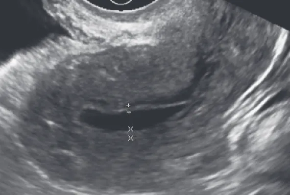
The endometrium should be analyzed for thickness, focal abnormalities, echogenicity, and the presence of fluid or masses in the cavity. The thickest part of the endometrium should be meas-ured perpendicular to its longitudinal plane in the anteroposterior diameter from echogenic to echogenic border (Figure 1). The adjacent hypoechoic myometrium and fluid in the cavity should be excluded (Figure 2). Assessment of the endometrium should allow for variations expected with phases of the menstrual cycle and with hormonal supplementation.5–7It should
be reported if the endometrium is not adequately seen in its entirety or is poorly defined. Sonohysterography may be a useful adjunct to evaluate the patient with abnormal uterine bleeding or to further clarify an abnormally thickened endometrium. (See the AIUM Practice Parameter for the Performance of Sonohysterography.) If the patient has an intrauterine contra-ceptive device, its location should be documented.
5
www.aium.org
Figure 1. Measurement of endometrial thickness. The endometrial thickness is measured in its thickest
portion from echogenic to echogenic border (calipers) perpendicular to the midline longitudinal plane of the uterus.
The addition of 3-dimensional to 2-dimensional ultrasound (transabdominal, transvaginal, transperineal, and/or transrectal) can be helpful in many circumstances, including but not lim-ited to evaluating the relationship of masses with the endometrial cavity, identifying uterine congenital anomalies and a thickened and/or heterogeneous endometrium, and evaluating the location of an intrauterine device and the integrity of the pelvic floor.8,9
C. Adnexa, Including Ovaries and Fallopian Tubes
When evaluating the adnexa, an attempt should be made to identify the ovaries first, since they can serve as a major point of reference for assessing the presence of adnexal pathology. The ovarian size may be determined by measuring the ovary in 3 dimensions (width, length, and depth) on views obtained in 2 orthogonal planes.10Any ovarian abnormalities should be
documented.11–15
The ovaries may not be identifiable in some patients. This occurs most frequently before puberty, after menopause, or in the presence of a large leiomyomatous uterus. The adnexal region should be surveyed for abnormalities, particularly masses and dilated tubular structures. If an adnexal abnormality is noted, its relationship with the ovaries and uterus should be assessed. The size and sonographic characteristics of adnexal masses should be documented. Spectral, color, and/or power Doppler ultrasound may be useful to evaluate the vascular char-acteristics of pelvic lesions.15–19
6
Figure 2. Measurement of endometrium with fluid in the cavity. In the presence of endometrial fluid,
measurements of the 2 separate layers of the endometrium (calipers), excluding the fluid, are added to determine the endometrial thickness.
D. Cul-de-Sac
The cul-de-sac and bowel posterior to the uterus may not be clearly defined. This area should be evaluated for the presence of free fluid or a mass. If a mass is detected, its size, position, shape, sonographic characteristics, and relationship to the ovaries and uterus should be docu-mented. Differentiation of normal loops of bowel from a mass may be difficult if only a trans-abdominal examination is performed. A transvaginal examination may be helpful to distinguish a suspected mass from fluid and feces within the normal rectosigmoid colon.
VI. Documentation
Adequate documentation is essential for high-quality patient care. There should be a perma-nent record of the ultrasound examination and its interpretation. Images of all appropriate areas, both normal and abnormal, should be recorded. Variations from normal size should be accompanied by measurements. Images should be labeled with the patient identification, facility identification, examination date, and side (right or left) of the anatomic site imaged. An official interpretation (final report) of the ultrasound findings should be included in the patient’s medical record. Retention of the ultrasound examination should be consistent both with clinical needs and with relevant legal and local health care facility requirements.
Reporting should be in accordance with the AIUM Practice Parameter for Documentation of an Ultrasound Examination.
VII. Equipment Specifications
A sonographic examination of the female pelvis should be conducted with a real-time scanner, preferably using sector, curved linear, and/or endovaginal transducers. The transducer or scan-ner should be adjusted to operate at the highest clinically appropriate frequency, realizing that there is a trade-off between resolution and beam penetration.3
VIII. Quality Control and Improvement, Safety, Infection Control,
and Patient Education
Policies and procedures related to quality control, patient education, infection control, and safety should be developed and implemented in accordance with the AIUMStandards and Guidelines for the Accreditation of Ultrasound Practices.
Equipment performance monitoring should be in accordance with the AIUM Standards and Guidelines for the Accreditation of Ultrasound Practices.
IX. ALARA Principle
The potential benefits and risks of each examination should be considered. The ALARA (as low as reasonably achievable) principle should be observed when adjusting controls that affect the acoustic output and by considering transducer dwell times. Further details on ALARA may be found in the AIUM publication Medical Ultrasound Safety, Third Edition.
7
Acknowledgments
This parameter was revised by the AIUM in collaboration with the American College of Radiology (ACR), the American College of Obstetricians and Gynecologists (ACOG), the Society for Pediatric Radiology (SPR), and the Society of Radiologists in Ultrasound (SRU) according to the process described in the AIUM Clinical Standards Committee Manual.
ACR
ACOG
Marcela Bohm-Velez, MD, Chair Daniel M. Breitkopf, MD M. Stephen Ledbetter, MD, MPH Wendy R. Brewster, MD, PhD Henrietta Kotlus Rosenberg, MD John W. Seeds, MD
Jason M. Wagner, MD
AIUM
SPR
Beryl R. Benacerraf, MD Amy N. Dahl, MD Steven R. Goldstein, MD Kassa Darge, MD, PhD Elizabeth Puscheck, MD Lynn A. Fordham, MD Laurel Stadtmauer, MD
SRU
Rochelle F. Andreotti, MD Oksana H. Baltarowich, MD Anna S. Lev-Toaff, MD
AIUM Clinical Standards Committee
Joseph Wax, MD, Chair
John Pellerito, MD, Vice Chair
Bryann Bromley, MD Pat Fulgham, MD
Charlotte Henningsen, MS, RT, RDMS, RVT Alexander Levitov, MD
Vicki Noble, MD, RDMS Anthony Odibo, MD, MSCE David Paushter, MD Dolores Pretorius, MD Khaled Sakhel, MD Shia Salem, MD Jay Smith, MD Paula Woodward, MD
Original copyright 1995; revised 2014, 2009, 2006, 2004 Renamed 2015
References
1. Garel L, Dubois J, Grignon A, Filiatrault D, Van Vliet G. US of the pediatric female pelvis: a clinical per-spective. Radiographics 2001; 21:1393–1407.
2. Rosenberg, HK, Chaudhry H. Pediatric pelvic sonography. In: Rumack CM, Wilson SR, Charboneau JW, Levine D (eds). Diagnostic Ultrasound. 4th ed. Philadelphia, PA: Elsevier Mosby Inc; 2011:1923– 1981.
3. Stagno SJ, Forster H, Belinson J. Medical and osteopathic boards’ positions on chaperones during gynecologic examinations. Obstet Gynecol 1999; 94:352–354.
4. Ascher SM, Imaoka I, Lage JM. Tamoxifen-induced uterine abnormalities: the role of imaging. Radiology 2000; 214:29–38.
5. Bree RL, Bowerman RA, Bohm-Velez M, et al. US evaluation of the uterus in patients with post-menopausal bleeding: a positive effect on diagnostic decision making. Radiology 2000; 216:260–264. 6. Bree RL, Carlos RC. US for postmenopausal bleeding: consensus development and patient-centered
outcomes. Radiology 2002; 222:595–598.
7. Nalaboff KM, Pellerito JS, Ben-Levi E. Imaging the endometrium: disease and normal variants. Radiographics 2001; 21:1409–1424.
8. Fong K, Kung R, Lytwyn A, et al. Endometrial evaluation with transvaginal US and hysterosonography in asymptomatic postmenopausal women with breast cancer receiving tamoxifen. Radiology 2001; 220:765–773.
9. Benacerraf BR, Shipp TD, Bromley B. Which patients benefit from a 3D reconstructed coronal view of the uterus added to standard routine 2D pelvic sonography? AJR Am J Roentgenol 2008; 190:626– 629.
10. Cohen HL, Tice HM, Mandel FS. Ovarian volumes measured by US: bigger than we think. Radiology 1990; 177:189–192.
11. Brown DL, Zou KH, Tempany CM, et al. Primary versus secondary ovarian malignancy: imaging find-ings of adnexal masses in the Radiology Diagnostic Oncology Group Study. Radiology 2001; 219:213– 218.
12. Jarvela IY, Sladkevicius P, Kelly S, Ojha K, Nargund G, Campbell S. Three-dimensional sonographic and power Doppler characterization of ovaries in late follicular phase. Ultrasound Obstet Gynecol 2002; 20:281–285.
13. Kinkel K, Hricak H, Lu Y, Tsuda K, Filly RA. US characterization of ovarian masses: a meta-analysis. Radiology 2000; 217:803–811.
14. Sato S, Yokoyama Y, Sakamoto T, Futagami M, Saito Y. Usefulness of mass screening for ovarian car-cinoma using transvaginal ultrasonography. Cancer 2000; 89:582–588.
15. Levine D, Brown DL, Andreotti RF, et al. Management of asymptomatic ovarian and other adnexal cysts imaged at US: Society of Radiologists in Ultrasound consensus conference statement. Radiology 2010; 256:943–954.
16. Funt SA, Hann LE. Detection and characterization of adnexal masses. Radiol Clin North Am 2002; 40:591–608.
17. Kaakaji Y, Nghiem HV, Nodell C, Winter TC. Sonography of obstetric and gynecologic emergencies, part II: gynecologic emergencies. AJR Am J Roentgenol 2000; 174:651–656.
18. Laing FC, Brown DL, DiSalvo DN. Gynecologic ultrasound. Radiol Clin North Am 2001; 39:523–540. 19. Polat P, Suma S, Kantarcy M, Alper F, Levent A. Color Doppler US in the evaluation of uterine vascular
abnormalities. Radiographics 2002; 22:47–53.
9