Iranian Journal of Basic Medical Sciences
ijbms.mums.ac.ir
Chronic effects of aerobic exercise on gene expression of
LOX‐1 receptor in the heart of rats fed with high fat diet
Simin Riahi
1, Mohammad Taghi Mohammadi
2*, Vahid Sobhani
1, Mansureh Soleimany
31 Exercise Physiology Research Center, Baqiyatallah University of Medical Sciences, Tehran, Iran
2 Department of Physiology and Biophysics, Faculty of Medicine, Baqiyatallah University of Medical Sciences, Tehran, Iran 3 Department of Anatomy, Faculty of Medicine, Iran University of Medical Sciences, Tehran, Iran
A R T I C L E I N F O A B S T R A C T Article type:
Original article Objective(s):complications, which is upregulated in numerous pathological conditions. Since exercise has beneficial Lectin‐like low density lipoprotein receptor (LOX‐1) has pivot role in vascular effects in prevention of hyperlipidemic complications, present study examined protective effects of aerobic exercise through reduction of LOX‐1 expression in heart during dyslipidemia.
Materials and Methods: Four groups of rats were used (N=25): Normal, Normal and exercise, High fat
and High fat and exercise. High fat diet (HFD) was made by adding 10% animal oil, 2% cholesterol and 0.5% colic acid to standard rodent chow. Exercise protocol consisted of swimming 1 hr/day, and 5 days/week for 8 weeks. Plasma lipids were evaluated at the end of experiment, 48 hr after final session of exercise. At the end, rats were sacrificed and heart was removed for determination of malondialdehyde (MDA) content, and LOX‐1 expression.
Results:HFD meaningfully changed lipid profile (>50%), but chronic exercise had no significant effects
on lipid profile. LOX‐1 expression was significantly increased in heart of rats fed with HFD, while swimming exercise considerably reduced gene expression of LOX‐1. MDA content was significantly enhanced in rats fed with HFD (4.37±0.6 nmol/mg, P<0.01) compared to normal group (1.56±0.48 nmol/mg), whereas swimming exercise decreased MDA level of heart in rats fed with HFD (2.28±0.32, P<0.01).
Conclusion:Findings indicated that swimming exercise is able to diminish heart expression of LOX‐1
receptor concomitant reduction of oxidative stress. Since these parameters are involved in generation of dyslipidemic complications, swimming exercise is a good candidate to reduce these complications. Article history: Received: Dec 9, 2014 Accepted: Mar 16, 2015 Keywords: Dyslipidemia LOX‐1 receptor Oxidative stress Swimming exercise
►Please cite this article as:
Riahi S, Mohammadi MT, Sobhani V, Soleimany M. Chronic effects of aerobic exercise on gene expression of LOX‐1 receptor in the heart of rats fed with high fat diet. Iran J Basic Med Sci 2015; 18:805‐812.
Introduction
Dyslipidemia and physical inactivity are two main factors that are involved in the development of atherosclerosis and their related complications like cardiovascular diseases (CVD) (1). Findings of recent studies show that coronary atherosclerosis begins at early age, and one in six people has atherosclerotic lesions (2). Dyslipidemia, which is characterized by high concentration of triglyceride, total cholesterol and low‐density lipoprotein cholesterol (LDL‐C), and low concentration of high‐density lipoprotein cholesterol (HDL‐C), is important risk factor in coronary diseases (3). Low levels of HDL‐C and high level of non‐HDL‐C are profile of atherogenic lipid phenotypes that are main risk factors for atherosclerosis (4, 5). Increment of free radicals production in endothelium of arteries is another
action of dyslipidemia that intensifies atherosclerosis progression (6). Based on recent findings, there is a strong association between plasma levels of oxidative stress and atherogenic lipoproteins (7). One of the most important toxic effects of reactive oxygen specious (ROS), is the production of oxidize low‐density lipoprotein (oxLDL) (8). It is noteworthy that oxLDL contributes to prompt lipid deposition and inflammatory response within blood vessels wall (9). Increment of oxLDL and other dyslipidemia‐ related factors activate lectin‐like low density lipoprotein (LOX‐1) receptor that has important role in coronary artery disease in chronic hyperlipidemic
conditions (8). Upregulation of this receptor is involved in initiation and progression of athero‐
sclerosis and its related complications like heart attacks (10). Finally, LOX‐1 receptor overexpression induces
*Corresponding author: Mohammad Taghi Mohammadi. Department of Physiology and Biophysics, School of Medicine Baqiyatallah University of Medical
Iran J Basic Med Sci, Vol. 18, No. 8, Aug 2015
806
endothelial dysfunction and vascular wall
inflammation that progress plaque formation in arteries wall (11). Therefore, inhibition of LOX‐1 expression may help to prevent from atherosclerosis. In addition to pharmacological treatment and diet, exercise is recommended for reduction of risk factors that involve in coronary vascular disease. In patient with coronary vascular diseases, exercise may reduce the mortality rate (12). Exercise is also important in prevention of atherosclerosis. Regular exercise not only has important role in the prevention of atherosclerosis and coronary vascular disease, but also reduces the size of previously formed lesions (13). However, in patient with coronary diseases, 3 months exercise results in moderate improvement on lipid profile (14). Changes of lipid profile in response to exercise depend on modality, intensity, frequency and duration of each session and how long exercise has been performed (15). Epidemiological studies show some beneficial effects of physical activity on the reduction of CVD in diabetic patients (16). Regular moderate‐intensity exercise also reduces the rate of mortality in patients with long‐suffering CVD (17). The results of a study indicated that the prevalence of atherosclerosis in people with low cardiopulmonary fitness is more than individuals with high cardiopulmonary fitness (18). Also, it is demonstrated that moderate aerobic exercise reduced the size of preexisting atherosclerotic lesions in LDL‐C receptor knockout mice fed with high fat diet (19). As well, it is shown that exercise training reduces the severity of atherosclerosis in apolipoprotein E knockout mice via nitric oxide (20).
In another study, also Okabe et al demonstrated that
swimming exercise reduces the severity of atherosclerosis in apolipoprotein E deficient mice by antioxidant effects (21). The results of a meta‐ analysis demonstrated that exercise increases HDL‐C levels, and there is a dose‐response relationship between the level of physical activity and serum HDL‐C concentration (22).
Although the reduction of cardiovascular mortality and morbidity due to exercise is obvious, the exact mechanisms that exercise prevents atherosclerosis lesion formation is unknown. Therefore, in the present study we examined the beneficial effects of exercise on CVD through alterations of LOX‐1 receptor expression and free radicals production in heart of rats fed with high fat diet.
Materials
and
Methods
Animals
Male Wistar rats (200±20 g) were purchased from pasture institute laboratory. Rats were kept under observation for one week before the experiment to be acclimatized with environmental condition. During the experiment, all animals were kept in
standard polyester cage (2 rats in each cage) in a
room with standard temperature (22±2 °C) and
humidity (%55±5) with 12 hr light/dark cycle and free access to water and food. All protocols of the study were approved by the institutional animal ethics committee of Baqiyatallah University of Medical Sciences, which follows the NIH (National Institutes of Health) guidelines for care and use of animals.
Experimentalgroupsanddesign
Rats were randomly divided into four groups (n=5 per group): Normal rats (Normal) are healthy animals that remained sedentary, trained normal rats (Normal&E) are healthy animals that were doing exercise for 8 weeks, high fat diet rats (HFD) are animals that remained sedentary and fed with high fat diet, and trained high fat diet rats (HFD&E) are animals that were doing exercise for 8 weeks and fed with high fat diet.
High fat diet was prepared by adding 2% cholesterol powder (Merck, Germany), 0.5% colic acid powder (Merck, Germany) and 10% animal oil to standard rodent chow (23).
Exerciseprotocol
In the present study, we used endurance swimming as a model of exercise intervention. The training included daily moderate intensity of swimming in 8 weeks, which could induce cardiac hypertrophy (24). The rats in training groups were performing swimming for 1 hr in the morning in a rubber swimming tank with dimension of 55×100×60 cm. The water depth was
enough to prevent from resting and eliminate bobbing behavior. The tank filled with tap water that was sufficient for 6 rats to swim, simultaneously. Water temperature was fixed at 32±2 °C to prevent from
hypothermia. The exercise program in first week of training was begun with acclimatization to water and swimming bath. In first day, rats swam for 10 min. Then, duration of training was added daily 10 min until each rat could swim continuously for 60 min. In subsequent weeks, rats swam daily one hour per day for five times a week (1 hr/day; 9:00–11:00 AM from Saturday to Wednesday) (25). The control groups (Normal and HFD) remained sedentary in the swimming tank when it was filled with tap water in depth that animal’s paws reached to the bottom of tank
and its head was out of water.After each session, the
animals were dried and kept in warm place to prevent from hypothermia stress.
Initial, weekly and final body weights were determined for all rats by digital scale in a certain day of week. Weight gain was calculated as percent of initial weight.
Forty eight hour after last training session, the rats were sacrificed with an overdose of chloral hydrate. Blood (5 ml) was draw from heart, and after
20 min, the blood clot was centrifuged at 4,000× rpm for 10 min at 4 °C. Heart was removed and after
washing with cold phosphate buffered saline (PBS) and snap freezing in liquid nitrogen, stored in ‐80 ºC for evaluation of gene expression and measurement of NOx and malondialdehyde (MDA) levels.
Assessment of serum concentrations of triglyceride,cholesterol,HDL‐CandLDL‐C
Collected blood samples were centrifuged at
10,000×g for 10 min at 4 °C. The supernatant (serum)
was then used to measure the concentration of triglyceride, cholesterol, LDL‐C, and HDL‐C in serum by using available commercial kit (Zeistchemy, Iran) according to the manufacturer’s protocol.
Evaluationofgeneexpression
Gene expression of LOX‐1 receptor was determined with mRNA measurement of this gene by semi quantitative technique of reverse transcriptase‐ polymerase chain reaction (RT‐PCR). Total RNA was extracted from 50 mg of heart tissue using the RNA
extraction kit (Topagene kavosh, Iran) according to the manufacturer’s protocol. The quantity and quality of the extracted RNA samples were
determined by spectrophotometry at 260 and 280 nm. Complementary DNA (cDNA) was synthesized from 3 μl of total RNA using the Revert Aid First Strand cDNA Synthesis Kit (BIONEER). Expression of a β‐actin housekeeping gene was used as a reference for the level of target gene expression. Two µl of cDNA was
amplified with PCR kit (BIONEER) according to the manufacturer’s protocol, and appropriate primers
were used for LOX‐1 (Forward: 5’‐TTTAGGACCAGGGGC
GTTTC‐3’, Reverse: 5’‐GGAGATGGACCCAAGTCGTG‐3’)
and β‐actin (Forward: 5’‐CCACACCCGCCACCAGTTCG‐
3’, Reverse: 5’‐CTAGGGCGGCCCACGATGGA‐3’) genes
that were designed by Gene Runner software. The products of PCR‐amplified samples were visualized on 1.5% agarose gel using ethidium bromide. The gel images were digitized by using a Gel Doc (Kiagene, Iran), and the images of the stained sections were taken.
Evaluationofoxidativeandnitrosativestress
The fractions of thawed tissue samples were weighed and homogenized medium (phosphate buffer, 0.1 mol, pH 7.4) were added to them. After homogenizing the tissues on ice by electric homogenizer, samples were centrifuged (20 min at
4 °C and 4000 rpm) and the supernatant (heart
cytosolic extract) was removed and stored in ‐80 °C for
analysis of NOx and MDA levels.
MDAassay
Amount of lipid peroxidation was measured by assessment of MDA formation using thiobarbituric acid (TBA) assay. Trichloroacetic acid (2.5 ml, 20%),
sulfuric acid (2.5 ml, 0.05 Molar) and TBA (3 ml, 0.2 g/dl) were added to cytosolic extract (500 μl), and vortexed. The mixture was placed in a water bath at 95 °C for 30 min. After cooling in runny water, 2 ml n‐
butanol was added and vigorously vortexed. Following centrifugation, the absorbance of the upper orange color phase was determined at 532 nm. Finally, tetraethoxypropane was used to prepare a standard curve, and results were expressed as nmol/mg protein.
NOxassay
The NOx in the ‘cytosolic extract’ was measured by the colorimetric reaction of the Griess reagent. 0.5 ml of cytosolic extract was deproteinized by adding 0.1 ml of zinc sulfate solution and centrifuged for 20
min at 4000 rpm and 4 °C to separate supernatant for
NOx assay. To reduce nitrate to nitrite, 0.1 ml of supernatant (as sample) or pure water (as blank) or sodium nitrate (as standard) was mixed with 0.1 ml vanadium III chloride. 0.05 ml sulfanilamide (0.01 %) and 0.05 ml N‐[1‐naphthyl] ethylenediamin dihydrochloride (NED, 0.01 %) were incubated for 30 min in dark place at 37 °C. Finally, the absorbance
of solution was determined at 540 nm. Nitrite concentration was determined from a standard curve generated from absorbance of each sodium nitrate solution (17). Finally, the nitrite‐nitrate levels were expressed as nmol/mg protein.
Protein content in the cytosolic extract was determined with Bradford method spectrophoto‐
metrically at 595 nm. Bovine serum albumin was used as a standard, and the concentration of protein was expressed as mg/ml. Amount of MDA and NOx in each sample were normalized to the cytosolic protein concentration and the results were expressed as nmol/mg of the cytosolic protein (nmol/mg protein).
Statisticalanalyses
The results were expressed as the Means±SEM. All statistical comparisons were performed using one‐way analysis of variance (ANOVA) and Tukey test as Post hoc. All states P<0.05 was considered as significant difference.
Results
Serum concentrations oftriglyceride, cholesterol, LDL‐CandHDL‐C
Serum concentration of triglyceride is shown in Table 1 in four experimental groups. In normal and normal trained rats, triglyceride concentration was 99±8 mg/dl and 104±14 mg/dl, respectively. These data indicate that exercise did not alter this value. High fat diet in HFD group increased the serum concentration of triglyceride (136±15 mg/dl), while exercise same as normal rats did not change this value in HFD group (135±24 mg/dl).
There were not any significant differences in serum levels of HDL‐C in four experimental groups.
Iran J Basic Med Sci, Vol. 18, No. 8, Aug 2015
808
Figure1. The curve shows weight gain of sedentary (normal and high fat diet (HFD) groups) and exercised rats (normal&E and HFD&E) after 8 weeks swimming exercise. Values are express as percentage of weight gaining in proportion weight of first week. All values are presented as mean±SEM
These data indicate that exercise or high fat diet could not alter the serum concentration of HDL‐C (Table 1).
Serum levels of cholesterol and LDL‐C in normal rats were 74±9 and 47±8 mg/dl, respectively. Exercise in normal trained rats did not alter these values (97±11 and 42±11 mg/dl). High fat diet significantly increased the serum levels of cholesterol and LDL‐C in HFD group compared with normal group, P<0.05 (164±20 and 123±24 mg/dl, respectively). Exercise in HFD group did not change these values, significantly (197±29 and 181±38 mg/dl, respectively).
Bodyweight
The weight gain of sedentary (normal and HFD groups) and exercised rats (normal and E and HFD and E) during experiment is shown in Figure 1. There was gradual increment of weight gain in all groups during experiment without significant difference.
GeneexpressionofLOX‐1protein
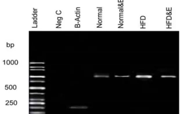
Figures 2 and 3 show the LOX‐1 gene expression of heart cells in different groups of our experiment.
Figure3. The curve shows the density of LOX‐1 mRNA that was normalized relative to density of β‐actin gene for different groups All values are presented as Mean±SEM
* Significantly different from normal group (P<0.01) $ Significantly different from high fat diet group (P<0.05)
Figure2. Picture shows the mRNA content of lectin‐like oxidized low‐density lipoprotein (LOX‐1) receptor in cardiac cells of different experimental groups. The mRNA bands were formed on agarose gel (1.5 %) that visualized with ethidiume bromide staining method. Qualifying assessments indicated that gene expression of LOX‐1 protein have been increased in high fat diet group compared other groups
Based on formed bands, the gene of LOX‐1 protein has expressed in heart cells of sedentary normal rats. Exercise in normal rats significantly attenuated the gene expression of LOX‐1 receptor
(P<0.05). On the other hands, high fat diet
significantly upregulated the expression of this gene
within the heart cells of sedentary HFD group (P<0.01). Whereas, swimming exercise significantly reduced the gene expression of this protein in heart cells of exercised HFD rats (P<0.05).
Parametersofoxidativeandnitrosativestress
Figure 4 shows the MDA content of heart tissue, as index of oxidative stress, in different experimental groups. MDA concentration in normal group was 1.56±0.48 nmol/mg protein and exercise did not change it in normal rats after 8 weeks swimming (1.42±90.11 nmol/mg protein). High fat diet in HFD group significantly increased the MDA content of heart tissue (4.37±0.6 nmol/mg protein, P<0.05). Training for 8 weeks significantly reduced the MDA content of heart tissue in rats fed with high fat diet (2.28±0.32 nmol/mg protein, P<0.01).
Figure4. Effects of 8 weeks swimming exercise on malondialdehyde (MDA) content in cardiac tissue. MDA values are indicated as nmol/mg protein. All values are presented as Mean±SEM
* Significantly different from normal group (P<0.05) $ Significantly different from high fat diet group (P<0.01)
Table1. Representative changes of blood triglyceride, cholesterol, LDL and HDL (mg/dl) in sedentary (normal and HFD groups) and
exercised groups (normal & E and high fat diet & E) after 8 weeks swimming exercise All values are presented as mean±SEM
* Significant difference from normal group (P<0.05) ** Significant difference from normal group (P<0.01)
Figure5. Effects of 8 weeks swimming exercise on NOx (nitrite and nitrate) content in cardiac tissue. NOx values are indicated as nmol/mg protein. All values are presented as mean±SEM
Total NOx content (nitrite and nitrate) of heart cells in normal rats was 5.15±0.43 nmol/mg protein. Swimming did not change the NOx content of normal trained rats after 8 weeks exercising (5.30±0.93 nmol/mg protein). However, HFD decreases NOx content of heart tissue (3.5±1.24 nmol/mg protein). Swimming for 8 weeks prevents this reduction in trained rats fed with HFD. However, these alterations are not statistically significant (Figure 5).
Discussion
Recent studies have shown that overexpression of LOX‐1 receptor plays an important role in pathogenesis of atherosclerosis (8, 26). This receptor causes oxidative stress and inflammatory responses in vascular wall by activating several intracellular signaling pathways that involve in most cardiovascular complications of dyslipidemic situations (8). In the present study, we showed some beneficial effects of swimming exercise on dyslipidemic condition via inhibition of LOX‐1 receptor. The findings of our study indicated that swimming exercise attenuated the gene expression of LOX‐1 receptor in situation of high blood cholesterol and LDL‐C (Figures 2, 3). On the other hands, this type of exercise decreased the parameters of oxidative stress in heart tissue following dyslipidemic condition (Figure 4). Based on our findings, it is concluded that swimming exercise is able to reduce oxidative stress of heart tissue possibly via reduction
of LOX‐1 receptor expression. These beneficial actions of exercise will be useful to prevent atherosclerosis in risky states such as dyslipidemic conditions.
LOX‐1 receptor has a pivot role in all stages of atherosclerosis development (beginning, progression and interruption of atherosclerotic plaques) (27). This receptor is upregulated in different pathological situations (8). Chen et al demonstrated that high cholesterol diet in rabbit induced the overexpression of LOX‐1 receptor in aorta (28). Oxidative stress is another factor that induces the overexpression of this receptor in vessel walls (29). Overexpression of LOX‐1 receptor cause endothelial dysfunction, vascular inflammation and plaque formation (11). Therefore, inhibition of this receptor will prevent atherosclerosis development. Dyslipidemia and oxidative stress are key factors in induction and development of atherosclerosis (6). In the present study, we showed that the LOX‐1 receptor was overexpressed in normal rats fed with high fat diet (Figures 2, 3). On the other hands, the concentration of MDA, as index of ROS, was significantly increased in heart of these rats (Figure 4). Upregulation of LOX‐ 1 receptor activates NADPH‐oxidase enzyme in cell membrane, which produces ROS (8). This enzyme is the main source of ROS production in different pathological situations like atherosclerosis (30). Also, there is a positive feed‐back between oxidative stress and LOX‐1 receptor activation. Oxidative stress augments the expression of LOX‐1 receptor, and overexpression of this receptor increases the concentration of tissue ROS (6). Szostak and Laurant demonstrated that physical inactivity can induce NADPH‐oxidase that enhances ROS production (31). Therefore, based on results of this study, it is suggested that dyslipidemia has stimulated the overexpression of LOX‐1 receptor accompanied with oxidative stress in heart tissue.
In the present study, swimming exercise for 8 weeks did not significantly change the lipid profile of HFD group, which had been altered after feeding with high fat diet (Table 1). Thomas et al showed that treadmill exercise 5 days per week for 16 to 20 weeks has no effect on atherogenic lipoprotein profile in swine fed with high fat diet
Groups Triglyceride (mg/dl) Cholesterol(mg/dl) (mg/dl)LDL (mg/dl)HDL Normal 99± 8 74±9 47±8 10±1 Normal & E 104± 14 97± 11 53 ±6 8±1 HFD 136±15 164±20* 123±24* 9±2 HFD&E 135±24 197±29** 181±38* 13±2
Iran J Basic Med Sci, Vol. 18, No. 8, Aug 2015
810
(24). Exercise can reduce hyperlipidemia and hypercholesterolemia when it accompanied with diet limitations (32). However, in our study the rats exercised without any diet limitation. Also, the authors believed that the rats of HFD and E group (the rats taking high fat diet and doing exercise) may have used extra food (High fat diet) because of exercise and more energy consumption. The response of the lipid profile to exercise or training program is dependent on the mode of exercise, its intensity and frequency, duration of each session and the duration of training (33). These findings are in agreement with our results that swimming exercise did not reduce blood cholesterol and LDL‐C concentrations.
In the present study, swimming exercise attenuated the gene expression of LOX‐1 receptor of HFD group in hyperlipidemic condition (Figures 2 and 3). This reduction is maybe due to the reduction of oxygen radicals (Figure 4). Based on previous findings, ROS can induce LOX‐1 gene expression in diabetic and dyslipidemic conditions (8). Since in our study, exercise decreased the ROS content of heart tissue (MDA content), perhaps the reduction of these free radicals is the main reason in reducing the level of LOX‐1 gene expression. Regular exercise reduces the tissue oxidative stress by different mechanisms. The Karabulut et al indicated that regular exercise intensify antioxidant defense system by increasing superoxide dismutase enzyme expression and
activity (34). Wanget al demonstrated that 4 weeks
walking exercise with moderate intensity in overweight or obese individual could prevent from oxidation of LDL and formation of oxLDL (35). Also, regular exercise diminishes the activity and content of NADPH‐oxidase enzyme (31). LOX‐1 receptor itself stimulates ROS production via NADPH‐oxidase activation that reduction of this receptor could reduce heart oxidative stress (8). Finally, we suggest other probable mechanisms that exercise can reduce LOX‐1 receptor by them that need to discover by new research.
Hypercholesterolemia leads to impairment in aerobic capacity, and this impairment happens due to reduction in NO bioavailability that reduces endothelium‐dependent relaxation (36). On the other hands, NO is the main factor in cardiovascular health, and its deficiency is the earliest sign of atherosclerosis (31). Based on recent findings, endothelial NO synthase (eNOS) content and activity has reduced by the induction of LOX‐1 receptor (8). In this study, high‐fat diet considerably decreased NOx content (nitrite and nitrate) of heart tissue, as index of NO metabolites (Figure 5). Induction of LOX‐ 1 receptor and reduction of eNOS enzyme could be one reason of this reduction. Also based on previous studies, negative correlation exists between NO content and MDA levels (as index of oxygen free
radicals) in cardiac cell (37). It is clear that NO combines with oxygen free radicals, especially superoxide anion and forms very poisonous compound. This reaction can diminish NO bioavailability that is harmful for health of heart function (38). However, in exercised rats that fed with high fat diet, the content of NO in heart was same as normal rats (Figure 5). This event can happen by several reasons. First, the expression of LOX‐1 receptor gene has been reduced by exercise (Figures 2, 3), which restores eNOS activity. Second, exercise has decreased the heart ROS content, which has enhanced NO bioavailability (Figure 4). Third, regular exercise increases NO bioavailability by
induction of eNOS expression (31). Thompson et al
showed that exercise increases endothelium dependent dilation in coronary artery in patient with coronary diseases (39). Therefore, these findings confirm that physical activity improves endothelial function via increasing eNOS activity and NO bioavailability.
Conclusion
Based on our findings, swimming exercise can diminish heart expression of LOX‐1 receptor in accompany with reduction of oxidative stress without any lipid profile alterations. It is appeared that reduction of LOX‐1 receptor expression by exercise is the main mechanism of heart oxidative stress reduction and improving vascular function by NO increment during hyperlipidemic situation. Since the LOX‐1 and oxygen free radicals are important in generation of dyslipidemia complications, swimming exercise is a good candidate to reduce these variables.
Acknowledgment
The authors are cordially appreciating department of Exercise Physiology Research Center (Baqiyatallah University of Medical Sciences) and the financial support of Vice Chancellor for Research, the University of Baqiyatallah Medical Sciences, Tehran, Iran. The results described in this paper were part of PhD thesis.
References
1. Kelley GA, Kelley KS. Efficacy of aerobic exercise on coronary heart disease risk factors. Prev Cardiol 2008; 11:71‐75.
2. Tuzcu EM, Kapadia SR, Tutar E, Ziada KM, Hobbs RE,
McCarthy PM, et al. High prevalence of coronary
atherosclerosis in asymptomatic teenagers and young adults evidence from intravascular ultrasound. Circulation 2001; 103:2705‐2710.
3. Lahoz C, Mostaza J, Tranche S, Martin‐Jadraque R,
Mantilla M, López‐Rodriguez I, et al. Atherogenic
dyslipidemia in patients with established coronary artery disease. Nutr Metab Cardiovasc Dis 2012; 22:103‐ 108.
4. Rubenfire M, Brook RD, Rosenson RS. Treating mixed hyperlipidemia and the atherogenic lipid
phenotype for prevention of cardiovascular events. Am J Med 2010; 123:892‐898.
5. Nejat A, Mirbolouk M, Mohebi R, Hasheminia M,
Tohidi M, Saadat N, etal. Changes in lipid measures
and incident coronary heart disease: Tehran Lipid & Glucose Study. Clin Biochem 2014; 47:1239‐1244. 6. Rizzo M, Kotur‐Stevuljevic J, Berneis K, Spinas G,
Rini GB, Jelic‐Ivanovic Z, et al. Atherogenic
dyslipidemia and oxidative stress: a new look. Transl Res 2009; 153:217‐223.
7. Jakus V. The role of free radicals, oxidative stress and antioxidant systems in diabetic vascular disease. Bratisl Lek Listy 2000; 101:541–551.
8. Yan M, Mehta JL, Zhang W, Hu C. LOX‐1, oxidative stress and inflammation: a novel mechanism for diabetic cardiovascular complications. Cardiovasc Drugs Ther 2011; 25:451‐459.
9. Adameova A, Xu Y, Duhamel T, Tappia P, Shan L, Dhalla N. Anti‐atherosclerotic molecules targeting oxidative stress and inflammation. Curr Pharm Des 2009; 15:3094‐3107.
10. Dominguez JH, Mehta JL, Li D, Wu P, Kelly KJ,
Packer CS, et al. Anti‐LOX‐1 therapy in rats with
diabetes and dyslipidemia: ablation of renal vascular and epithelial manifestations. Am J Physiol Renal Physiol 2008; 294:F110‐9.
11. Akhmedov A, Rozenberg I, Paneni F, Camici GG,
Shi Y, Doerries C, etal. Endothelial overexpression of
LOX‐1 increases plaque formation and promotes
atherosclerosis in vivo. Eur Heart J 2014; 35:2839‐
2848.
12. Trejo‐Gutierrez JF, Fletcher G. Impact of exercise on blood lipids and lipoproteins. J Clin Lipidol 2007; 1:175‐181.
13. Joseph B. Physical activity in prevention and treatment of coronary disease: the battle line is in exercise vascular cell biology. Med Sci Sports Exerc 2004; 36:352‐62.
14. Brochu M, Poehlman ET, Savage P, Fragnoli‐Munn K, Ross S, Ades PA. Modest effects of exercise training alone on coronary risk factors and body composition in coronary patients. J Cardiopulm Rehabil 2000; 20:180‐188.
15. Kannan U, Vasudevan K, Balasubramaniam K, Yerrabelli D, Shanmugavel K, John NA. Effect of Exercise Intensity on Lipid Profile in Sedentary Obese Adults. Journal of J Clin Diagn Res 2014; 8:8‐10. 16. Koivula RW, Tornberg AB, Franks PW. Exercise and diabetes‐related cardiovascular disease: systematic review of published evidence from observational studies and clinical trials. Curr Diab Rep 2013; 13:372‐ 380.
17. Blair SN, Kampert JB, Kohl HW, Barlow CE,
Macera CA, Paffenbarger RS, et al. Influences of
cardiorespiratory fitness and other precursors on cardiovascular disease and all‐cause mortality in men and women. JAMA 1996; 276:205‐210.
18. Iemitsu M, Fujie S, Murakami H, Sanada K,
Kawano H, Gando Y, etal. Higher cardiorespiratory
fitness attenuates the risk of atherosclerosis associated with ADRB3 Trp64Arg polymorphism. Eur J Appl Physiol 2014: 114:1‐8.
19. Ramachandran S, Penumetcha M, Merchant NK, Santanam N, Rong R, Parthasarathy S. Exercise reduces preexisting atherosclerotic lesions in LDL
receptor knock out mice. Atherosclerosis 2005; 178:33‐38.
20. Shimada K, Kishimoto C, Okabe TA, Hattori M,
Murayama T, Yokode M, et al. Exercise training
reduces severity of atherosclerosis in apolipoprotein E knockout mice via nitric oxide. Circ J 2007; 71:1147‐1151.
21. Okabe TA, Shimada K, Hattori M, Murayama T,
Yokode M, Kita T, et al. Swimming reduces the
severity of atherosclerosis in apolipoprotein E deficient mice by antioxidant effects. Cardiovasc Res 2007; 74:537‐545.
22. Mann S, Beedie C, Jimenez A. Differential effects on cholesterol and lipid profile of physical activity, aerobic exercise, resistance training and combined exercise modalities: A review and synthesis. Sports Med 2014; 44:211‐221.
23. Heidarian E, Jafari‐Dehkordi E, Seidkhani‐Nahal A. Effect of garlic on liver phosphatidate phosphohydrolase and plasma lipid levels in hyperlipidemic rats. Food Chem Toxicol 2011; 49:1110‐1114.
24. Thomas TR, Pellechia J, Rector RS, Sun GY, Sturek MS, Laughlin MH. Exercise training does not reduce hyperlipidemia in pigs fed a high‐fat diet. Metabolism 2002; 51:1587‐1595.
25. Teerapornpuntakit J, Dorkkam N, Wongdee K, Krishnamra N, Charoenphandhu N. Endurance
swimming stimulates transepithelial calcium
transport and alters the expression of genes related to calcium absorption in the intestine of rats. Am J Physiol Endocrinol Metab 2009; 296:775‐786. 26. Ishino S, Mukai T, Kume N, Asano D, Ogawa M,
Kuge Y, et al. Lectin‐like oxidized LDL receptor‐1
(LOX‐1) expression is associated with atherosclerotic plaque instability‐‐analysis in hypercholesterolemic rabbits. Atherosclerosis 2007; 195:48‐56.
27. Kataoka H, Kume N, Miyamoto S, Minami M,
Moriwaki H, Murase T, etal. Expression of lectinlike
oxidized low‐density lipoprotein receptor‐1 in human atherosclerotic lesions. Circulation 1999; 99:3110‐3117.
28. Chen H, Li D, Sawamura T, Inoue K, Mehta JL. Upregulation of LOX‐1 expression in aorta of hypercholesterolemic rabbits: modulation by losartan. Biochem Biophys Res Commun 2000; 276:1100‐1104. 29. Nagase M, Ando K, Nagase T, Kaname S, Sawamura T, Fujita T. Redox‐Sensitive Regulation of LOX‐1 Gene Expression in Vascular Endothelium. Biochem Biophys Res Commun 2001; 281:720–725. 30. Mehta JL, Chen J, Hermonat PL, Romeo F, Novelli G. Lectin‐like, oxidized low‐density lipoprotein receptor‐1 (LOX‐1): a critical player in the development of atherosclerosis and related disorders. Cardiovasc Res 2006; 69:36‐45.
31. Szostak J, Laurant P. The forgotten face of regular physical exercise: a natural anti‐atherogenic activity. Clin Sci 2011; 121:91‐106.
32. Hardman AE. Interaction of physical activity and diet: implications for lipoprotein metabolism. Public Health Nutr 1999; 2:369‐376.
33. Kraus WE, Houmard JA, Duscha BD, Knetzger KJ,
Wharton MB, McCartney JS, et al. Effects of the
amount and intensity of exercise on plasma lipoproteins. N Engl J Med 2002; 347:1483‐1492.
Iran J Basic Med Sci, Vol. 18, No. 8, Aug 2015
812
34. Karabulut AB, Kafkas ME, Kafkas AS, Onal Y, Kiran TR. The effect of regular exercise and massage on oxidant and antioxidant parameters. Indian J Physiol Pharmacol 2013; 57:378‐383.
35. Wang JS, Chow SE, Chen JK, Wong MK. Effect of exercise training on oxidized LDL‐mediated platelet function in rats. Thromb Haemost 2000;83:503‐508. 36. Niebauer J, Maxwell AJ, Lin PS, Tsao PS, Kosek J,
Bernstein D, et al. Impaired aerobic capacity in
hypercholesterolemic mice: partial reversal by exercise training. Am J Physiol 1999; 276:1346‐1354.
37. Kojda G, Harrison D. Interactions between NO and
reactive oxygen species: pathophysiological
importance in atherosclerosis, hypertension, diabetes and heart failure. Cardiovasc Res 1999; 43:652‐671. 38. Raij L. Nitric oxide in the pathogenesis of cardiac disease. J Clin Hypertens 2006; 8:30‐39.
39. Thompson MA, Henderson KK, Woodman CR,
Turk JR, Rush JW, Price E, etal. Exercise preserves
endothelium‐dependent relaxation in coronary arteries of hypercholesterolemic male pigs. J Appl Physiol 2004; 96:1114‐1126.