10-19-2016
Tanscriptomic Study of the Soybean-Fusarium
virguliforme Interaction Revealed a Novel
Ankyrin-Repeat Containing Defense Gene, Expression of
Whose during Infection Led to Enhanced
Resistance to the Fungal Pathogen in Transgenic
Soybean Plants
Micheline N. Ngaki
Iowa State University, [email protected]
Bing Wang
Iowa State UniversityBinod B. Sahu
Iowa State UniversitySubodh K. Srivastava
Iowa State UniversityMohammad S. Farooqi
Iowa State UniversityThis Article is brought to you for free and open access by the Agronomy at Iowa State University Digital Repository. It has been accepted for inclusion in Agronomy Publications by an authorized administrator of Iowa State University Digital Repository. For more information, please contact
Follow this and additional works at:
http://lib.dr.iastate.edu/agron_pubs
Part of the
Agronomy and Crop Sciences Commons
,
Plant Biology Commons
, and the
Plant
Breeding and Genetics Commons
The complete bibliographic information for this item can be found at
http://lib.dr.iastate.edu/
agron_pubs/201
. For information on how to cite this item, please visit
http://lib.dr.iastate.edu/
howtocite.html
.
Revealed a Novel Ankyrin-Repeat Containing Defense Gene, Expression
of Whose during Infection Led to Enhanced Resistance to the Fungal
Pathogen in Transgenic Soybean Plants
Abstract
Fusarium virguliformecauses the serious disease sudden death syndrome (SDS) in soybean. Host resistance to this pathogen is partial and is encoded by a large number of quantitative trait loci, each conditioning small effects. Breeding SDS resistance is therefore challenging and identification of single-gene encoded novel resistance mechanisms is becoming a priority to fight this devastating this fungal pathogen. In this
transcriptomic study we identified a few putative soybean defense genes, expression of which is suppressed duringF.virguliformeinfection. TheF.virguliformeinfection-suppressed genes were broadly classified into four major classes. The steady state transcript levels of many of these genes were suppressed to undetectable levels immediately followingF.virguliformeinfection. One of these classes contains two novel genes encoding ankyrin repeat-containing proteins. Expression of one of these genes,GmARP1, duringF.virguliforme
infection enhances SDS resistance among the transgenic soybean plants. Our data suggest thatGmARP1is a novel defense gene and the pathogen presumably suppress its expression to establish compatible interaction.
Disciplines
Agronomy and Crop Sciences | Plant Biology | Plant Breeding and Genetics
Comments
This article is from PLoS ONE 11(10): e0163106.https://doi.org/10.1371/journal.pone.0163106. Posted with permission.
Creative Commons License
This work is licensed under aCreative Commons Attribution 4.0 License.
Authors
Micheline N. Ngaki, Bing Wang, Binod B. Sahu, Subodh K. Srivastava, Mohammad S. Farooqi, Sekhar Kambakam, Sivakumar Swaminathan, and Madan K. Bhattacharyya
Tanscriptomic Study of the
Soybean-Fusarium virguliforme Interaction Revealed
a Novel Ankyrin-Repeat Containing Defense
Gene, Expression of Whose during Infection
Led to Enhanced Resistance to the Fungal
Pathogen in Transgenic Soybean Plants
Micheline N. Ngaki, Bing Wang, Binod B. Sahu¤a, Subodh K. Srivastava¤b, Mohammad S. Farooqi¤c, Sekhar Kambakam, Sivakumar Swaminathan, Madan K. Bhattacharyya*
Department of Agronomy, Iowa State University, Ames, IA, United States of America
¤a Current address: National Institute of Technology, Rourkela, India
¤b Current address: 317C Biosystems research complex, Clemson University, Clemson, South Carolina, 29634, United States of America
¤c Current address: Center for Agricultural Bioinformatics, IASRI, Pusa Campus, New Delhi, India
Abstract
Fusarium virguliforme causes the serious disease sudden death syndrome (SDS) in soy-bean. Host resistance to this pathogen is partial and is encoded by a large number of quan-titative trait loci, each conditioning small effects. Breeding SDS resistance is therefore challenging and identification of single-gene encoded novel resistance mechanisms is becoming a priority to fight this devastating this fungal pathogen. In this transcriptomic study we identified a few putative soybean defense genes, expression of which is sup-pressed during F. virguliforme infection. The F. virguliforme infection-supsup-pressed genes were broadly classified into four major classes. The steady state transcript levels of many of these genes were suppressed to undetectable levels immediately following F. virguli-forme infection. One of these classes contains two novel genes encoding ankyrin repeat-containing proteins. Expression of one of these genes, GmARP1, during F. virguliforme infection enhances SDS resistance among the transgenic soybean plants. Our data sug-gest that GmARP1 is a novel defense gene and the pathogen presumably suppress its expression to establish compatible interaction.
Introduction
Soybean [Glycine max(L.) Merr.] is an economically important crop. Sudden death syndrome (SDS) is one of the most serious soybean diseases and a major cause of soybean yield losses in
a11111
OPEN ACCESS
Citation: Ngaki MN, Wang B, Sahu BB, Srivastava
SK, Farooqi MS, Kambakam S, et al. (2016) Tanscriptomic Study of the Soybean-Fusarium
virguliforme Interaction Revealed a Novel
Ankyrin-Repeat Containing Defense Gene, Expression of Whose during Infection Led to Enhanced Resistance to the Fungal Pathogen in Transgenic Soybean Plants. PLoS ONE 11(10): e0163106. doi:10.1371/journal.pone.0163106
Editor: Hon-Ming Lam, Chinese University of Hong
Kong, HONG KONG
Received: March 14, 2016 Accepted: September 4, 2016 Published: October 19, 2016
Copyright:©2016 Ngaki et al. This is an open access article distributed under the terms of the
Creative Commons Attribution License, which permits unrestricted use, distribution, and reproduction in any medium, provided the original author and source are credited.
Data Availability Statement: All relevant data are
within the paper and its Supporting Information files. RNA Sequences are deposited at NCBI Gene Expression Omnibus (GEO) under accession number: GSE86201.
Funding: This study was supported by the USDA
NIFA (grant no. 2013-68004-20374) and Iowa Soybean Association.
the United States as well as in South American countries [1–3]. In North America, it is caused by the soil-borne fungus,Fusarium virguliformeO’Donnell and T. Aoki (formerlyF.solani
(Mart.) Sacc. f. sp.glycines); whereas in South America, it is caused by fourFusariumspp.,F.
virguliforme,F.tucumaniae,F.brasiliense, andF.cuneirostrum[4,5]. Of the fourF.spp.,F.
tucumaniaeis the major causal agent of SDS in South America [4].F.virguliformeis asexually propagated, whereasF.tucumanieis sexually propagated. Recently, it has been shown thatF.
tucumaniaecarries two idiomorphs at theMATlocus, whereasF.virguliformecarries only one [6].
F.virguliformeis a hemi-biotrophic fungus that remains in soil. It attacks roots and pro-duces root rot symptoms [7,8]. The pathogen has never been detected in the aboveground dis-eased plants. In infected roots, it produces fungal toxins including FvTox1 that cause foliar SDS [9–13]. Additional candidate toxins have been detected in xylem sap ofF.virguliforme -infected soybean plants [14].
Gene expression profiling using RNA sequencing has facilitated understanding the molecu-lar basis of plant-pathogen interactions. Such studies have revealed interesting novel genes and pathways modulated following pathogen infections, and their myriad of responses to overcome pathogen attacks [15–18].
Transcriptome profiles of soybean and other crops infected with pathogens have brought new insights in our understanding of host-pathogen interactions. For instance, Moy and col-leagues [19] reported that defense and pathogenesis-related protein genes were strongly induced while lipoxygenases and peroxidases genes were strongly repressed during infection of soybean with the oomycete pathogenPhytophthora sojae. Following inoculation of soybean withF.virguliforme, many defense-related genes were up-regulated in the partially resistant soybean recombinant inbred line 23 (RIL23), whereas these genes were either unchanged or down-regulated in the SDS susceptible cultivar, ‘Essex’ [20]. Defense-related genes have been shown to be induced in both resistant and susceptible soybean cultivars followingF. virguli-formeinfection [21].
In the US, although SDS was first detected in Arkansas only in 1971, it has now spread throughout the soybean growing areas of the North Central United States and Canada [22–25] and is becoming a serious threat to soybean production. The disease has been reported to cause soybean yield losses valued over 100 million dollars [2]. Options for managing SDS are limited. Use of resistant cultivars has been the most effective method of managing this disease. Unfortu-nately, SDS resistance is partial and governed by a large number of QTL, each contributing a small effect [26–29]. To date, more than 40 QTL for SDS resistance have been reported [30]. As a result, development of SDS resistant soybean lines by combining a large number of QTL by hybridization is not trivial; and therefore, identification of novel single major genes confer-ring SDS resistance is becoming essential. Unfortunately, it is very unlikely that there are any natural major genes in managing this emerging disease problem. Therefore, development of transgenic soybean lines with manipulated expression of candidate or known defense genes is becoming very urgent for controlling SDS. Earlier it has been demonstrated that transgenic approaches can effectively reduce the yield losses caused by pathogens [31–34].
We hypothesize that pathogens suppress defense-related genes to overcome potent host defense mechanisms to establish in host cells, multiply and spread. To our knowledge, no attempt has been made to alter the expression of down-regulated putative host defense genes to enhance disease resistance in transgenic plants. This study was undertaken primarily to uncover candidate defense-related genes repressed duringF.virguliformeinfection and to determine if altered expression of such a gene can enhance resistance againstF.virguliforme. We examined the expression profile of soybean genes in roots of young etiolated seedlings infected withF.virguliformeconidial spore suspensions or treated with sterile water. We
Competing Interests: The authors have declared
observed that following inoculation withF.virguliforme, transcripts of more genes were up-regulated than down-up-regulated. We altered the expression of one member, of a family of two down-regulated genes,GmARP1andGmARP2, encoding ankyrin repeat-containing proteins duringF.virguliformeinfection in transgenic soybean plants. Several independent transgenic soybean plants showing induced expression ofGmARP1exhibited enhanced SDS resistance. Our study suggests that (i)F.virguliformesomehow suppresses defense-related genes to cause susceptibility and (ii)GmARP1encoding ankyrin repeats containing protein is a defense gene.
Materials and Methods
Plant materials, treatments, and growth conditions
For RNA-sequencing (RNA-seq) experiment, soybean seeds of cultivar ‘Williams 82’ were sown in vermiculite and grown under the dark for 10 days according to Bhattacharyya and Ward [35]. Etiolated seedlings were inoculated with either water (water treatment) orF. virgu-liformeconidial spores (infection) at a concentration of 107spores ml-1. Root samples were harvested at different time-points, 3, 5, 10 and 24 days following inoculation withF. virguli-formeor treatment with sterile water [10]. We grouped the samples in four categories: S1, a pooled sample of equal amounts RNAs isolated from roots collected 3 and 5 days following water treatment; S2, a pooled sample of equal amounts RNAs isolated from roots collected 10 and 24 days following water treatment; S3, a pooled sample of equal amounts RNAs isolated from roots collected 3 and 5 days following inoculation withF.virguliforme; S4, a pooled sam-ple of equal amounts RNAs isolated from roots collected 10 and 24 days following inoculation withF.virguliforme. For three independent RT-PCR experiments, RNA samples were prepared from roots harvested 8 h, 12 h, 1 d, 3 d, and 5 d following either treatment with water or inocu-lation withF.virguliforme.
DNA isolation, plasmid vector construction, and soybean transformation
Three root specific and infection inducible promoters (Prom) and theGmARP1gene (S1and
S2Figs) were amplified from soybean cv. Williams 82 DNA. Prom 1 (Glyma18g47390) was dis-covered in our lab (B.B. Sahu and M.K. Bhattacharyya, unpublished); Prom 2 (Glyma10g31210) and Prom 3 (Glyma20g36300) are two root specific promoters, reported earlier (http://www. oardc.ohio-state.edu/SURE/GmROOT/GmRoot.htm). Genomic DNA was isolated using a modified CTAB extraction method [36] adapted from Doyle and Doyle [37]. Promoter sequences were amplified using the following pairs of primers:Prom1F-Prom1R;Prom2F
-Prom2R;Prom3F-Prom3R(S1 Table). The sequence ofGmARP1was amplified using the prim-ers GmARP1G-F and GmARP1G-R. In these primprim-ers, sequences in bold font indicate cloning sites. The binary vector pTF102 [38] (S3 Fig) was used to create threeGmARP1transgenes:
Prom1-GmARP1,Prom2-GmARP1, andProm3-GmARP1as follows. First, the CaMV 35S pro-moter was removed from pTF102 by digesting withXbaI and replaced it with any of the three new promoters. The restriction site for cloningGmARP1(BstXI) was inserted at the 3’-end of the promoter primers. Next, we excised theGUSgene and CaMV 35S terminator (containing the Poly(A+) signal) by digesting withBstXI and theHindIII. The CaMV 35S terminator was reinserted with addition of theBstXI restriction site at the 5’-end-specific PCR primer, and cloned in theBstXI andHindIII sites. Finally, the created vectors were digested withBstXI and theGmARP1sequence including 84 nucleotides upstream of the ATG start codon and 45 nucleotides beyond the TAA stop codon was inserted. The constructs were cloned in to Escher-ichia colistrain DH10B and sequenced to confirm their identity. The constructs were then transferred by electroporation intoAgrobacterium tumefaciensstrain EH101 for transforma-tion of Williams 82 at the Plant Transformatransforma-tion Facility, Iowa State University. R0transgenic
soybean plants carryingGmARP1transgenes were maintained in a greenhouse. R1seeds were
harvested for further characterization in growth chambers and then under field conditions.
Infection assays of transgenic soybean plants
Evaluation of transgenic plants in growth chambers. For inoculation of transgenic soy-bean plants,F.virguliformeMont-1 was grown on 1/3 potato dextrose agar (PDA) plates for three to five weeks. We prepared the inocula on sorghum grains and mixed with a 1:1 mixture of sand and soil in a 1:20 inoculum: soil ratio for sowing soybean seeds [39]. To assess the responses of the R1progenies toF.virguliformeinfection, we conducted three independent
inoculation experiments as follows. We evaluated responses of 15 to 30 R1progenies toF.
vir-guliformeinfection by sowing 3 seeds in a 237-ml Styrofoam cup containing the inocula mixed soil and sand mixture. The cups were then placed in growth chamber maintained at 22–23°C and 16 h light and 8 h dark. The light intensity was 350 μE/m2/s. The plants were watered daily.
Foliar symptoms were scored 4 weeks following planting in a 1 to 7 scale, modified from previously published protocols [40–42]. Plants were considered resistant if they showed symp-toms of scores 1 and 2 with sympsymp-toms of slight yellowing. The plants were classified as suscep-tible when disease scores were 3 to 7 characterized by severe chlorosis to necrosis. For
molecular analysis, roots of infected plants were harvested and frozen in liquid nitrogen. Chlo-rophyll contents in leaves of infected plants were used as a measure of foliar symptoms. Extrac-tion and estimaExtrac-tion of chlorophyll contents were conducted according to [43]. Extent of root rots was visually evaluated and root resistance to the pathogen was calculated in percentage of healthy roots with no obvious blackening caused by necrosis and rotting.
Field evaluation of transgenic plants with SDS pathogen. A field test of transgenic soy-bean plants was carried out in the Hinds Research Farm, Iowa State University located in north of Ames, Iowa between June 11 and October 30, 2015. Each transgenic line carryingGmARP1
transgenes were grown in two replications along with the SDS resistant cultivar, MN1606, and the SDS susceptible transgene recipient line, Williams 82. Seeds of individual genotypes were mixed withF.virguliformeNE305S inoculum grown on sorghum grains during planting with a push planter. At 1 to 2-trifoliate stage, all transgenic lines were sprayed with basta herbicide (glufosinate at a 250 mg/L concentration) mixed with 0.1% Tween 20 twice with an interval of two days (S2 Table). DNA samples were harvested from twelve plants that showed resistance to the herbicide. The plants were heavily irrigated in the last week of August that followed by heavy rains. SDS symptoms appeared following heavy rainfall and flood. Individual plants were scored on September 11th, 22nd, 30th, and October 7th, based on a scale of 1 to 9, with 1 being symptomless to 9 for severe symptoms with death of soybean plants (S3 Table) (www. siu.edu/~soybean).
RNA extraction, RNA sequencing, sequence assembly and alignment of
reads to Glycine max reference genome
Total RNA samples were extracted from root tissues using the SV Total RNA isolation system (Promega, Madison, WI, USA) following the protocol provided by the manufacturer. The amount and the quality of RNAs in each sample were determined using a spectrophotometer and running on formaldehyde agarose gels, respectively. RNA sequencing was conducted on an Illumina HiSeq 2500 instrument at the DNA Facility, Iowa State University. The sequences were first processed for quality check using FASTX tool-kit. They were then indexed on the soybean reference genome using the open source Bowtie 2 tool [44]. The processed files were aligned to corresponding predicted high confidence coding sequences of theGlycine max
reference genome to calculate RPKM values using Bowtie program and generated SAM (Sequence Alignment/Map) output files for each condition using unix script command [45]. For GO annotation, sequences of differentially expressed genes (DEG) were extracted from
Soyabase.orgthrough scripts and Phytozome [46]. The assigned biological function to the DEG was categorized further based on their molecular functions, biological processes and cel-lular component.
Semi-quantitative RT-PCR amplification
cDNA synthesis was conducted using the M-MLV reverse transcriptase following the instruc-tions of the manufacturer from two μg of total RNAs in each sample (Promega, Inc., Madison, WI, USA). Approximately 200 to 500 bp cDNA fragments were amplified by PCR using gene specific primers for five soybean genes. PCR was conducted for 25 cycles using the following condition: Step 1, 94°C for 2 min; Step 2 94°C for 30 sec; Step 3, at annealing temperature of 60°C for 30 sec; Step 4, extension for 1 min at 72°C; Step 5, repeated cycles 2 through 4 for 24 more times; Step 6, final extension step of 10 min at 72°C. For the three independent RT-PCR experiments of Fusarium-infected and water-treated roots, gene specific primers of each of the four selected genes were used to determine their transcript levels in infected and non-infected roots (S4 Table). Expression of soybean levels was quantified by analyzing the scanned gels car-rying electrophoresed RT-PCR products with the ImageJ program (http://imagej.nih.gov/ij/) [47].
For expression analysis ofGmARP1transgenes among transgenic plants,GmARP1-specific forward (GmARP1-RT-F) and reverse primer specific to the poly(A+) signal of transgenes (RT-pTF102-R) were used to determine the expression levels of theGmARP1transgenes (S1 Table).
Transgene copy number analysis by qPCR
Genomic DNA was extracted from young leaves of 12 transgenic plants for each line. We used approximately 50 mg of lyophilized leaf tissues for DNA extraction at the Iowa State University DNA Facility using the fully automated system, Autogen Autogenprep 740 DNA extraction robot (AutoGen, MA, USA). DNA quantity in each sample was determined using a nanodrop spectrophotometer, and diluted to 20 ng per μl for qPCR reaction.
qPCR was conducted on a Biomark HD system using the 192.24 Taqman CNV protocol (Fluidigm, South San Francisco, CA, USA). Two Taqman assays were designed, thebargene (target) and the reference gene (an endogenous single copy gene,Glyma.05G014200). Reporter/quencher dyes were FAM/MGB-NFQ forbarand VIC/TAMRA for the reference gene. Data were analyzed using a Biomark HD data collection software and the copy number for thebargene was calculated.
Results
Identification of differentially expressed soybean genes following
F. virguliforme infection
Ten day-old seedlings of cultivar Williams 82 were either treated with water (water treatment) or infected withF.virguliformeisolates. In order to monitor the expression of genes during infection, roots tissues were harvested at different time periods: (i) S1, early time period (ETP) of pooled root samples, 3 and 5 days following water treatment; (ii) S2, late time period (LTP) of pooled root samples, 10 and 25 days following water treatment; (iii) S3, ETP of pooled root samples, 3 and 5 days followingF.virguliformeinfection; (iv) S4, LTP of pooled root samples,
10 and 24 days followingF.virguliformeinfection. Total RNA samples were extracted from the root tissues and sequenced using Illumina HiSeq 2500 (Illumina, San Diego, CA) and deposited in GEO (accession GSE86201).
The deep-transcript sequencing experiment was conducted only once. We therefore, con-sidered the genes showing at least 10-fold or more changes in transcript levels between infected and control tissues as the differentially expressed genes (DEGs). Furthermore, we considered only those genes as DEGs that have shown to contain at least five sequence reads or fragments per kilobase pair exon sequences in at least one of the treatments considered for comparison. RPKM (reads per kilobase of exon model per million mapped reads) values for individual genes were calculated to normalize the expression levels of individual genes and were used in calculating the fold changes. To validate the transcriptomic data, we conducted three indepen-dent biological replications of an RT-PCR experiment for four soybean genes that were repressed followingF.virguliformeinfection.
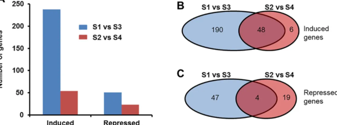
Pairwise comparison of the expression levels of soybean genes in inoculated roots with those of corresponding water treated roots during ETP- or LTP revealed 314 DEGs that
showed10-fold change (FC). In identifying DEGs, we considered only those genes that showed to contain at least 5 sequence reads per kilobase pair exon sequences in at least one of the treat-ments considered for comparison. We detected transcripts for 54,305 of the predicted soybean genes [48,49]. We found more DIGs in roots of ETP than that in LTP (Fig 1A). In infected roots of both ETP and LTP, there were more up-regulated genes than the down-regulated ones (Fig 1). During ETP, 289 genes were differentially expressed between infected and water-treated roots with FC10. The majority of these DIGs (238; 82%) were induced; only 54 genes (18%) were repressed in the infected roots of ETP as compared to the water treated root tissues (Fig 1B and 1C;Table 1;S1andS2Datasets). In the infected root tissues of LTP, of the 77 DIGs, 54 (70%) were up-regulated and 23 (30%) were down-regulated (Fig 1B and 1C;Table 2,S3 Dataset).
Functional classification of soybean genes induced in roots following F.
virguliforme infection
We used public transcriptomic databases such as SoyBase [50](http://soybase.org/) and Phyto-zome (phytozome.jgi.doe.gov), PFAM, and National Center for Biotechnology Information
Fig 1. Distribution of differentially expressed genes in soybean roots in response to F. virguliforme infection. A. Total number of genes differentially (with FC10) regulated by F. virguliforme infection. B. Number of genes up-regulated in the infected roots at early and late time-periods. C. Number of genes repressed in the infected roots at early and late time-periods. S1, pooled RNA samples prepared from roots harvested 3 and 5 days following water treatment; S2, pooled RNA samples prepared from roots harvested 10 and 24 days following water treatment; S3, pooled RNA samples prepared from roots harvested 3 and 5 days following F. virguliforme infection; S4, pooled RNA samples prepared from roots harvested 10 and 24 days following F. virguliforme infection.
Table 1. Genes down-regulated (with FC>10) in soybean roots during early time period following infection with F. virguliforme. The full list is
reported inS2 Dataset.
Locus ID RPKM Fold change P-value Functional Annotation
S1 S3
Glyma.01g171600 12.38 0.05 254.8 1.12E-06 SAM dependent carboxyl methyltransferase Glyma.02g054200 37.71 0.17 225.5 9.33E-15 SAM dependent carboxyl methyltransferase Glyma.07g098500 19.19 0.13 144.4 6.39E-08 copper transport protein atox1-related Glyma.08g163600 1.29 0.02 74.4 2.13E-04 pre-mrna processing protein prp39-related Glyma.09g022800 193.8 2.63 73.7 7.33E-15 peroxidase
Glyma.10g185900 8.01 0.11 71.5 2.34E-09 sieve element occlusion protein Glyma.03g024300 1199.19 17.48 68.6 2.51E-06 glycosyl hydrolases family 18 Glyma.03g024200 1496.7 22.9 65.4 3.13E-05 glycosyl hydrolases family 18 Glyma.03g024400 1102.06 16.91 65.2 3.53E-07 glycosyl hydrolases family 18 Glyma12g12470* 10.06 0.16 62.1 3.05E-10 ankyrin repeat-containing Glyma.10g266500 1.34 0.02 60.5 4.50E-04 plant protein of unknown function Glyma03g02810* 56.25 1.01 55.8 1.27E-13 glycoside hydrolase
Glyma.03g025000 53.91 0.97 55.5 3.73E-13 glycosyl hydrolases family 18 Glyma03g02818* 32.43 0.58 55.5 5.20E-13 unknown
Glyma03g02843* 45.41 0.82 55.2 1.66E-13 unknown
Glyma.06g294400 7.77 0.16 47.7 1.73E-09 ankyrin repeat-containing Glyma.13g113100 11.65 0.27 43.8 6.61E-10 flavin-containing monooxygenase Glyma.03g024800 64.4 1.53 42 1.09E-11 glycosyl hydrolases family 18
Glyma.07g204900 6.09 0.16 39 6.94E-07 lipase (class 3); alpha/beta-hydrolases superfamily protein Glyma.14g032000 13.31 0.34 38.7 1.23E-04 unknown
Glyma.06g013200 29.27 0.82 35.7 8.37E-12 protein of unknown function (DUF2775) Glyma.03g116300 108.09 3.21 33.7 2.86E-10 ubiquitin specific protease family C19-related Glyma.06g300100 4.26 0.14 31.3 1.30E-05 transcription factor; MYB-related
Glyma.01g115500 40.93 1.35 30.4 3.42E-08 unknown Glyma.10g094800 6.29 0.22 29 2.13E-05 unknown
Glyma.14g051600 18.92 0.84 22.6 2.54E-07 copper transport protein atox1-related Glyma.13g205400 73.71 3.36 21.9 2.33E-07 AT1G11655
Glyma.14g061800 2.07 0.11 19.6 2.28E-04 HXXXD-type acyl-transferase family protein Glyma.18g182800 6.03 0.32 18.6 5.34E-06 2-deoxyglucose-6-phosphate phosphatase 2 Glyma18g39500* 1.59 0.09 18.4 1.90E-05 NADPH oxidase; flavin adenine dinucleotide binding Glyma14g05621* 4.93 0.3 16.3 2.87E-04 copper transport protein atox1-related
Glyma.01g115700 4.63 0.29 15.9 4.71E-06 plant protein of unknown function
Glyma.03g061200 18.63 1.2 15.5 1.60E-08 plant protein of unknown function (DUF247) Glyma07g18225* 24.51 1.59 15.4 6.82E-06 SGNH hydrolase-type esterase superfamily protein Glyma.11g216700 6.2 0.4 15.4 6.85E-05 unknown
Glyma.03g141900 20.64 1.37 15.1 5.40E-06 unknown Glyma.16g197300 11.65 0.8 14.6 2.34E-04 unknown Glyma.16g197400 11.65 0.8 14.6 2.34E-04 unknown Glyma.08g189600 1.31 0.09 14.5 1.73E-04 lipoxygenase Glyma.02g083500 351.22 26.37 13.3 4.78E-06 extensin
Glyma10g25800* 10.86 0.87 12.5 9.47E-06 serine-threonine protein kinase; disease resistance/LRR family Glyma.19g144500 28.35 2.31 12.3 1.52E-04 unknown
Glyma.06g149600 4.36 0.36 12.2 2.94E-04 nodulin MtN21 /EamA-like transporter family protein Glyma.09g023000 542.08 44.79 12.1 4.68E-04 peroxidase
Glyma0466s00200 63.01 5.4 11.7 5.39E-08 unknown
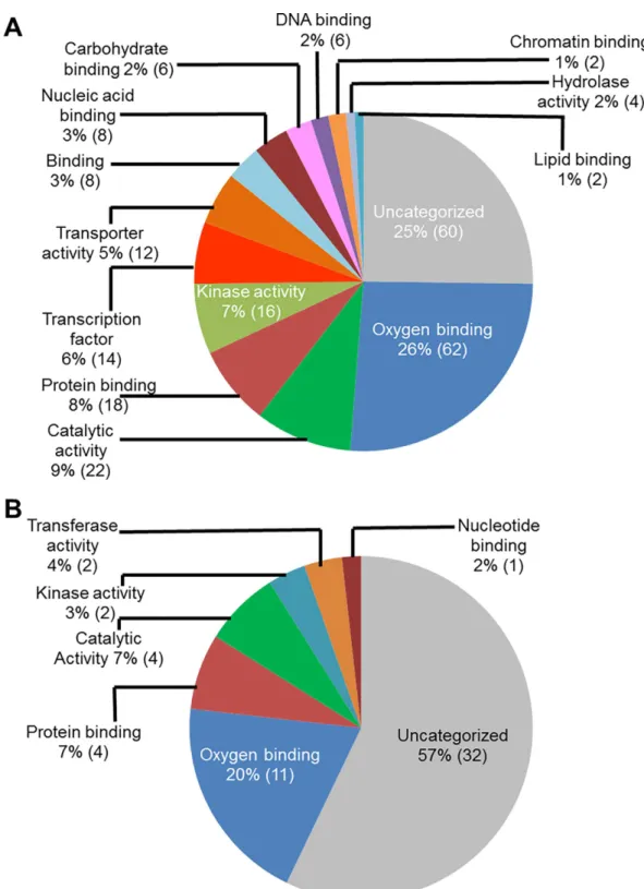
(NCBI) for assignment of the gene models associated with the infection-induced genes, and also for their functional annotations. Functional classification of these genes based on their putative molecular functions indicated that a majority of the genes (62 genes during ETP and 11 during LTP) had putative oxygen binding properties (Fig 2). A large number of infection-Table 1. (Continued)
Locus ID RPKM Fold change P-value Functional Annotation
S1 S3
Glyma.09g099900 10.49 0.93 11.2 3.07E-05 serine/threonine protein kinase Glyma.20g173800 13.19 1.2 11 1.25E-04 protein-tyrosine phosphatase 1 Glyma.12g225700 6.51 0.59 11 4.85E-04 unknown
Glyma.20g215500 32.66 3.02 10.8 3.71E-08 unknown
Glyma.01g002300 3.63 0.34 10.7 1.63E-04 cation transport protein Glyma.08g285400 6.34 0.61 10.4 2.88E-05 glycosyl hydrolase family 28 S1, roots tissues 3 and 5 days following water treatment; S3, root tissues 3 and 5 days following infection.
*, Predicted genes in the old soybean genome sequence version [Glyma.Wm82.a1.v1.1 (Gmax1.01)].
doi:10.1371/journal.pone.0163106.t001
Table 2. Genes down-regulated at late time period in soybean roots infected with F. virguliforme.
Locus ID RPKM Fold change P-value Functional Annotation
S2 S4
Glyma.06g300000 27.9 0.1 187.4 5.81E-05 MYB-related; myb binding domain
Glyma.02g054200 33.6 0.4 93.7 4.79E-05 SAM dependent carboxyl methyltransferase Glyma.07g234100 187.1 3.5 53.9 1.24E-05 uncharacterized protein
Glyma.15g082200 170.5 4 42.6 8.53E-07 cysteine proteinase cathepsin F Glyma.16g038100 943.1 23.5 40.1 4.11E-06 uncharacterized protein Glyma13g11969* 662.7 20.4 32.5 3.43E-06 uncharacterized protein Glyma.09g022800 93.9 3.2 29.2 6.42E-06 peroxidase
Glyma.09g163800 434.4 15.3 28.3 6.47E-06 trypsin and protease inhibitor; endopeptidase inhibitor Glyma.03g024400 664.4 23.5 28.3 3.92E-05 hydrolase activity
Glyma.10g232100 2129.7 77.8 27.4 8.57E-05 uncharacterized protein
Glyma.06g298700 639.1 23.6 27 1.72E-05 wound-induced protein; wound-responsive Glyma.02g303200 211.4 8.1 26 7.51E-06 uncharacterized protein
Glyma.03g024300 635.2 25.7 24.7 9.18E-05 hydrolase activity Glyma17g03850* 32802 1367.3 24 6.10E-05 uncharacterized protein
Glyma.16g178000 262.1 11 23.8 1.21E-05 lipid-transfer/copper transport protein atox1-related Glyma.11g224900 472.4 20.4 23.2 1.20E-05 uncharacterized protein
Glyma.17g039400 2205.5 96.7 22.8 2.26E-05 uncharacterized protein
Glyma.13g282200 117.7 5.4 21.7 2.30E-05 wound-induced protein; wound-responsive Glyma.13g282400 82.9 3.9 21 3.73E-05 wound-induced protein; wound-responsive Glyma13g42850* 1375.5 69.2 19.9 5.80E-05 uncharacterized protein
Glyma.12g048000 393.6 21.3 18.4 5.90E-05 uncharacterized protein Glyma.01g210500 50.7 2.8 18.3 3.52E-05 oligopeptide transporter-related Glyma.03g082100 197.6 13.1 15.1 9.50E-05 metallothion binding
S2, roots tissues 10 and 24 days following water treatment; S4, root tissues 10 and 24 days following infection.
*, Predicted genes in the old soybean genome sequence version [Glyma.Wm82.a1.v1.1 (Gmax1.01)].
Fig 2. Classes of genes up-regulated in roots infected with F. virguliforme as compared to the roots treated with water. A. A total of 238 genes were induced (with a FC10) in the pooled RNA sample of roots harvested 3 and 5 days following F. virguliforme infection as compared to the water control; and were classified based on their putative molecular functions. B. A total of 54 genes were induced in pooled RNA sample of roots harvested 10 and 25 days following F. virguliforme infection as compared to the water control; and were classified based on their putative molecular functions.
induced genes (60 genes during ETP and 32 in LTP) encode proteins with unknown molecular functions.
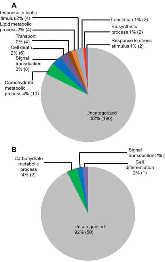
We investigated the infection-induced genes for their putative biological processes (Fig 3). Again a large portion of genes induced during ETP (82%) and in LTP (92%) encodes proteins with unknown functions. The next category of genes induced during ETP (Fig 3A) includes 10 genes (4%) involved in carbohydrate metabolic processes. In addition, eight genes (3%) involved in the signal transduction, six (2.5%) in cell death, four (2%) responsive to stress and stimuli, four (2%) are transporters, and four (2%) involved in the lipid metabolism processes. During LTP, only four genes (8%) were assigned with a biological process and are likely involved in the carbohydrate metabolic process, signal transduction, and cell differentiation (Fig 3B).
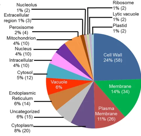
We conducted GO analyses of the up-regulated genes for determining the putative sub-cel-lular locations of their encoded proteins. A large number of the genes (24%; 58 genes), up-regu-lated in infected-roots during ETP, most likely encode cell wall proteins (Fig 4A). Among the rest, 34 genes (14%) encode membrane bound proteins, 26 genes (11%) encode plasma mem-brane associated, 20 genes (8%) encode cytoplasmic proteins, and 14 genes (6%) encode endo-plasmic reticulum proteins (Fig 4A). Similarly, the majority of the genes up-regulated in infected roots during LTP, 29 genes (37%) encode extracellular proteins and nine genes (12%) encode cell wall proteins (Fig 4B).
We observed that 48 of the 54 genes induced in the infected roots of LTP were induced also during ETP (Fig 1B;S1andS3Datasets). Functionally, 10 of the 50 genes up-regulated at both ETP and LTP belong to the cytochrome P450 CYP2 subfamily protein involved in the synthesis of defense metabolites [51]. Seven of these genes encode RmlC-like cupins with a nutrient res-ervoir activity; five genes encode peroxidases probably associated with signaling [52]; four genes encode receptor-like kinases (RLKs) presumably to modulate the induction of immunity [53]; three genes are members of the thaumatin family of pathogenesis-related protein that were shown to be induced duringF.virguliformeinfection [21], and three encode chitinase-related proteins involved in plant defense mechanisms [54,55]. Among the genes induced only in infected roots during LTP include: (i) a cupin-like gene, (ii) two defense/pathogenesis-related genes, and (iii) a gene with unknown function (S3 Dataset).
Functional classification of soybean genes repressed in roots infected
with F. virguliforme
We observed that steady-state transcript levels of 51 soybean genes were decreased (with a FC
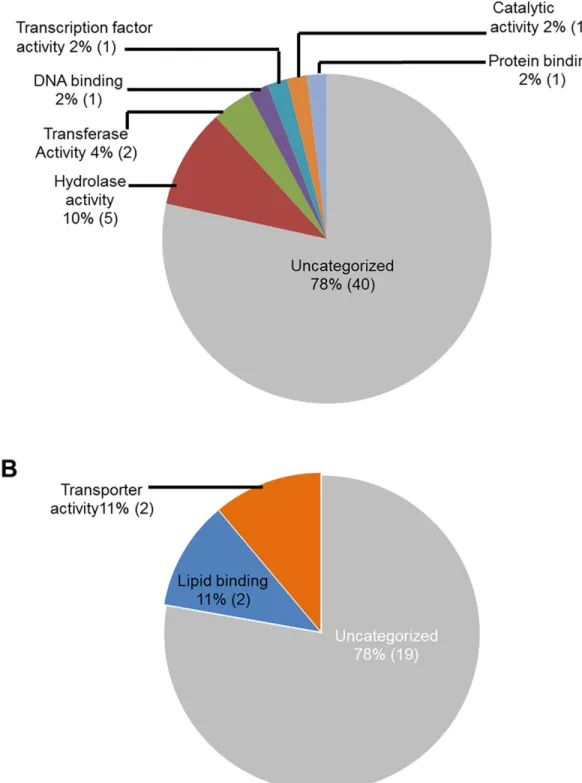
10) inF.virguliforme-infected roots as compared to that in the water control roots (Fig 1; Tables1and2;S2 Dataset). We investigated the possible function of these infection-repressed genes for (i) molecular functions, (ii) biological processes and (iii) cellular locations through gene ontology (GO) analyses and results are presented in Figs5–7.
GO analyses for molecular functions revealed that a large proportion of the down-regulated soybean genes do not show any identity to previously characterized genes and thus could be novel genes. For example, 40 genes (78%) with reduced transcript levels during ETP and 19 genes (78%) during LTP did not show homology to any functionally characterized genes (Fig 5). Infection-suppressed soybean genes encode proteins, most of which possess hydrolase and transferase activities at ETP (10% and 4% respectively) and lipid binding and transporter activ-ities at LTP (11% for each category). Some of the repressed genes encode transferases, catalytic proteins, and transcription factors (Fig 5).
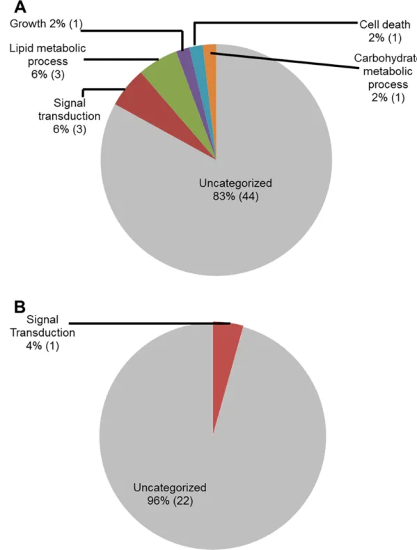
Reclassification of the down-regulated genes through GO analyses for biological processes again revealed that majority of the genes (83% in ETP and 96% in LTP) did not show identify
Fig 3. Classes of genes up-regulated in roots infected with F. virguliforme as compared to the roots treated with water. A. A total of 238 genes were induced (with a FC10) in the pooled RNA sample of roots harvested 3 and 5 days following F. virguliforme infection as compared to the water control. Induced genes were
to any genes with known biological processes. Down-regulated soybean genes with known bio-logical processes during ETP include four (8%) genes for primary metabolic processes (lipid and carbohydrate metabolism), three (6%) for signal transduction, two (4%) for growth and cell death (Fig 6).
To shed light on the possible function of the down-regulated genes, we conducted GO anal-yses for putative sub-cellular location. Surprisingly, a significant proportion (15%) of the down-regulated genes encode plasma membrane proteins especially during ETP. These pro-teins may be involved in regulating signaling process during infection (Fig 7). Again, a large number of the genes (20 [39%] genes during and 14 [64%] in LTP) were found not to show similarity to any known functionally characterized genes.
Of the 51 genes repressed in soybean roots during ETP, only 15 with FC15 continued to be suppressed during LTP following infection (Fig 1C; Tables1and2,S2 Dataset). These genes includeGlyma.02g054200encoding a SAM (salicylic acid methyltransferase) dependent car-boxyl methyltransferase,Glyma.15g082200encoding a cysteine proteinase Cathepsin F, Gly-ma.13g282200andGlyma.13g282400encoding wound-induced proteins,Glyma.16g178000
encoding a lipid transfer protein, andGlyma.06g294400andGlyma.12g111500encoding ankyrin-repeat containing proteins.
Validation of transcriptomic data for a few selected genes repressed
during SDS infection
To validate down-regulation of a few selected genes during infection, we performed a semi-quantitative reverse transcriptase polymerase chain reaction (RT-PCR) for one member from each of the four selected classes of down-regulated genes (Table 1). They are: (i)
Gly-ma.01g171600encoding an uncharacterized salicylate o-methyltransferase (SAM)-like protein, we termedGmSAM1, most likely involved in the conversion of salicylic acid to the volatile methyl salicylate, a plant defense signal [56] for defending attack of necrotrophic pathogens includingF.virguliforme; (ii)Glyma12g12470encoding an uncharacterized ankyrin repeat-containing protein, we termed it asGmARP1;(iii)Glyma.10g185900encodes a sieve element-occlusion protein (SEO)-like protein (http://www.uniprot.org;http://www.phytozome.net); and (iv)Glyma.10g094800encoding an uncharacterized, putative transmembrane protein [46] (http://www.phytozome.net).
RNA samples of soybean roots harvested 8 and 12 h and 1, 2, 3 and 5 d post inoculation withF.virguliformeor treated with water were considered for RT-PCR. The results of the RT-PCR analysis confirmed the observation of the RNA seq experiment for the four selected genes of interest (Fig 8). RT-PCR data also suggested that the down-regulation of three of the four selected genes,Glyma.01g171600,Glyma.10g094800, andGlyma12g12470, is very rapid with little or no detectible transcript levels observed 8 h post inoculation.
Alteration in expression of a F. virguliforme-repressed gene GmARP1
showed foliar SDS resistance
We hypothesized thatF.virguliformedown-regulates transcription of some of the defense-related genes to cause susceptibility in soybean. To test this hypothesis, we induced the expres-sion ofGmARP1(Glyma12g12470) in transgenic soybean plants followingF.virguliforme
classified based on their putative biological processes. B. A total of 54 genes were induced (with a FC10) in pooled RNA sample of roots harvested 10 and 25 days following F. virguliforme infection as compared to the water control. Induced genes were classified based on their putative biological processes.
Fig 4. Classes of genes up-regulated in roots infected with F. virguliforme as compared to the roots treated with water. A. A total of 238 genes were induced (with a FC10) in pooled RNA sample of roots harvested 3 and 5 days following F. virguliforme infection as compared to the water control. Induced genes were
infection.Glyma.12g111500andGlyma.06g294400are two highly similar genes that encode ankyrin repeats containing proteins with 76% identity (S4andS5Figs), but with no identity to any functionally characterized ankyrin repeat-containing genes includingGmNPR1[57]. GmARP1 protein contains 218 residues and slightly larger than all known ankyrin repeat-con-taining proteins including ANK superfamily with 71–199 residues; ANK_2 with 81–171; ANK_4 with 145–199 residues.
We used threeF.virguliforme-infection inducible promoters to induce the expression of
GmARP1duringF.virguliformeinfection. Three fusion genes were generated by ligating
GmARP1individually to these three promoters (S1A Fig) and used to transform soybean culti-var ‘Williams 82’. R0plants were grown in a greenhouse and analyzed for integration of
GmARP1transgenes by genomic PCR (S1B Fig).
R1progenies of individual transformants (R0) carrying any of the three fusionGmARP1
genes were inoculated withF.virguliformeMont-1 isolate in a growth chamber (Fig 9A–9E). Approximately 30 to 50% of the R1plants showed no symptoms to slight yellowing with
dis-ease scores 1 and 2. The rest showed severe disdis-ease symptoms with interveinal to severe chloro-sis and necrochloro-sis with disease scores 3 to 7 (Fig 9B). On the contrary, susceptible R1plants
presumably lacking a functional transgene exhibited typical SDS foliar symptoms from the sec-ond week of infection and were severely diseased by the end of the fourth week following inoc-ulation, with severe chlorosis and necrosis (disease scores 3 to 7), and reduced chlorophyll content as compared to the resistant R1progeny plants (Fig 9C). Moreover, resistant transgenic
plants showed increased root weight as compared to the susceptible R1progenies implying root
resistance of those resistant R1progenies to the pathogen (Fig 9D) and is supported by the
lev-els of root resistance observed among the R1progenies (Fig 9E). Molecular characterization of
the infected R1plants indicated that expression of foliar and root SDS resistance among the R1
progenies of multiple transformants was associated with the expression ofGmARP1transgenes (Fig 9F). No transcript of the endogenousGmARP1gene was detected duringF.virguliforme
infection.
We evaluated whetherGmARP1transgenes could provide enhanced SDS resistance in transgenic plants under field conditions. We observed that 70 to 100% of the transgenic R1
plants descended from six independent transgenic R0plants showed enhanced SDS resistance
under field conditions and did not develop SDS symptoms. Non-trangenic Williams 82 control plants developed severe SDS symptoms including chlorosis and necrosis of leaves that caused total defoliation of plants before maturity stage (Fig 10A and 10C;S6 Fig). The transgene copy number assay conducted using qPCR revealed that all six transgenic lines contain at least one copy of the transgene, sufficient to enhance resistance againstF.virguliforme(Table 3;S4 Data-set). The line Prom2-ARP1-9 carrying single copyProm2-ARP1transgene showed 100% resis-tant R1progenies; whereas, Prom2-ARP1-9 line carrying on the average 4 copies of the same
Prom2-ARP1transgene showed the lowest number SDS resistant plants (70%).
Discussion
Study of the steady state transcriptomes using next-generation sequencing is a powerful method for revealing candidate genes that may play roles in host-pathogen interactions. Alter-ations of the expression of defense-related genes have been reported in plants that are infected by fungi, particularly byFusariaspecies [18,58]. Transcriptomic studies have revealed that
classified based on their putative cellular components. B. A total of 54 genes were induced (with a FC10) in pooled RNA sample of roots harvested 10 and 25 days following F. virguliforme infection as compared to the water control; and were classified based on their putative cellular components.
Fig 5. Classes of genes down-regulated in roots infected with F. virguliforme as compared to the roots treated with water. A. A total of 51 genes were repressed (with a FC10) in pooled RNA sample of roots harvested 3 and 5 days following F. virguliforme infection as compared to the water control; and were classified based on their putative molecular functions. A total of 23 genes were repressed (with a FC10) in pooled RNA sample of roots harvested 10 and 25 days following F. virguliforme infection as compared to the water control; and were classified based on their putative molecular functions.
Fig 6. Classes of genes down-regulated in roots infected with F. virguliforme as compared to the roots treated with water. A. A total of 51 genes were repressed (with a FC10) in pooled RNA sample of roots harvested 3 to 5 days following F. virguliforme infection as compared to the water control; and were classified based on their putative biological processes. B. A total of 23 genes were repressed in pooled RNA sample of roots harvested 10 and 25 days following F. virguliforme infection as compared to the water control; and were classified based on their putative biological processes.
more host genes are induced and fewer repressed following infection with fungal pathogens [19,21,58,59]. The majority of the induced genes are defense related, and it has been shown that overexpression of defense genes induced during infection in transgenic plants could enhance resistance to pathogens [32,60].
Here we report GO analyses of differentially expressed soybean genes identified by compar-ing transcriptomes of soybean roots followcompar-ingF.virguliformeinfection with those of water treated soybean roots. A large number of the genes induced in soybean roots infected withF.
virguliformeencode cell wall and plasma membrane proteins (Fig 4;S1 Dataset) and are pre-sumably involved in generating protective barriers against the invading pathogens. A majority of proteins encoded by these genes have binding activities. It is known that proteins and enzymes that bind other proteins and metabolites are implicated in various metabolic func-tions. For instance, DNA binding proteins have been shown to recognize specific promoter sequences of the target defense genes for regulating defense mechanisms [61,62].
A set of infection-induced genes are involved in carbohydrate metabolism. Besides being primary metabolites in plant cells, sugars are also known as signals for plant induced responses to pathogen attacks [63,64]. Many of the genes induced in roots followingF.virguliforme infec-tion encode enzymes with catalytic or kinase activities. Some of the kinases are receptor kinases for receiving and transmitting signals [65] and are induced in roots during fungal attack [33,54]. Our data suggest that defense-related transcripts are induced following infection pre-sumably to defend soybean against theF.virguliformeattack.
In the Arabidopsis-F.oxysporuminteraction a large number of genes were repressed as opposed to induction as observed in most plant-pathogen interactions [66]. Despite a large number of soybean genes were up-regulated uponF.virguliformeinfection, in our analysis of the soybean-F.virguliformeinteraction revealed that 70 soybean genes were suppressed follow-ing infection. Of these, only four genes were down-regulated in both ETP and LTP followfollow-ingF.
virguliformeinfection (Fig 1C;S2 Dataset).
Among the genes down-regulated followingF.virguliformeinfection, seven soybean genes encode glycosyl hydrolases (Table 1). Of these three genesGlyma.03g024400,
Gly-ma.03g024300, andGlyma.03g024200encoding the uncharacterized glycosyl hydrolase family 18 were highly expressed during ETP of the water treated root tissues (RPKM>1,102). The expression of these genes was strongly suppressed uponF.virguliformeinfection (FC>65) (Table 1). Members of the glycosyl hydrolase 18 with high identity to some pathogen related (PR) genes are not only implicated in metabolic processes of cell wall, but also considered to have defense and signaling functions [67–70]. The glycosyl hydrolases encoded by Gly-ma.03g024400,Glyma.03g024300, andGlyma.03g024200are 100% identical and contain a GH18_chitinase-like domain; therefore most likely they function in plant defense against path-ogens with chitin molecules [71,72]. Four additional soybean genes,Glyma03g02810, Gly-ma.03g025000 Glyma.03g024800, andGlyma.08g285400encoding glycosyl hydrolases with moderate expression levels (FC from 6 to 56), were also repressed during ETP followingF. vir-guliformeinfection.
Transcript levels of two genesGlyma.09g022800andGlyma.09g023000encoding uncharac-terized peroxidases were strongly down-regulated followingF.virguliformeinfection (Table 1). Fig 7. Classes of genes down-regulated in roots infected with F. virguliforme as compared to the roots treated with water. A. A total of 51 genes were repressed (with a FC10) in pooled RNA sample of roots harvested 3 to 5 days following
F. virguliforme infection as compared to the water control; and were classified based on their putative cellular components.
B. A total of 23 genes were repressed in pooled RNA sample of roots harvested 10 and 25 days following F. virguliforme infection as compared to the water control; and were classified based on their putative cellular components.
The two proteins are 64% identical and contain a secretory peroxidase domain. Some peroxi-dases genes are also shown to bePRgenes [73] and involved in plant defense against
pathogens.
In our transcriptomic study, we included transcripts of roots of seedlings 3 and 5 days fol-lowingF.virguliformeinfection or water treatment for ETP. In the confirmatory RT-PCR experiment, we included roots of seedlings 8, 12 and 24 hours in addition to 3 and 5 days fol-lowing infection or water treatment. Transcripts of all four genes included in the RT-PCR study were not detectable after 12 h followingF.virguliformeinfection (Fig 8;Table 1). Of these four genes,Glyma.10g185900(Fig 6A and 6B;Table 1;S2 Dataset) encodes a sieve ele-ment-occlusion protein (SEO), which is a structural protein implicated to play a role in plant defense [74,75]. Our data suggest that somehowF.virguliformesuppressed the expression of this gene to avoid any barrier arisen from this protein in the roots.
More than half of plant genes encode proteins of unknown functions [76]. We observed down-regulation of many genes encoding proteins of unknown functions during both ETP and LTP (FC>10; Tables1and2). During LTP, four genes have RPKM values in water treated roots over 1,000 (1,375 to 32,802) and are strongly repressed (FC20) inF.virguliforme
infected roots. The most highly expressed,Glyma17g03850(RPKM in water treated roots 32,802 and repressed after infection to FC = 24.0), is an orphan gene ofGlycine max[77] (http://www.greenphyl.org/) with no annotated functional domain. This gene is only co-expressed withGlyma.10G216000(correlation 0.87,www.phytozome.com) that encodes a gib-berellin-regulated protein 2 involved in gibberellic acid mediated signaling pathway [78]. Simi-larly, infection-suppressed geneGlyma.10g232100(RPKM 2,130 in water treated roots vs. FC 27 following infection) encoding an unknown protein also co-expressed with the gibberellin-regulated protein 2. Finally,Glyma.17g039400(RPKM 2,206 in water treated roots vs. FC 24 following infection). It is highly co-expressed with a fasciclin-like arabinogalactan protein gene (correlation 0.88,www.phytozome.com) implicated in the cell-wall composition [79]. Thus, these genes with unknown functions could be involved in plant immunity. Future studies on these four genes might yield new insights about the soybean-F.virguliformeinteraction.
Aknyrin repeat-containing proteins widely exist in plants [80]. We identified fiveGmARP1
homologs inG.sojaandPhaseolus vulgarisexhibiting62% identities among them, specifi-cally in the ankyrin domain (S7 Fig). Two to more than 20 ankyrin-repeats involved in pro-tein-protein interactions could be present in ankyrin-repeat containing proteins [81–83]. Ankyrin-repeats are involved in many metabolic processes including defense against pathogens and signaling [81,84–86]. The ankyrin repeat-containing ACD6 is implicated in salicylic acid signaling in Arabidopsis [87]. The ankyrin repeat-containing protein CaKR1 inCapsicum annuumis responsive to both abiotic and biotic stresses [88]. Ankyrin repeat-containing pro-tein NPR1 modulates plant immunity cooperatively with transcription factors [89]. In Arabi-dopsis, BDA1 containing ankyrin repeats has been shown to regulate immunity [90]. Similarly, Fig 8. Reduced expression levels of soybean genes following F. virguliforme infection as compared to the water control. A. Expression levels of four selected soybean genes following water treatment at early
(S1: 3 and 5 d) and late time (S2: 10 and 24 d) periods and F. virguliforme infection at early (S3: 3 and 5 d) and late time (S4: 10 and 24 d) periods. B. RT-PCR analyses of the selected soybean genes. RT-PCR products of each of the four selected soybean amplified from RNAs of root tissues harvested 8 and 12 h, and 1, 2, 3, 5 days following (i) water treatment or (ii) infection with the F. virguliforme Mont-1. The results presented here are from one of three independent experiments showing similar results. Glyma12g12470 is from the Glyma.Wm82.a1.v1.1 version of the soybean genome sequence. Other three genes are from the recent version of the soybean genome sequence (Glyma.Wm82.a2v1). Elf1b, elongation factor 1-βencoded by Glyma02g44460. C–F. Quantified expression levels of four selected genes. Gel pictures of the three biological replications are presented inS8 Fig.
Mou and colleagues [60] reported that overexpression of the aknyrin repeat-containing protein geneOsPIANK1enhanced immunity againstMagnaporthe oryzaein transgenic rice.GmARP1 Fig 9. Expression of GmARP1 enhances SDS resistance in transgenic soybean plants. R1plants were tested for resistance to F. virguliforme under
growth chamber conditions. A. Root phenotype of a resistant (R) and a susceptible (S) R1progeny of a transformant, Prom2-ARP1-7, carrying the Prom2-GmARP1 fusion gene. B. Enhanced foliar SDS resistance among R1progenies. W82, the SDS susceptible line Williams 82; MN1606, the SDS
resistant line. C. Chlorophyll content per individual R1progeny carrying GmARP1 of three independent transformants. ‘Resistant’ and ‘Susceptible’ classes
are defined as in (A). D. Average root weight of R1progeny of three independent transformants. ‘Resistant’ and ‘Susceptible’ classes are defined as in A. E.
Enhanced root resistance among R1progenies. Extent of root resistance to the pathogen was expressed in percent; e.g., 100%, healthy roots with no
obvious blackening caused by necrosis and rotting due to infection of F. virguliforme. F. Expression of GmARP1 transgenes. Two random SDS resistant and susceptible R1progenies from each R0line were analyzed. Top panel, resistant plants (two representatives from each line). Bottom panel, susceptible
plants (two representatives from each line). Red arrow, GmARP1; black arrow, ELF1b internal control.*, significantly different at p<0.01. Results are means
±SE of three independent experiments.
(Glyma12g12470)has been shown to have a protein binding function and putatively localized to plasma membrane [80]. Here we have shown that the steady state transcript level of this gene is not detectable immediately afterF.virguliformeinfection (Fig 8). Expression of this gene using infection-inducible promoters in transgenic plants enhanced resistance to the SDS pathogen, indicating that like other known aknyrin repeat-containing protein genes,GmARP1
is involved in soybean defense against this fungal pathogen.
Conclusions
Our transcriptomic study revealed many genes that are induced, whereas expression of only a few genes including a previously uncharacterizedGmARP1gene is strongly suppressed by
F.virguliformeinfection. Many of the genes that are suppressed during infection might be defense-related and, are somehow suppressed by pathogens to establish the compatible interaction or to cause susceptibility. This hypothesis was tested by expressingGmARP1
duringF.virguliformeinfection in transgenic soybean lines. We observed that expression of
GmARP1led to induction of foliar SDS resistance. HowGmARP1regulates immunity against SDS pathogen is yet to be determined. It is also unknown how the gene is down-reg-ulated byF.virguliformeinfection. Our transgenic study suggests that altered expression of pathogen-repressed host genes could be a suitable strategy in engineering disease resistance in crop plants.
Fig 10. Expression of GmARP1 enhances SDS resistance in transgenic soybean plants. R1plants were
tested for resistance to F. virguliforme under field conditions. A. Average disease scores gathered at four scoring dates for R1progenies of each transgenic line carrying GmARP1 under the control of Prom1, Prom2, or Prom3
promoters. n, number of R1plants. B. Average disease scores of Williams 82 (susceptible control) and MN1606
(resistant control). n, number of plants and results in A and B are means±SE of n plants. C. Percentage of R1
plants resistant to F. virguliforme.
Supporting Information
S1 Fig. Generation of GmARP1 transgenes and R0transgenic soybean plants.
(DOCX)
S2 Fig. GmARP1 Sequences used in the transgenic study. (DOCX)
S3 Fig. Development of a binary construct for transformation of soybean. (DOCX)
S4 Fig. Screenshots of the GmARP1 gene in the new and the old versions of genome sequence assemblies (www.soybase.org).
(DOCX)
S5 Fig. Sequence analysis of two ankyrin-repeat containing proteins downregulated follow-ing F. virguliforme infection.
(DOCX)
S6 Fig. Phenotype of transgenic soybean plants carrying GmARP1 fusion genes in a field trial.
(DOCX)
S7 Fig. Sequence analysis of five ankyrin-repeat containing GmARP1 (Glyma12g12470) homologs.
(DOCX)
S8 Fig. RT-PCR analysis of three independent replicates for the four selected genes. (DOCX)
Table 3. Average GmARP1 transgene copy numbers among the transgenic lines.
Genotype Transgene copy number Number of plants Average copy number (±SE)
1 7 Prom1-ARP1-5 2 4 2 (0.06) 3 4 1 3 3 2 Prom2-ARP1-3 4 3 4 (0.18) 5 1 6 1 7 2 2 7 Prom2-ARP1-7 3 1 3 (0.14) 4 2 5 2 1 9 Prom2-ARP1-9 2 2 1 (0.05) 3 1 Prom3-ARP1-3 2 10 2 (0.02) 3 2 1 7 Prom3-ARP1-11 2 4 2 (0.06) 3 1 doi:10.1371/journal.pone.0163106.t003
S1 Table. List of primers used for PCR and RT-PCR (enzyme sites highlighted). (DOCX)
S2 Table. Number of plants resistant to basta for GmARP1 transgenic lines used in the field trial.
(DOCX)
S3 Table. Method of foliar SDS scoring in the field (www.siu.edu/~soybean). (DOCX)
S4 Table. List of primers used for RT-PCR of five selected genes in the infected soybean roots.
(DOCX)
S1 Dataset. Genes up-regulated in soybean roots infected with F. virguliforme at early time period.
(XLSX)
S2 Dataset. Genes down-regulated at early time period in soybean roots infected by F.
vir-guliforme.
(XLSX)
S3 Dataset. Genes up-regulated in soybean roots infected by F. virguliforme at late time period.
(XLSX)
S4 Dataset. Transgene copy number analysis by qPCR. (XLSX)
Acknowledgments
This work was supported by the National Institute of Food and Agriculture (NIFA), United States Department of Agriculture (Grant no. 2013-68004-20374) and Iowa Soybean Associa-tion. We are thankful to David Grant for reviewing the manuscripts, Jordan Baumbach and Yang Yang for their contributions to managing soybean transgenic plants (gaining USDA per-mit, planting, watering and harvesting). We are also thankful to the Iowa State University (ISU) Plant Transformation Facility for generation of soybean transgenic plants and ISU DNA Facility for sequencing the transcript samples and preparing DNA from leaf samples.
Author Contributions
Conceptualization: MKB. Data curation: SKS. Formal analysis: MSF MNN SKS. Funding acquisition: MKB. Investigation: MNN BBS SK SS BW. Methodology: MNN SM SS SKS. Project administration: MKB. Resources: BBS.Software: SKS. Supervision: MKB. Validation: MNN. Visualization: MNN.
Writing – original draft: MNN.
Writing – review & editing: MKB SKS MSF BBS SS SK BW.
References
1. Wrather JA, Anderson TR, Arsyad DM, Gai J, Ploper LD, Porta-Puglia A, et al. Soybean disease loss estimates for the top 10 soybean producing countries in 1994. Plant Dis. 1997; 81: 107–110.
2. Wrather JA, Koenning SR. Estimates of disease effects on soybean yield in United States 2003 to 2005. J Nematol. 2006; 38: 173–180. PMID:19259444
3. Koenning SR, Wrather JA. Suppression of soybean yield potential in the continental United States by plant diseases from 2006 to2009. Plant Health Progress. 2010. doi: 101094/PHP-2010-1122-01-RS.
4. Aoki T, O’Donnell K, Homma Y, Lattanzi A. Sudden-death syndrome of soybean is caused by two mor-phologically and phylogenetically distinct species within the Fusarium solani species complex- F.
virgu-liforme in North America and F. tucumaniae in South America. Mycologia. 2003; 95: 660–684. PMID:
21148975
5. Aoki T, O’Donnell K, Scandiani MM. Sudden death syndrome of soybean in South America is caused by four species of Fusarium: Fusarium brasiliense sp. nov., F. cuneirostrum sp. nov., F. tucumaniae, and F. virguliforme. Mycoscience. 2005; 46: 162–183.
6. Hughes TJ, O’Donnell K, Sink S, Rooney AP, Scandiani MM, Luque A, et al. Genetic architecture and evolution of the mating type locus in fusaria that cause soybean sudden death syndrome and bean root rot. Mycologia. 2014; 106: 686–697. doi:10.3852/13-318PMID:24891421
7. Njiti VN, Suttner RJ, Gray LE, Gibson PP, Lightfoot DA. Rate reducing resistance to Fusarium solani f. sp. Phaseoli underlies field resistance to soybean sudden death syndrome. Crop Sci. 1997; 37: 132– 138.
8. Gao X, Jackson K, Lambert K, Li S, Niblack T, Hartman G. Detection and quantification of Fusarium
solani f.sp. glycines in soybean roots with real-time quantitative polymerase chain reaction. Plant Dis.
2004; 88: 1372–1380.
9. Jin H, Hartman GL, Nickel CD, Widholm JM. Phytotoxicity of culture filtrates from Fusarium solani, the causal agent of sudden death syndrome of soybean. Plant dis. 1996; 80: 922–927.
10. Brar HK, Swaminathan S, Bhattacharyya MK. The Fusarium virguliforme toxin FvTox1 causes foliar sudden death syndrome-like symptoms in soybean. Mol Plant-Microbe Interact. 2011; 24: 1179–1188. doi:10.1094/MPMI-12-10-0285PMID:21635141
11. Brar HK, Bhattacharyya MK. Expression of a single-chain variable-fragment antibody against a
Fusar-ium virguliforme toxin peptide enhances tolerance to sudden death syndrome in transgenic soybean
plants. Mol Plant-Microbe Interact. 2012; 25: 817–824. doi:10.1094/MPMI-12-11-0317PMID:
22397408
12. Pudake RN, Swaminathan S, Sahu BB, Leandro LF, Bhattacharyya MK. Investigation of the Fusarium
virguliforme fvtox1 mutants revealed that the FvTox1 toxin is involved in foliar sudden death syndrome
development in soybean. Current Genet. 2013; 59: 107–117.
13. Chang HX, Domier LL, Radwan O, Yendrek CR, Hudson ME, Hartman GL. Identification of multiple phytotoxins produced by Fusarium virguliforme including a phytotoxic effector (FvNIS1) associated with sudden death syndrome foliar symptoms. Mol Plant-Microbe Interact. 2016; 29: 96–108. doi:10. 1094/MPMI-09-15-0219-RPMID:26646532
14. Abeysekara NS, Bhattacharyya MK. Analyses of the xylem sap proteomes identified candidate
Fusar-ium virguliforme proteinacious toxins. PLoS One. 2014; 9: e93667. doi:10.1371/journal.pone. 0093667PMID:24845418
15. Zou J, Rodriguez-Zas S, Aldea M, Li M, Zhu J, Gonzalez DO, et al. Expression profiling soybean response to Pseudomonas syringae reveals new defense-related genes and rapid HR-specific down-regulation of photosynthesis. Mol Plant-Microbe Interact. 2005; 18: 1161–1174. doi: 10.1094/MPMI-18-1161PMID:16353551
16. Zabala G, Zou J, Tuteja J, Gonzalez DO, Clough SJ, Vodkin LO. Transcriptome changes in the phenyl-propanoid pathway of Glycine max in response to Pseudomonas syringae infection. BMC Plant Biol. 2006; 6: 26. doi:10.1186/1471-2229-6-26PMID:17083738
17. Yuan JZ, Zhu MX, Lightfoot DA, Iqbal MJ, Yang JY, Meksem K. In silico comparison of transcript abun-dances during Arabidopsis thaliana and Glycine max resistance to Fusarium virguliforme. BMC Geno-mics. 2008; 9 (Suppl 2): S6. doi:10.1186/1471-2164-9-S2-S6PMID:18831797
18. D’Ippo´lito S, Martin LM, Salcedo MF, Atencio HM, Casalongue CA, Godoy AV, Fiol DF. Transcriptome profiling of Fusarium solani f. sp. eumartii-infected potato tubers provides evidence of an inducible defense response. Physiol Mol Plant Path. 2010; 75: 3–12.
19. Moy P, Qutob D, Chapman BP, Atkinson I, Gijzen M. Patterns of gene expression upon infection of soybean plants by Phytophthora sojae. Mol Plant-Microbe Interact. 2004; 17: 1051–1062. doi:10. 1094/MPMI.2004.17.10.1051PMID:15497398
20. Iqbal MJ, Yaegashi S, Ahsan R, Shopinski KL, Lightfoot DA. Root response to Fusarium solani f. sp.
glycines: temporal accumulation of transcripts in partially resistant and susceptible soybean. Theor
Appl Genet. 2005; 110: 1429–1438. doi:10.1007/s00122-005-1969-9PMID:15815926
21. Radwan O, Liu Y, and Clough SJ. Transcriptional analysis of soybean root response to Fusarium
virgu-liforme, the causal agent of sudden death syndrome. Mol Plant-Microbe Interact. 2011; 24: 958–972.
doi:10.1094/MPMI-11-10-0271PMID:21751852
22. Roy K, Rupe JC. Sudden death syndrome of soybean. Plant Dis. 1997; 81: 259–266.
23. Rupe JC. Frequency and pathogenicity of Fusarium solani recovered from soybeans with sudden death syndrome. Plant Dis. 1989; 73: 581–584.
24. Anderson TR, Tenuta A. First report of Fusarium solani f. sp. glycines causing sudden-death syndrome of soybean in Canada. Plant Dis. 1998; 82: 448.
25. Leandro LF, Tatalovic N, Luckew A. Soybean sudden death syndrome—Advances in knowledge and disease management. CAB Rev. 2012; 7: 1–14.
26. Stephens PA, Nickell CD, Kolb FL. Genetic-analysis of resistance to Fusarium solani in soybean. Crop Sci. 1993; 33: 929–930.
27. Njiti VN, Meksem K, Iqbal MJ, Johnson JE, Kassem M, Zobrist KF, et al. Common loci underlie field resistance to soybean sudden death syndrome in Forrest, Pyramid, Essex, and Douglas. Theor Appl Genet. 2002; 104: 294–300. doi:10.1007/s001220100682PMID:12582700
28. Kassem MA, Ramos L, Leandro LF, Mbofung GYC, Hyten DL, Kantartzi SK, et al. The ‘PI 438489B’ by ‘Hamilton’ SNP-based genetic linkage map of soybean [Glycine max (L.) Merr.] identified quantitative trait loci that underlie seedling SDS resistance. J Plant Gen Sci. 2012; 1: 18–30.
29. Wen Z, Tan R, Yuan J, Bales C, Du W, Zhang S, et al. Genome-wide association mapping of quantita-tive resistance to sudden death syndrome in soybean. BMC Genomics. 2014; 15: 809. doi:10.1186/ 1471-2164-15-809PMID:25249039
30. Swaminathan S, Abeysekara NS, Liu M, Cianzio SR, Bhattacharyya MK. Identification of quantitative trait loci underlying the sensitivity of soybean to the Fusarium virguliforme toxins that induce foliar soy-bean sudden death syndrome in soysoy-bean. Theor Appl Genet. 2016; 129: 495–506. doi:10.1007/ s00122-015-2643-5PMID:26678962
31. Kesarwani M, Azam M, Natarajan K, Mehta A, Datta A. Oxalate decarboxylase from Collybia velutipes. Molecular cloning and its overexpression to confer resistance to fungal infection in transgenic tobacco and tomato. J Biol Chem. 2000; 275: 7230–7238. PMID:10702293
32. Cunha WG, Tinoco MLP, Pancoti HL, Ribeiro RE, Aragão FJL. High resistance to Sclerotinia
sclero-tiorum in transgenic soybean plants transformed to express an oxalate decarboxylase gene. Plant
Path. 2010; 59: 654–660.
33. Rey T, Nars A, Bonhomme M, Bottin A, Huguet S, Balzergue S, Jardinaud MF, Bono JJ, Cullimore J, Dumas B, Gough C, Jacquet C. NFP, a LysM protein controlling Nod factor perception, also intervenes in Medicago truncatula resistance to pathogens. New Phytologist. 2013; 198: 875–886. doi:10.1111/ nph.12198PMID:23432463
34. Catanzariti AM, Lim GTT, Jones DA. The tomato I-3 gene: a novel gene for resistance to Fusarium wilts disease. New Phytol. 2015; 207: 106–118. doi:10.1111/nph.13348PMID:25740416 35. Bhattacharyya MK, Ward EWB. Resistance, susceptibility and accumulation of glyceollin I-III in
soy-beans inoculated with Phytophthora megasperma f. sp. glycinea. Physiol Mol Plant Pathol. 1986; 29: 227–237.
36. Ngaki MN, Louie GV, Philippe RN, Manning G, Pojer F, Bowman ME, et al. Evolution of the chalcone-isomerase fold from fatty-acid binding to stereospecific catalysis. Nature. 2012; 485: 530–533. doi:10. 1038/nature11009PMID:22622584