eScholarship@UMMS
eScholarship@UMMS
Cancer Concepts: A Guidebook for theNon-Oncologist Radiation Oncology
2015-09-17
Pediatric Oncology Principles
Pediatric Oncology Principles
Christopher P. KeukerUniversity of Massachusetts Medical School
Let us know how access to this document benefits you.
Follow this and additional works at: https://escholarship.umassmed.edu/cancer_concepts
Part of the Cancer Biology Commons, Neoplasms Commons, Oncology Commons, Pediatrics Commons, and the Radiology Commons
Repository Citation Repository Citation
Keuker CP. (2015). Pediatric Oncology Principles. Cancer Concepts: A Guidebook for the Non-Oncologist.
https://doi.org/10.7191/cancer_concepts.1018. Retrieved from https://escholarship.umassmed.edu/ cancer_concepts/18
Creative Commons License
This work is licensed under a Creative Commons Attribution-Noncommercial-Share Alike 4.0 License.
This material is brought to you by eScholarship@UMMS. It has been accepted for inclusion in Cancer Concepts: A Guidebook for the Non-Oncologist by an authorized administrator of eScholarship@UMMS. For more information, please contact [email protected].
Christopher Keuker, MD, FAAP
Summary and Key Points
1. The incidence, distribution, treatment, and survivorship of childhood cancer differ in many ways from malignancies found mainly in adults. 2. It is important to be able to recognize typical clinical presentation of
common childhood hematologic and solid tumor malignancies.
3. Chemotherapy, radiation therapy, and surgery all have critical roles to play in the treatment of common childhood cancers and improvements in each have improved survival rates.
4. Treatment and outcomes of childhood cancer differ with the age of a child.
5. At least 328,000 Americans alive today are survivors of childhood cancer with increased health care needs.
Perspective
Cancer is a rare disease among children less than 20 years of age with an age-adjusted incident rate of approximately 16 per 100,000.1 However, cancer is “second” only to accidents (i.e. unintentional injuries) and homicides/suicides as a cause of death in children between 1 and 19 years of age.1
Cancer treatment and survival for children differ widely from adults in many ways. Chemotherapy treatment in children may be more intense and frequent because dose-limiting toxicities are reached at higher doses than in adults who commonly have more health issues and co-morbidities than children. Childhood and adult cancers also differ biologically which may be relevant to therapeutic response and survival differences. Most adult cancers are carcinomas derived from epithelial tissue, whereas a majority of pediatric cancers derive from developing tissue of mesodermal and ectodermal embryonic origin such as bone marrow, nerve tissue, lymph nodes, bone, and muscle. Experience has
shown that “blastic” cancers that arise from primitive tissues are more responsive to therapy, especially chemotherapy, than carcinomas.5 Treatment outcomes for children and adolescents with cancer have improved dramatically over the last 3 decades with 82% of children ages 0-19 years during 2001-2007 surviving 5 years after diagnosis of cancer compared to only 61% during the period 1975-1977.2 Table 1 illustrates the statistically significant improvement of relative survival rates of children with cancer over 25 years and differences in survival between different age groups. This progress is a result of advances in multimodal and combination drug therapies and supportive care, participation in systematic cooperative group clinical trials, and recognition that “children are not small adults.” Indeed, the results of treatment of children with cancer have been shown to be superior when specialized care is directed by a pediatric cancer center3 and studies have shown that adolescents with acute lymphoblastic leukemia had a better outcome when treated on pediatric cooperative group trials versus adult cooperative group trials.4
Citation: Keuker CP. Pediatric Oncology Principles. In: Pieters RS, Liebmann J, eds. Cancer Concepts: A Guidebook for the Non-Oncologist. Worcester, MA: University of Massachusetts Medical School; 2014. doi:. 10.7191/cancer_concepts.1018.
This project has been funded in whole or in part with federal funds from the National Library of Medicine, National Institutes of Health, under Contract No. HHSN276201100010C with the University of Massachusetts, Worcester.
Copyright: All content in Cancer Concepts: A Guidebook for the Non-Oncologist, unless otherwise noted, is licensed under a Creative Commons Attribution-Noncommercial-Share Alike License, http://creativecommons.org/licenses/by-nc-sa/4.0/
Toxicities of cancer treatments including chemotherapy, radiation therapy, and surgical therapies, however, may have more significant and irreversible effects on children than on adults because of the active growth and development of most organ systems during childhood and adolescence. Radiation therapies, for example, that may represent standard of care for adult cancer, may be contraindicated for children because of the effects on the developing brain and growing bones.
Fortunately, evolution of successful multimodality treatments for children with cancer allows for reduction of intensity and duration of individual therapies including the use of more conservative radiation and surgical procedures and chemotherapy to minimize the late effects of these therapies. This concept is highlighted by contemporary protocols for treatment of extremity sarcomas combining multidrug adjuvant chemotherapy to treat occult disseminated disease and decrease the size of primary tumor and allowing for improved local control therapy utilizing limb salvage surgery enhanced with radiation therapy. This concept is also seen in protocols for treatment of low stage Hodgkin lymphoma in children with multidrug chemotherapy and reduced intensity involved field radiation therapy.
Long term toxicities or late effects of cancer therapies that may be more significant in children include growth of muscles and bones, dental abnormalities such as tooth agenesis, hearing loss, learning deficits, heart problems, lung problems, and psychosocial disorders including social withdrawal and educational problems.
The Children’s Oncology Group (COG) has published a web-based resource for physicians called “Long-Term Follow-Up Guidelines for Survivors of Childhood, Adolescent, and Young Adult Cancers”
http://www.survivorshipguidelines.org. Acute Lymphoblastic Leukemia (ALL)
Leukemia is the most common cancer diagnosed in children representing about 25% of all cancer cases among those younger than 20 years of age. There are multiple types of leukemia. Table 2 illustrates the frequency distribution of leukemia types, in children.
Clinical signs and symptoms of ALL in children are non-specific and manifestations of the failure of normal hematopoesis, immune suppression, and visceral organ infiltration; hepatosplenomegaly is the most common finding. ALL may present with hilar and mediastinal
adenopathy. (Table 3) Duration of symptoms in children presenting with ALL may vary from days to less commonly months.
Laboratory findings in children with ALL include abnormal complete blood count (CBC) with pancytopenia, elevated sedimentation rate, and identifiable blast cells on peripheral blood smear which may be present. Isolated thrombocytopenia is a rare event in ALL6 and total white blood cell count (WBC) is commonly not significantly elevated (Table 4).
Radiologic findings in ALL include mediastinal mass on chest x-ray in 5 – 10% of all cases and 75% of cases of T-cell ALL, skeletal abnormalities (diffuse or localized) on plain radiographs in 50% of cases,
organomegaly (liver, spleen, kidneys), and increased uptake on radionuclide bone scans (Figure 1).
Figure 1. Whole Body Tc-99m MDP Bone Scan of a 15 year old male with ALL presenting with fever and back pain showing tracer activity in L2 and L3 vertebrae. University of Massachusetts Medical School.
Diagnosis of ALL requires examination of peripheral blood and bone marrow aspiration with/without bone marrow biopsy for morphology (Figure 2), immunophenotyping, and cytogenetic (karyotype, FISH) and molecular analyses. Bone marrow examination to evaluate for leukemia is indicated in a child with abnormal CBC and “blast forms”, abnormal CBC and unexplained adenopathy, and/or hepatosplenomegaly and pancytopenia.
Figure 2. Bone Marrow Aspiration Smear from a 3 year old male presenting with ALL (CBC with WBC of 9,600/mm3 (neutrophil count 480/mm3), hemoglobin 9.4g/dL, and platelets 130,000/mm3) showing hemodilute bone marrow and infiltration of L1 lymphoblasts. [ ] (60x) University of Massachusetts Medical School.
Risk stratification of factors for survival in childhood ALL is based on age and WBC at diagnosis (favorable outcome for ages 1-9 years and WBC < 50,000/mm3), genetic/molecular subtype of ALL (e.g. presence of t(9;22) translocation is a very high risk feature), and early treatment response (i.e. residual leukemic blasts in bone marrow at ‘day 8’ or ‘day 15’ and minimal residual disease measured by multiparameter flow cytometry at ‘day 29’ of induction chemotherapy).
Successful therapy for ALL in children requires multidrug chemotherapy given in phases: induction, intensification or consolidation, and
maintenance as well as treatment or prophylaxis of extramedullary
disease sites- central nervous system (CNS) and testes in boys.
Treatment or prophylaxis of CNS disease is done with intrathecal
chemotherapy. Cranial irradiation is reserved for those children with excessive CNS disease at diagnosis. Boys with persistent testicular disease receive testicular radiation.
Brain/Central Nervous System Tumor
Central nervous system (CNS) tumors represent 16.7 % of all cancers in children less than 20 years of age with an annual incidence rate of 30 per million. CNS tumors are the most common solid tumor cancers in children.1
Astrocytic tumors, including all grades of astrocytomas and glioblastoma multiforme, account for 52% of all CNS cancer in children less than 20 years of age, followed in frequency by primitive neuroectodermal tumors including medulloblastoma (21%), other gliomas including oligodendrogliomas (15%), ependymomas (9%), craniopharyngiomas, and choroid plexus tumors.
Over 90% of CNS cancers in children are located in the brain. Unlike adults, a majority of brain tumors in children occur infratentorially in the cerebellum and brainstem and present with diplopia, gait problems, nystagmus, and weakness. Supratentorial tumors in the frontal, temporal, parietal, and occipital lobes and thalamic/basal ganglia present with localizing focal and nonlocalizing (change in affect, energy level, motivation, and behavior, sexual precocity, delayed puberty, growth failure, somnolence, and autonomic abnormalities) signs and symptoms and seizures. These include increased intracranial pressure (headache, vomiting, and papilledema) and are usually late findings of a child with a brain tumor (Figure 3).
Figure 3. MRI of Brain with well-defined tumor in posterior fossa and hydrocephalus of supratentorial ventricles [] in an 8 year old male with medulloblastoma presenting with 1 week of morning headache and vomiting. University of Massachusetts Medical School.
Initial staging of a brain tumor requires imaging of brain and spinal cord to evaluate for spinal metastases with magnetic resonance imaging (MRI). Brain tumors in children are treated with combined-modality
therapy. Complete surgical resection, if possible, is preferred. Adjuvant chemotherapy and radiation therapy are given after recovery from surgery. Low grade tumors may be treated with complete surgical resection and observation. Radiation therapy is usually restricted in children under 3 years of age because of neurotoxicity, however, recent advances in radiation therapy technology, primarily in three-dimensional image-based conformal treatment fields, appear to decrease the morbidity associated with radiation.
Non-Hodgkin Lymphoma and Hodgkin Lymphoma
Lymphomas account for 15.5% of all cancers in children less than 20 years of age with an annual incidence of 26 per million making them the third most frequent cancer in children (lymphoma is the most common cancer in adolescents 15-19 years of age).1
Lymphomas are malignancies of lymphoid cells (B or T lymphocytes) and are classified as either Hodgkin disease (HD) or non-Hodgkin lymphoma (NHL). HD is an unusual lymphoma in which the malignant or Reed-Sternberg cells appear to originate from a B-lymphoid cell and compose only a small percentage of the tumor.
NHL is more common in younger children and HD more common in adolescents. HD has a bimodal age distribution with peak incidence in the 3rd and 6th decade of life and is rarely diagnosed before 5 years of age. The most common histologic subtype of HD in children is the nodular sclerosis subtype (70%).
In children, almost all of NHL is of high grade, unlike adults where low and intermediate grade lymphomas are more common. The predominant histopathologic subtypes of NHL are anaplastic large cell lymphoma, Burkitt and Burkitt-like (small noncleaved cell) lymphoma, diffuse large B-cell lymphoma, and lymphoblastic lymphoma (precursor T- or precursor B-cell), each with distinctive molecular and biological characteristics. The lymphoblastic lymphomas are indistinguishable histologically from their acute lymphoblastic leukemia counterparts.
Lymphoma typically presents with painless localized or regional lymph node enlargement and tumor mass (Figure 4). Extranodal involvement is much more common in NHL. Constitutional or ‘B’ symptoms occurring in 30% of patients with HD include unexplained fever, weight loss, and drenching night sweats.
Figure 4. CXR Showing Mediastinal Adenopathy in a 13 year old male with HD presenting with several month history of neck adenopathy. University of Massachusetts Medical School.
Evaluation for lymphoma includes:
• Complete blood count and sedimentation rate (ESR)
• Bone marrow aspiration and biopsy
• Radiology staging with
• Chest/abdomen/pelvis computed tomography
• Chest x-ray
• Positron emission tomography (PET) or gallium scan
• Bone scan if indicated
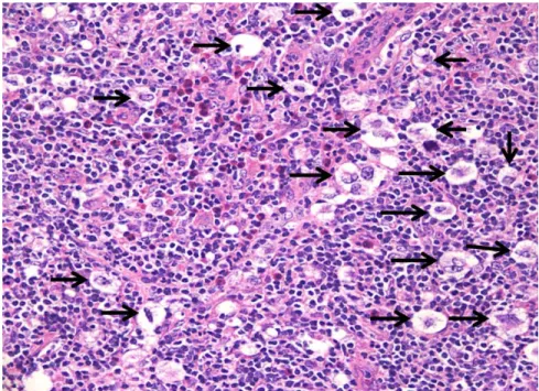
Diagnosis is made by surgical incisional biopsy (Figures 5A & 5B); fine-needle aspiration biopsy may not be adequate for diagnosis of lymphoma in children. Lymph node biopsies should be sent for routine histology, immunohistochemistry, molecular/cytogenetic analysis, and immunophenotyping by flow cytometry.
Figure 5a. Incisional Lymph Node Biopsy of Neck shows effacement of lymph node with fibrous bands [] in a 13 year old male with several month history of neck adenopathy without ‘B’ symptoms, ESR 79, and CXR with mediastinal mass. (Hematoxylin-eosin stain; 2x) University of Massachusetts Medical School.
Figure 5b. Incisional Lymph Node Biopsy of neck shows lacunar cells (Reed-Sternberg cell variant caused by cytoplasmic shrinking during formalin fixation) consistent with a diagnosis of nodular sclerosing HD in same patient. [] (Hematoxylin-eosin stain; 60x) University of Massachusetts Medical School.
Treatment for NHL includes multiagent chemotherapy ± central nervous system prophylaxis based on tumor stage for less than 1 year except for lymphoblastic lymphoma which requires longer maintenance chemotherapy. HD is treated with multiagent chemotherapy for less than 6 months and involved field radiation therapy.
Neuroblastoma
Sympathetic nervous system tumors represent 5.4% of all cancers in children less than 20 years of age with an annual incidence rate of 7 per million in children less than 15 years of age; approximately 95% of these tumors are neuroblastoma and ganglioneuroblastoma.1 Most cases of neuroblastoma are diagnosed before 4 years of age and neuroblastoma
is the most common cancer of infancy and the most common extracranial solid tumor cancer in children.
The 5-year relative survival rate from 1985-1994 for infants with neuroblastoma is 83% compared with 55% for children 1 to 4 years of age and reflects the favorable biological features of neuroblastoma occurring in infants.1
Neuroblastoma is an embryonal malignant neoplasm arising from primitive adrenergic neural crest cells in the adrenal medulla, sympathetic ganglia, and sympathetic paraganglia. Neuroblastoma can arise from any site along the sympathetic nervous system chain, adrenal gland, and organ of Zuckerkandl in the pelvis. Most primary tumors are found in the abdomen (60%), adrenal gland (35%), and retroperitoneum (25%), and thorax in the posterior mediastinum (15%). Histologically, neuroblastoma is one of the “small, round blue cell tumors” of childhood. Amplification of MYCN proto-oncogene occurs in 20% to 25% of neuroblastomas and is a reliable marker of aggressive clinical behavior.7 The presenting signs and symptoms of neuroblastoma depend on the location of the primary tumor, presence of metastatic disease (bone marrow, bone, liver, and skin), and paraneoplastic effects (e.g. opsomyoclonus and intractable secretory diarrhea) seen in a minority of children. Paraspinal thoracic and retroperitoneal tumors may present with spinal cord compression representing anoncologic emergency, cervical tumors may present with Horner syndrome, and orbital bone metastatic disease may present with periorbital ecchymoses (“raccoon eyes”) and proptosis.
Laboratory findings in a child with neuroblastoma include elevation of the urinary catecholamine metabolites vanillylmandelic (HVA) and homovanillic acid (VMA). Serum LDH and ferritin or the complete blood count may show evidence of metastatic bone marrow disease. Plain radiographs may show mass with calcifications (Figure 6).
Figure 6. Plain Abdominal Radiograph showing large right upper quadrant mass with calcifications [] in a 2 year old female with unfavorable histology, N-MYC amplified metastatic neuroblastoma of right adrenal gland presenting with abdominal swelling and fever; random urine sample showed elevated HVA and VMA. University of Massachusetts Medical School.
Initial evaluation and staging of neuroblastoma requires computed tomography or magnetic resonance imaging of the primary tumor and suspected metastatic sites, bone scan, metaiodobenzylguanidine (MIBG) scan, and bilateral bone marrow biopsies/aspirates. Surgical incisional biopsy is usually required to confirm diagnosis and obtain adequate tissue for genetic and molecular biology studies essential for determining therapy.
Neuroblastoma has remarkable clinical heterogeneity and therapy is risk-stratified based on age, tumor stage, MYCN status, histopathologic classification, and other biologic factors that are under study. Low-risk patients are usually treated with surgery alone and high-risk patients are treated with multimodal treatment including dose intensified and high-dose chemotherapy with autologous stem cell support, radiation therapy for bulky residual disease, and biologic therapy with 13-cis retinoic acid.
Osteosarcoma/Ewing Bone Sarcoma
Bone tumors represent 5.6 % of all cancers in children less than 20 years of age with an annual incidence rate of 8.7 per million. Osteosarcoma and Ewing sarcoma (EWS) account for 56% and 34% respectively of malignant bone tumors in children. The incidence of bone cancer is characterized by a steady rise in rate beginning at 11 years of age with a peak incidence at 15 years of age coinciding with the adolescent growth spurt.1
The 5-year survival rates from 1985-1994 for osteosarcoma and Ewing bone sarcoma were 63% and 58% respectively; children with EWS of the pelvis have a poor survival outcome.1
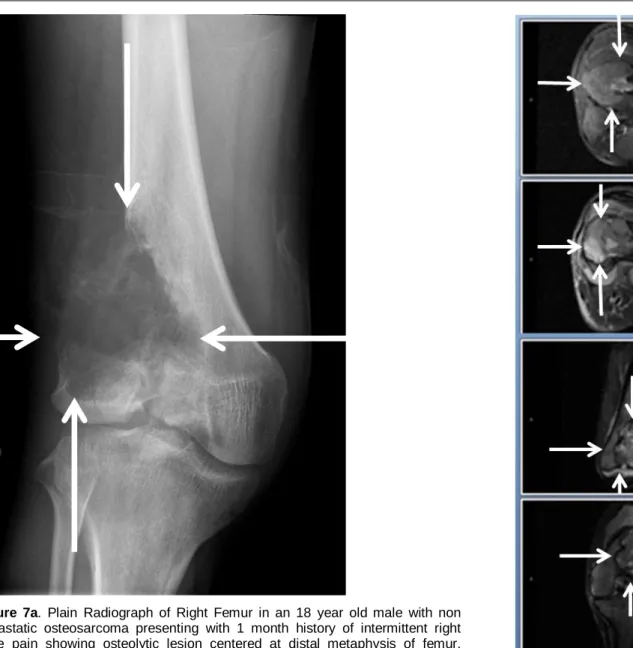
Osteosarcoma arises from primitive bone mesenchymal cells and usually occurs at the metaphyses of tubular bones. The most frequent primary sites are distal femur and proximal tibia. (Figures 7A & 7B) EWS is believed to arise from cells of neural crest origin and usually occurs at the diaphyses of tubular bones and flat bones. The most frequent primary sites are pelvis, femur, tibia, and humerus. (Figure 8) About 95% of patients with EWS have a t(11;22) or t(21;22) genetic translocation involving the EWS gene on chromosome 22. Clinically detectable metastases to other bones and lung (and bone marrow in cases of EWS) are found in about 20 - 25% of patients at time of diagnosis.8
Figure 7a. Plain Radiograph of Right Femur in an 18 year old male with non metastatic osteosarcoma presenting with 1 month history of intermittent right knee pain showing osteolytic lesion centered at distal metaphysis of femur. University of Massachusetts Medical School.
Figure 7b. MRI of Right Femur in same patient showing a large intramedullary lesion. University of Massachusetts Medical School.
Figure 8. MRI of Pelvis evidencing soft tissue mass arising from right ilium in a 10 year old female with metastatic Ewing sarcoma (FISH evaluation positive for rearrangement of EWS gene) presenting with 6 months history of right hip pain, weight loss, fever, and elevated LDH. University of Massachusetts Medical School.
Bone tumors present with pain or swelling in a bone or joint and an associated mass. Laboratory findings may include elevated serum lactate dehydrogenase (LDH) and alkaline phosphatase.
Initial evaluation and staging of osteosarcoma/Ewing bone sarcoma requires anatomic imaging of primary tumor with magnetic resonance imaging (MRI) and plain radiographs. Metastatic sites are evaluated with a chest computed tomography scan (CT) and functional whole body imaging (bone scan or positron emission tomography (PET)). Surgical incisional biopsy is preferred over needle biopsy to confirm diagnosis and obtain tissue for genetic and molecular biology studies.
Osteosarcoma/Ewing bone sarcoma in children are treated with combined-modality therapy with neoadjuvant chemotherapy to decrease the size of primary tumor followed by local control therapy (surgery for osteosarcoma; surgery and/or radiation therapy for EWS) and adjuvant
consolidative chemotherapy after local therapy.
Wilms Tumor
Renal tumors represent 4.4 % of all cancers in children less than 20 years of age with an annual incidence rate of 6.2 per million. Nephroblastoma or Wilms tumor (WT) account for 92% of renal cancer in children. WT is the most common renal cancer in children and approximately 75% of cases are diagnosed before 5 years of age.1
WT is an embryonic neoplasm arising from primitive kidney cells or nephrogenic rests and may occur in a few children as part of a genetic syndrome (e.g. Beckwith-Wiedemann syndrome, WAGR syndrome). Histologic classification of Wilms tumors is based on extent of anaplasia. The classic symptom of WT is a painless abdominal mass often noted by a parent while bathing or dressing their child (Figure 9). WT may present with change in bowel habits, abdominal pain, vomiting, and weight loss. Signs of hypertension, microscopic hematuria, and metastatic disease to the lungs may also be seen at diagnosis.
Figure 9. CT of Abdomen with large mass arising from right kidney in a 2 year old male with Stage 1 WT with favorable histology presenting with painless mass in right upper quadrant. University of Massachusetts Medical School.
Initial evaluation and staging of WT requires computed tomography (CT) or magnetic resonance imaging of the primary tumor, chest CT, and plain radiograph of the chest and sometimes ultrasound of renal vessels to evaluate for tumor thrombus. Unilateral radical nephrectomy and tumor excision is indicated at time of diagnosis unless tumor should be considered unresectable by the surgeon.
WT is treated with multi-agent chemotherapy and radiation therapy to primary and metastatic tumor. Radiation therapy is omitted in patients with WT without anaplastic histology who have complete resection of tumor (Stage 1 and 2) at time of diagnosis.
Rhabdomyosarcoma
Soft tissue sarcomas represent 7.4 % of all cancers in children less than 20 years of age with an annual incidence rate of 11 per million. Rhabdomyosarcoma (RMS) is the most common soft tissue sarcoma and accounts for 40% of these cancers with an incidence of 4.3 per million in children less than 20 years of age.1 The 5-year survival rates from 1985-1994 for RMS was 64%; embryonal RMS has a superior outcome compared to alveolar RMS subtype.1
RMS arises from striated muscle mesenchymal cells and can occur at any site. (Figure 10) The 2 most common histologic subtypes are embryonal RMS and alveolar RMS. Embryonal RMS is the most common subtype in children accounting for 75% of cases. However, the relative percentage of embryonal RMS subtype decreases with increasing age and alveolar RMS occurs more commonly than embryonal RMS at extremity sites. Most cases of alveolar RMS are characterized by recurring genetic translocations involving the FKHR
gene on chromosome 13 while embryonal RMS generally has a loss of heterzygosity at chromosome 11p15.5. Clinically detectable metastases to regional lymph nodes, lungs, bone, and bone marrow are found in about 15 percent of patients at time of diagnosis.8
Figure 10. MRI of Abdomen and Pelvis in an 8 year old male with non-metastatic embryonal rhabdomyosarcoma presenting with 1 week history of left leg pain and limp showing large heterogenous retroperitoneal soft tissue mass in left side of pelvis. University of Massachusetts Medical School.
Soft tissue sarcomas can present with a painless soft-tissue mass. RMS of the orbit may present with proptosis.
Initial evaluation and staging of RMS requires imaging of primary tumor with magnetic resonance imaging or computed tomography and imaging to evaluate for metastatic disease with chest computed tomography and whole body bone scan. Surgical incisional biopsy or total excisional (35% of children have resectable tumors at diagnosis) biopsy, when possible, is preferred to confirm diagnosis and obtain tissue for genetic and molecular biology studies. Surgical sampling of ipsilateral inguinal or axillary lymph nodes is required in patients with RMS of the extremities. RMS is treated with multi-agent chemotherapy and radiation therapy along with surgery to primary and metastatic tumor. RMS tumors that are initially unresectable are treated with neoadjuvant chemotherapy to decrease the size of primary tumor followed by surgical resection (if
feasible), local control radiation therapy, and consolidative chemotherapy after local control therapy.
Summary
Cancer is a rare disease in children; however, it is the leading cause of disease-related mortality among children 1 to 19 years of age. The most common childhood cancers (leukemias and lymphomas, osteosarcoma and Ewing sarcoma, Wilms tumor, and rhabdomyosarcoma), excluding those arising from the brain/central and sympathetic nervous system, originate from mesenchymal cells and, unlike the more common epithelial carcinomas in adults, are associated with spontaneous chromosomal translocations generating fusion oncogenes. Outcomes for children with cancer have improved significantly over the last 25 years as a result of disease risk stratification and multidisciplinary and multimodal approach to disease therapy. Advances in childhood cancer treatment have meant that at least 328,000 of the more than 10 million cancer survivors that are alive in the United States were originally diagnosed with cancer under the age of 21.
However, this success has a price, as cancer treatment in children may cause health problems that may develop years after successful treatment and are called late effects. Because of this, childhood cancer survivors require close, life-long follow-up. The Childhood Cancer Survivor Study (CCSS) was started in 1993 to better understand these late effects. http://ccss.stjude.org/
Thought Questions
1. Which of the following variables is usually least predictive of survival in children with cancer: age at diagnosis, stage of cancer, biologic characteristics of cancer cell, early response to treatment? List 1 or 2 single predominant prognostic factors of cure rate for the following childhood cancers: acute lymphoblastic leukemia, neuroblastoma, and Wilms tumor.
Your answer:
Expert Answer
2. A 3 year old presents with right-sided flank pain and a right abdominal mass. A 63 year old presents with right-sided flank pain and a right abdominal mass. CT scans in each patient show large right renal masses. How are these masses likely to be different from each other? How are the causes of these masses different?
Your answer:
3. Why have survival rates for most childhood cancer improved more than those for most adult cancers?
Your answer:
Expert Answer
4. As children with cancer survive their original tumor and live into adulthood, they may develop new cancers. Some new cancers in survivors of childhood malignancies are clearly related to previous treatment (e.g., breast cancer in women who received chest irradiation in adolescence). However, some new cancers in these patients may be due to an underlying susceptibility to developing cancer. How could you distinguish between these possibilities in a patient?
Your answer:
5. Why do physicians and members of the lay community consider risks of severe late effects acceptable after treatment of childhood
cancer? Your answer:
Expert Answer
Glossary
Adenopathy- Abnormal lymph nodes
Autologous stem cell support- Use of autologous hematopoietic stem cells to rescue patients from profound myelosuppression or myeloablation that has been caused by chemotherapy or radiation Chemotherapy consolidation- Use of chemotherapy in a patient whose tumor is in remission in order to improve the chance of cure
Chemotherapy intensification- Use of increasing doses of chemotherapy Chemotherapy maintenance- Use of ongoing courses of chemotherapy to lower risk of recurrence in a patient whose cancer is in remission Ectodermal embryonal tissue- One of the three primary germ layers in the embryo; differentiates to form the nervous system.
Extramedullary leukemia- Leukemia in sites other than bone marrow, e.g. central nervous system, testes (in boys)
Hematopoesis- Production of blood cells
Hepatosplenomegaly- Enlarged liver and spleen
Induction chemotherapy- Initial chemotherapy that is used to treat a patient’s cancer; so-called because the goal is to “induce” a remission Intrathecal chemotherapy- Chemotherapy delivered directly into the cerebrospinal fluid
Leukemia- Malignancy of hematopoietic cells
Mesenchymal- Refers to cells that develop into connective tissue, blood vessels, and lymphatic tissue
Mesodermal- Embryonal tissue pertaining to or derived from the middle layer of the three primary germ layers of the embryo which derive connective tissue, bone and cartilage, muscle, blood and blood vessels, lymphatics and lymphoid organs, pleura, pericardium, peritoneum, kidney, and gonads
Neoadjuvant chemotherapy- Chemotherapy given prior to surgical resection of tumor
Pancytopenia- Reduced levels of multiple hematopoietic cell lines; as opposed to anemia (reduced red blood cells), leucopenia (low white blood cell levels), or thrombocytopenia (low platelet levels)
Prophylactic treatment- Treatment (prophylaxis) to prevent the development of a disease
WAGR syndrome- Wilms tumor, aniridia, genitourinary abnormalities, and mental retardation/intellectual disability syndrome that affect the development of multiple body systems
References
1. Ries LAG, Smith MA, Gurney JG, Linet M, Tamra T, Young JL, Bunin GR (eds). Cancer Incidence and Survival among Children and Adolescents: United States SEER Program 1975-1995, National Cancer Institute, SEER Program. NIH Pub. No. 99-4649. Bethesda, MD, 1999.
2. Howlader N, Noone AM, Krapcho M, Garshell J, Miller D, Altekruse SF, Kosary CL, Yu M, Ruhl J, Tatalovich Z,Mariotto A, Lewis DR, Chen HS, Feuer EJ, Cronin KA (eds). SEER Cancer Statistics Review, 1975-2012, National Cancer Institute. Bethesda, MD,
http://seer.cancer.gov/csr/1975_2012/, based on November 2014 SEER data submission, posted to the SEER web site, April 2015. 3. Guidelines for the pediatric cancer center and role of such centers in
diagnosis and treatment. American Academy of Pediatrics Section Statement Section on Hematology/Oncology. Pediatrics. 1997; 99(1):139-41.
PubMed Abstract
4. Boissel N, Auclerc MF, Lhéritier V, Perel Y, Thomas X, Leblanc T, Rousselot P, Cayuela JM, Gabert J, Fegueux N, Piguet C, Huguet-Rigal F, Berthou C, Boiron JM, Pautas C, Michel G, Fière D, Leverger G, Dombret H, Baruchelet A. Should adolescents with acute lymphoblastic leukemia be treated as old children or young adults? Comparison of the French FRALLE-93 and LALA-94 trials. J Clin Oncol. 2003; 21(5):774-80.
5. Simone JV, Lyons J. The evolution of cancer care for children and adults. J Clin Oncol. 1998; 16(9):2904-5.
PubMed Abstract
6. Dubansky AS, Boyett JM, Falletta J, Mahoney DH, Land VJ, Pullen J, Buchanan G. Isolated thrombocytopenia in children with acute lymphoblastic leukemia: a rare event in a Pediatric Oncology Group Study. Pediatrics. 1989; 84(6):1068-71.
PubMed Abstract
7. Maris JM, Matthay KK. Molecular biology of neuroblastoma. J Clin Oncol. 1999; 17(7):2264-79.
PubMed Abstract
8. Arndt CA, Crist WM. Common musculoskeletal tumors of childhood and adolescence. N Engl J Med. 1999; 341(5):342-52.
PubMed Abstract
9. Mariotto AB, Rowland JH, Yabroff KR, et al. Long-term survivors of childhood cancers in the United States. Cancer Epidemiol Biomarkers Prev. 2009; 18(4):1033-40.


![Figure 5a. Incisional Lymph Node Biopsy of Neck shows effacement of lymph node with fibrous bands [] in a 13 year old male with several month history of neck adenopathy without ‘B’ symptoms, ESR 79, and CXR with mediastinal mass](https://thumb-us.123doks.com/thumbv2/123dok_us/442322.2551172/7.1188.54.529.94.578/figure-incisional-biopsy-effacement-fibrous-adenopathy-symptoms-mediastinal.webp)
![Figure 6. Plain Abdominal Radiograph showing large right upper quadrant mass with calcifications [] in a 2 year old female with unfavorable histology, N-MYC amplified metastatic neuroblastoma of right adrenal gland presenting with abdominal swelling an](https://thumb-us.123doks.com/thumbv2/123dok_us/442322.2551172/9.1188.66.1053.92.802/abdominal-radiograph-calcifications-unfavorable-histology-metastatic-neuroblastoma-presenting.webp)