Different in Older Adults?
Pradeep Suri, MD,
wz§David J. Hunter, MBBS, PhD,
wkCristin Jouve, MD,
wCarol Hartigan, MD,
wJanet Limke, MD,
wEnrique Pena, MD,
wLing Li, MPH,
wJennifer Luz, BA,
#and James Rainville, MD
wOBJECTIVES: To determine whether older adults (aged
60) experience less improvement in disability and pain with nonsurgical treatment of lumbar disk herniation (LDH) than younger adults (o60).
DESIGN: Prospective longitudinal comparative cohort study.
SETTING: Outpatient specialty spine clinic.
PARTICIPANTS: One hundred thirty-three consecutive patients with radicular pain and magnetic resonance–con-firmed acute LDH (89 younger, 44 older).
INTERVENTION: Nonsurgical treatment customized for the individual patient.
MEASUREMENTS: Patient-reported disability on the Oswestry Disability Index (ODI), leg pain intensity, and back pain intensity were recorded at baseline and 1, 3, and 6 months. The primary outcome was the ODI change score at 6 months. Secondary longitudinal analyses examined rates of change over the follow-up period.
RESULTS: Older adults demonstrated improvements in ODI (range 0–100) and pain intensity (range 0–10) with nonsurgical treatment that were not significantly different from those seen in younger adults at 6 month follow-up, with or without adjustment for potential confounders. Ad-justed mean improvement in older and younger adults were 31 versus 33 (P5.63) for ODI, 4.5 versus 4.5 (P5.99) for leg pain, and 2.4 versus 2.7 for back pain (P5.69). A greater amount of the total improvement in leg pain and back pain in older adults was noted in the first month of follow-up than in younger adults.
CONCLUSION: These preliminary findings suggest that the outcomes of LDH with nonsurgical treatment were not
worse in older adults (60) than in younger adults (o60). Future research is warranted to examine nonsurgical treat-ment for LDH in older adults.J Am Geriatr Soc 59:423– 429, 2011.
Key words: herniation; intervertebral disk displacement; geriatrics; outcomes
L
ower extremity pain in the setting of low back pain affects 12% of older men in the community-based pop-ulation1and 21% of older adults in retirement communi-ties.2Lumbar disk herniation (LDH) is a common cause of these symptoms and most typically manifests as a lumbo-sacral radicular syndrome: a combination of one or more of radicular pain, paresthesia, sensory changes, motor weak-ness, and impaired reflexes in the distribution of one or more lumbosacral spinal nerve roots in the lower extremity.3,4A classical dichotomy has been prominent in spine care whereby LDH is considered a clinical entity common mainly to younger adults, with a shift to a predominance of degen-erative lumbar spinal stenosis (LSS) in older adults.5 The view that LDH is rare in older adults is echoed in scientific reports and textbooks of spine care,6–9 but other reports caution that LDH in older adults is more common than pre-viously believed.10–12The prevalence of LDH in older adults is of particular importance because the outcomes with non-surgical treatment of LDH are favorable in the majority of individuals,13whereas dramatic improvements in LSS with nonsurgical treatment are seen less commonly.12Decompressive lumbar spinal surgery for LDH and LSS typically involves removal of a portion of the intervertebral disk (diskectomy), removal of the spinal lamina (laminec-tomy or lamino(laminec-tomy), or both. Rates of laminec(laminec-tomy and diskectomy in the Medicare population have shown steady increases over recent decades and exceed the rates of growth in younger populations.14Higher rates of increase in surgical procedures in older adults may be due to the increasing recognition that spine surgery in older adults can be performed safely in properly selected patients.11,15
Address correspondence to Pradeep Suri, Division of Research, New England Baptist Hospital, 125 Parker Hill Ave, Boston, MA 02130. E-mail: psuri@caregroup.harvard.edu
DOI: 10.1111/j.1532-5415.2011.03316.x
From theDepartment of Physical Medicine and Rehabilitation, Harvard Medical School, Boston, Massachusetts;wNew England Baptist Hospital, Boston, Massachusetts;zSpaulding Rehabilitation Hospital, Boston,
Massachusetts;§Veterans Affairs Boston Healthcare System, Boston,
Massachusetts;kNorthern Clinical School, University of Sydney, Sydney,
New South Wales, Australia; and#Tufts University School of Medicine,
Boston, Massachusetts.
JAGS 59:423–429, 2011 r2011, Copyright the Authors
Another explanation for increasing rates of surgical proce-dures may be related to the perception of clinicians that the outcomes of nonsurgical treatment of LDH are poorer in older than younger adults. Various reports in the surgical literature have suggested poor outcomes with nonsurgically treated LDH in older adults.16–18In a landmark study of LDH by Henrik Weber, older age was the only character-istic associated with a poor outcome at multiple follow-up time points.19Poor outcomes in older adults may be due to age-related histological and inflammatory changes in the lumbar intervertebral disk.8,9,20–22Furthermore, concom-itant age-related degenerative changes, such as a decrease in reserve spinal canal space due to osteoarthritic joint hyper-trophy, may impede the natural history of improvement typically seen in younger adults, although no prior study has examined the outcomes of LDH with nonsurgical treat-ment in older adults.
The current study was a prospective cohort study com-paring the outcomes of the nonsurgical treatment of LDH in adults aged 60 and older with those of adults younger than 60. The objective was to determine whether older adults experienced less improvement in back-related disability and pain over a 6-month follow-up period than younger adults. It was hypothesized that the outcomes of nonsurgical treat-ment of LDH in older adults would be poorer than the outcomes of treatment in younger adults. To characterize rates of recovery over time, longitudinal outcome data at multiple time points were used to conduct secondary an-alyses examining possible differences between older and younger adults.
METHODS Study Participants
Participants were recruited from a hospital-based outpa-tient spine center between January 2008 and March 2009. The institutional review board of New England Baptist Hospital approved the study. All consecutive patients aged 18 and older with lumbosacral radicular pain for less than 12 weeks were evaluated for participation. All patients un-derwent a standardized history and physical examination. Inclusion criteria were recent onset radicular pain (o12 weeks) in an L2, L3, L4, L5, or S1 dermatome, with or without neurological changes, and available magnetic res-onance imaging (MRI) demonstrating LDH corresponding to the neurological level and side suggested by the clinical presentation. Exclusion criteria were known pregnancy; severe active medical or psychiatric comorbidities that would limit study participation; the presence of significant central canal or neuroforaminal stenosis from reasons other than LDH as the likely cause of radicular pain; infectious, inflammatory, or neoplastic cause of radiculopathy; signifi-cant degenerative or isthmic spondylolisthesis suspected of contributing to symptoms; and prior lumbar spine surgery at the affected level. Some patients met clinical criteria for study participation but had not yet undergone MRI to con-firm whether LDH was present at the baseline evaluation. These patients were offered informed consent at the base-line evaluation for practical reasons but did not formally enter the study unless their subsequent MRI met study cri-teria. Subjects who received surgical treatment during the 6-month follow-up period were excluded from analysis.
Participant Demographics and Historical Features Information on participant age, sex, race, comorbidity, du-ration of symptoms, prior history of low back pain, prior lumbar spine surgery, tobacco use, employment status, and worker’s compensation status was prospectively collected. Race was categorized as white and nonwhite. Medical and psychiatric comorbidity burden was measured using the Self-Administered Comorbidity Questionnaire, which is widely used in orthopedic research and has previously demonstrated reliability and validity. Employment status was categorized as part-time employment, full-time em-ployment, student, retired, disabled, and unemployed. Physical Examination Characteristics
Each participant received a comprehensive physical exam-ination for the evaluation of lumbar radiculopathy from one of six board-certified physiatrists specializing in spine care. Specific physical examination tests received emphasis in this analysis because of their common usage or because deficits on these tests were felt to have greater clinical or functional importance. The provocative tests of the straight leg raise and the femoral stretch test are used commonly for the diagnosis of LDH. These tests elicit symptoms of neural tension affecting the low lumbar and midlumbar nerves roots, respectively, and have been well described previ-ously.23 Motor strength testing included functional tests designed to use the patients’ own body weight as the mea-sure of resistance, to provide a physical challenge sufficient to detect subtle losses in strength, and to facilitate repro-ducibility.24 Knee extension strength was measured using the single leg sit-to-stand test, and ankle plantarflexion strength was measured using the heel-raise test.24,25 Any deficits were further characterized using manual mus-cle testing.26
MRI Characteristics
MRI scans consisted at minimum of T1- and T2-weighted images of the lumbar spine in the sagittal and axial planes. For the purposes of this study, the recruiting physician, who in most cases also had access to the official report of the interpreting neuroradiologist or direct consultation with the neuroradiologist, evaluated features of LDH and nerve root impingement. The recruiting physician recorded her-niation level, nerve root impingement level, herher-niation morphology, and herniation location. Herniation level was classified as midlumbar (L1–L2, L2–L3, or L3–L4) or low lumbar (L4–L5 or L5–S1) disk herniation. Herniation mor-phology was classified as protrusion, extrusion, or seques-tration.27 Herniation location was classified as central (central, paracentral, or subarticular or lateral recess loca-tion) or foraminal (foraminal or extraforaminal localoca-tion).27 Outcomes
Patient-reported disability and pain intensity were recorded at the baseline clinic visit. The primary outcome of this study was patient-reported change in functional limitations and disability at 6-month follow-up, as measured according to the Oswestry Disability Index (ODI). The ODI is a condition-specific measure of disability that has been used extensively in prior studies of low back pain and radicu-lopathy and has demonstrated validity and reliability in
these contexts.28 ODI scores range from 0 to 100, with higher scores indicating greater disability The secondary outcomes of this study were change in leg pain and back pain at 6-month follow-up, as measured on a 0 to 10 visual analogue scale (VAS).29 Follow-up information was ob-tained using a mailed questionnaire at 1, 3, and 6 months. Each questionnaire consisted of the ODI, VAS for leg pain, VAS for back pain, and questions regarding nonsurgical and surgical treatments received.
Statistical Analysis
To characterize the study population at baseline, means and standard deviations were calculated for continuous vari-ables, medians and interquartile ranges (IQRs) for ordinal variables, and frequencies and proportions for categorical variables. The chi-square test was used for categorical vari-ables and the Studentt-test or Wilcoxon signed rank test for continuous variables to compare the baseline characteristics of older adults with those of younger adults. Because of small numbers in individual cells, the employment status categories of unemployed and student were combined as one category. Herniation morphology was dichotomized as protrusion versus extrusion or sequestration because of small numbers of individuals with sequestered disks. Bivar-iate associations between age group and 6-month change scores for ODI, VAS leg pain, and VAS back pain were then examined. Statistical significance was determined using a threshold ofP5.05. For associations that demonstrated at least a trend toward statistical significance (P.15) in the bivariate analyses, multivariate regression models were cre-ated including as covariates baseline characteristics that demonstrated a statistical trend toward between–age group differences (P.15) or were felt to have a conceptual basis for explaining the observed differences. The method of last value carried forward was used to account for missing out-come data. To examine whether age was related to outout-come when treated as a continuous variable, the multivariate an-alyses were repeated replacing age group with age in years. Last, given the absence of any prior literature on differences in rates of recovery from LDH according to age, secondary longitudinal analyses of outcomes were conducted accord-ing to age group at baseline and 1, 3, and 6 months, adjusting for covariates, using generalized estimating equations. All analyses were performed using SAS soft-ware, version 9.0 (SAS Institute, Inc., Cary, NC).
RESULTS
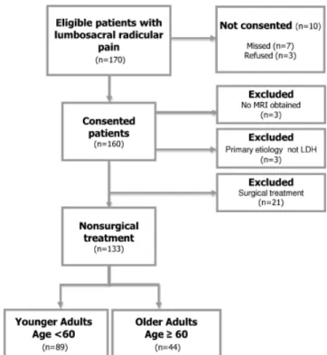
Figure 1 presents a flowchart of study recruitment. One hundred seventy patients were eligible to participate in this observational study. Seven of these were not offered in-formed consent because of failure on the part of the re-cruiting physicians, and an additional three patients refused to participate. Three of 160 consented patients experienced clinical improvement and did not undergo MRI, and three were excluded for having nerve root impingement due pri-marily to causes other than LDH. One hundred fifty-four subjects met initial criteria, including 106 in the younger group (o60) and 48 in the older group (60). Twenty-one subjects received lumbar decompression surgery and were excluded from this analysis of nonsurgical outcomes. Indi-viduals who underwent surgery were younger (48.413.2)
than those who did not (53.613.5; P5.05), were less likely to be retired, and were more likely to be disabled or unemployed. There were otherwise no demographic or clinical factors significantly associated with surgical treat-ment over the follow-up period (data not shown).
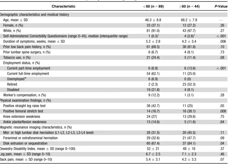
The mean age of the study sample was 53.613.5 (range for older adults 60–87; 60%, 60–69; 27% 70–79, 13% 80), 33% of participants were women, and 93.8% were white. People who were eligible to participate but were missed or refused were not materially different from study participants with respect to demographic features. Baseline characteristics of the study sample according to age group are presented in Table 1. Older adults had a higher comorbidity burden (median (IQR) 4 (2–6) vs 1 (0–3); P5o.001) and a shorter duration of symptoms (4.23.4 vs 5.22.8;P5.006) at clinical presentation than younger adults. Employment status was significantly different in older adults (Po.001). Some physical examination and MRI characteristics differed according to age group. A positive straight leg raise test was significantly less common in older adults, and conversely, a positive femoral stretch test was significantly more common in older adults. Mid-lumbar disk herniations and foraminal disk herniations were more common in older adults. Baseline ODI scores and VAS leg pain were comparable in younger and older adults, but baseline VAS back pain was slightly lower in older than younger adults (4.2 vs 5.4;P5.07).
Associations between age group and outcomes of non-surgical treatment at 6-month follow-up are presented in Table 2. Proportions of missing data for the outcomes of change in ODI, leg pain, and back pain were 8%, 4%, and 4%, respectively. There were no statistically significant bi-variate associations between age group and the primary outcome of ODI change score at 6 months. In multivariate analysis including the covariates of sex, race, employment status, prior lower back pain, tobacco history, comorbidity Figure 1. Flowchart of recruitment.
(Self-Administered Comorbidity Questionnaire), duration of symptoms, baseline ODI, herniation level, herniation location, and herniation morphology, the association be-tween age group and ODI remained nonsignificant. Up to 3% of data were missing for some covariates. Age group was not significantly associated with the secondary out-come of leg pain in bivariate analyses. In multivariate anal-ysis including all covariates used in the full model for ODI described above (with adjustment for baseline leg pain), age group continued not to be associated with leg pain change scores. Older adults showed significantly less improvement
in back pain than younger adults (2.04.1 vs 3.23.1; P5.04) in bivariate analysis, although older adults had re-ported less back pain at baseline than younger adults (Table 1). In multivariate analysis including all covariates used in the full models described above (with adjustment for base-line back pain), adjusted back pain improvement was not significantly different in older and younger adults (2.4 vs 2.7; P5.69). When the outcomes of change in ODI, leg pain, and back pain at 6 months were expressed instead as percentage change from baseline, age group was not sig-nificantly associated with any outcome in bivariate or Table 1. Baseline Characteristics of the Study Sample According to Age
Characteristic o60 (n589) 60 (n544) P-Value
Demographic characteristics and medical history
Age, meanSD 46.38.8 68.27.9 F
Female, n (%) 33 (37.1) 12 (27.2) .26
White, n (%) 81 (91.0) 43 (97.7) .27
Self-Administered Comorbidity Questionnaire (range 0–45), median (interquartile range) 1 (0.3)w 4 (2.6)w o.001
Duration of symptoms, weeks, meanSD 5.22.8 4.23.4 .006
Prior low back pain history, n (%) 61 (68.5) 36 (81.8) .10
Prior lumbar spine surgery, n (%) 6 (6.7) 4 (9.1) .73
Tobacco use, n (%) 21 (24.4) 5 (11.4) .08
Employment status, n (%)
Current part-time employment 6 (6.9) 6 (13.6) o.001
Current full-time employment 54 (62.1) 11 (25.0)
Unemployed 6 (6.9) 0 (0)
Retired 2 (2.3) 23 (52.3)
Disabled 19 (21.8) 4 (9.1)
Worker’s compensation, n (%) 9 (12.2) 1 (3.1) .28
Physical examination findings, n (%)
Positive straight leg raise test 38 (42.7) 11 (25) .05
Positive femoral stretch test 14 (16.7) 16 (38.1) .008
Knee extension weakness 24 (27) 13 (29.6) .75
Ankle plantarflexion weakness 13 (14.6) 5 (11.6) .64
Magnetic resonance imaging characteristics, n (%)
Mid- or high lumbar disk herniation (L1-L2, L2-L3, L3-L4 level) 28 (31.5) 20 (45.5) .11
Foraminal or extraforaminal herniation 29 (32.6) 21 (47.7) .09
Disk extrusion or sequestration 60 (67.4) 37 (84.1) .04
Oswestry Disability Index, meanSD (range 0–100) 5221 4818 .37
Leg pain, meanSD (range 0–10) 6.72.5 7.12.3 .46
Back pain, meanSD (range 0–10) 5.43.1 4.23.3 .07
Includes unemployed and student.
SD5standard deviation.
Table 2. Associations Between Age Group and Outcomes (Change Scores) at 6-Month Follow-Up
Outcome
Crude Associations Multivariate-Adjusted Associations
o60 60 P-Value o60 60 P-Value
Primary outcome: Oswestry Disability Index (range 0–100)
3722 3425 .63 33 31 .63
Secondary outcome
Leg pain (range 0–10) 5.03.0 5.23.0 .73 4.5 4.5 .99
Back pain (range 0–10) 3.23.1 2.04.1 .04 2.7 2.4 .69
Adjusted for sex, race, work status, prior lower back pain, tobacco history, comorbidities, duration of symptoms, baseline severity, midlumbar disk herniation,
multivariate analyses (data not shown). When age was treated instead as a continuous variable in a secondary analysis, age remained not significantly associated with change in ODI, leg pain, or back pain (data not shown).
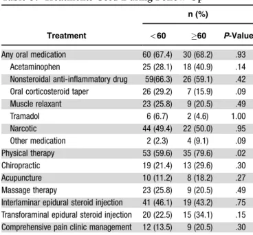
Table 3 describes treatments used by study participants over the 6-month follow-up period. Older adults used oral corticosteroid tapers less frequently than younger adults (16% vs 29%; P5.09) and physical therapy (79.6% vs 59.6%;P5.02) and transforaminal epidural steroid injec-tions (37.5% vs 22.6%; P5.06) more frequently. To ac-count for the influence of treatments received, secondary analyses were conducted of the associations between age group and 6-month change scores for disability and pain, adjusting for the use of oral corticosteroids, physical ther-apy, transforaminal epidural steroid injections, and baseline covariates. Accounting for these treatments did not mate-rially change the findings. No single treatment was signifi-cantly associated with outcomes for disability and pain (data not shown).
In longitudinal analyses, the outcomes of disability and pain were examined according to age group at 1, 3, and 6 months, adjusting for demographic and historical features that were significant in the primary analyses: comorbidity score, duration of symptoms, and work status. Specific her-niation characteristics were not adjusted for, because it was not desired to ‘‘adjust out’’ age-related anatomic factors. Figure 2 depicts outcome scores over time, adjusted for co-morbidity, duration of symptoms, and work status. Both groups demonstrated the largest improvements in ODI and pain scores over the first month of follow-up, with a slower rate of improvement thereafter. In longitudinal analyses, no interaction between age group and time was found for dis-ability on the ODI, indicating no differences in rates of improvement between older and younger individuals, al-though significant interactions between age group and time were noted for the outcomes of leg pain (P5.02) and back pain (P5.04). The meaning of this interaction can be easily appreciated using simple visual inspection of longitudinal trends for adjusted pain scores in Figure 2. The figure
dem-onstrates that although trajectories of improvement be-tween age groups were similar for the outcome of ODI, a greater amount of the total improvement in leg pain and back pain intensity in older adults was noted than in younger adults in the first month of follow-up.
DISCUSSION
The primary finding of this study was that older adults demonstrated improvements in disability and pain with nonsurgical treatment that were not significantly differ-ent from those seen in younger adults over 6 months of follow-up, with or without adjustment for potential con-founders. No prior studies of nonsurgical treatment of Table 3. Treatments Used During Follow-Up
Treatment
n (%)
o60 60 P-Value
Any oral medication 60 (67.4) 30 (68.2) .93 Acetaminophen 25 (28.1) 18 (40.9) .14 Nonsteroidal anti-inflammatory drug 59(66.3) 26 (59.1) .42 Oral corticosteroid taper 26 (29.2) 7 (15.9) .09 Muscle relaxant 23 (25.8) 9 (20.5) .49 Tramadol 6 (6.7) 2 (4.6) 1.00 Narcotic 44 (49.4) 22 (50.0) .95 Other medication 2 (2.3) 4 (9.1) .09 Physical therapy 53 (59.6) 35 (79.6) .02 Chiropractic 19 (21.4) 13 (29.6) .30 Acupuncture 10 (11.2) 8 (18.2) .27 Massage therapy 23 (25.8) 9 (20.5) .49 Interlaminar epidural steroid injection 41 (46.1) 19 (43.2) .75 Transforaminal epidural steroid injection 20 (22.5) 15 (34.1) .15 Comprehensive pain clinic management 12 (13.5) 9 (20.5) .30
Figure 2. Longitudinal outcomes adjusted for comorbidity bur-den, duration of symptoms, and work status.
MRI-confirmed acute LDH have used repeated assessment with validated outcome measures at fixed intervals.30–32 This has left a notable gap in the knowledge base regard-ing the course of improvement early in nonsurgically treated LDH. A secondary finding of this study was that, although rates of improvement in disability were not significantly different in older and younger adults, a greater amount of the total improvement in pain intensity occurred in the first month of follow-up in older adults. Neverthe-less, this difference with respect to rate of improvement in pain intensity was small and not likely to be clinically significant.
Although some authors have asserted that the out-comes of LDH with nonsurgical treatment are poor in older adults,16–18only one prospective study including nonsurgi-cally treated patients was found that has reported a negative influence of age on outcomes with LDH. In Weber’s ran-domized comparison of surgical and nonsurgical treatment of LDH, older age was found to be correlated with poor long-term outcome.19 The findings of the current study contradict that observation. The lack of concordance be-tween these and the previous findings may be because the earlier study, performed during a time when LDH was gen-erally considered to be a problem exclusively of younger adults, did not include adults aged 60 and older. To the contrary, the current study demonstrates that, in a nonsur-gical specialty spine clinic, the diagnosis of LDH is common in older adults, corroborating prior reports from surgical clinics. Almost one-third of all adult disk herniations pre-senting to this nonsurgical clinic during the study period were seen in adults aged 60 and older. The overly simplistic paradigm of LDH as a disorder primarily of younger adults and LSS as a disorder primarily of older adults may lead to misdiagnosis if relied upon heavily.
This study has limitations. First, the small sample size of this study may have limited statistical power to detect differences between the two age groups with respect to the study outcomes. For this reason, it was not possible to ex-amine whether different age cut points (o60) were associ-ated with clinical outcomes. Second, the findings may not be generalizable to older adults with severe bony stenosis in addition to stenosis secondary to LDH, because these adults may have been excluded because of the predefined study criteria. Despite this, many subjects in this sample had moderate bony stenosis at one or more levels. Third, this study does not allow assessment of whether differences in outcomes affecting older adults may have emerged after longer than 6 months of follow-up. This is highly unlikely, in light of multiple studies that document that the vast ma-jority of improvement in LDH occurs over the first 6 months of recovery.3,33Fourth, the influence of some im-portant psychological factors such as treatment expectancy, coping, self-efficacy, and fear avoidance beliefs were not examined in this study. Future studies may wish to examine the effects of these factors in older adults with LDH. Last, formal testing for cognitive impairments using a mental status examination was not performed on all subjects. Al-though subjects with severe medical or psychiatric comor-bidities that would limit study participation were excluded according to the study criteria, the unintentional inclusion of subjects with cognitive impairments is possible and could have affected the findings.
It is likely that many different factors contribute to the well-documented increase in the use of spinal decompres-sion procedures for older adults over recent decades. The current study offers no evidence to support the notion that outcomes of LDH with nonsurgical treatment are different in older and younger adults. Other explanations for in-creasing rates of spine surgery in older adults include an increasing prevalence of spinal disorders in the community, surgical advancements in patient selection and technique allowing safer procedures for older adults, and a lack of consensus on indications for surgery. Further research is warranted to investigate the reasons behind higher surgical rates and to determine whether this translates into better patient outcomes at a reasonable cost to society.
ACKNOWLEDGMENTS
The authors wish to thank the study participants for their time and effort.
Conflict of Interest:The authors declare that they have no potential financial and personal conflict of interests in the publication of this study.
Dr. Suri is funded by the Rehabilitation Medicine Sci-entist Training Program and the National Institutes of Health (K12 HD001097-12).
Author Contributions:Dr. Suri: study concept and de-sign, acquisition of data, analysis of data, interpretation of data, and drafting of the manuscript. Dr. Hunter: study concept and design, analysis of data, interpretation of data, and manuscript preparation. Drs. Jouve, Hartigan, Limke, and Pena and Ms. Luz: acquisition of data and manuscript preparation. Ms. Li: analysis of data, interpretation of data, and manuscript preparation. Dr. Rainville: study concept and design, acquisition of data, interpretation of data, and manuscript preparation. All authors were involved with critical revision of the manuscript for important intellectual content and approved the final version.
Sponsor’s Role:None. REFERENCES
1. Vogt MT, Cawthon PM, Kang JD et al. Prevalence of symptoms of cervical and lumbar stenosis among participants in the Osteoporotic Fractures in Men Study. Spine 2006;31:1445–1451.
2. Hicks GE, Gaines JM, Shardell M et al. Associations of back and leg pain with health status and functional capacity of older adults: Findings from the Re-tirement Community Back Pain Study. Arthritis Rheum 2008;59:1306–1313. 3. Peul WC, van Houwelingen HC, van den Hout WB et al. Surgery versus prolonged conservative treatment for sciatica. N Engl J Med 2007;356: 2245–2256.
4. Peul WC, van Houwelingen HC, van der Hout WB et al. Prolonged conser-vative treatment or ‘early’ surgery in sciatica caused by a lumbar disc herniat-ion: Rationale and design of a randomized trial [ISRCT 26872154]. BMC Musculoskelet Disord 2005;6:8.
5. Jonsson B, Stromqvist B. Influence of age on symptoms and signs in lumbar disc herniation. Eur Spine J 1995;4:202–205.
6. Bono CM. Lumbar disc herniations. In: Herkowitz HN, Garfin SR, Eismont FJ et al., editors. The Spine. Philadelphia: Elsevier, Inc., 2006, p 986. 7. Maistrelli GL, Vaughan PA, Evans DC et al. Lumbar disc herniation in the
elderly. Spine 1987;12:63–66.
8. Yasuma T, Koh S, Okamura T et al. Histological changes in aging lumbar intervertebral discs. Their role in protrusions and prolapses. J Bone Joint Surg 1990;72:220–229.
9. St.John TA, Handling MA, Daffner SD et al. The aging lumbar spine: The pain generator. In: Vaccaro A, Betz RR, Zeidman SM, editors. Principles and Prac-tice of Spine Surgery. Philadelphia, PA: Mosby, 2002, pp 83–95.
10. Akagi S, Saito T, Kato I et al. Clinical and pathologic characteristics of lumbar disk herniation in the elderly. Orthopedics 2000;23:445–448.
11. An HS, Vaccaro A, Simeone FA et al. Herniated lumbar disc in patients over the age of fifty. J Spinal Disord 1990;3:143–146.
12. Katz JN, Harris MB. Clinical practice. Lumbar spinal stenosis. N Engl J Med 2008;358:818–825.
13. McCulloch JA. Focus issue on lumbar disc herniation: Macro- and micro-discectomy. Spine (Phila Pa 1976) 1996;21:45S–56S.
14. Deyo RA, Mirza SK. Trends and variations in the use of spine surgery. Clin Orthop Relat Res 2006;443:139–146.
15. Best NM, Sasso RC. Outpatient lumbar spine decompression in 233 patients 65 years of age or older. Spine 2007;32:1135–1139; discussion 40. 16. Fujii K, Henmi T, Kanematsu Y et al. Surgical treatment of lumbar disc
her-niation in elderly patients. J Bone Joint Surg Br 2003;85:1146–1150. 17. Gembun Y, Nakayama Y, Shirai Y et al. Surgical results of lumbar disc
her-niation in the elderly. J Nippon Med Sch5Nihon Ika Daigaku zasshi 2001; 68:50–53.
18. Kulali A, von Wild K. Lumbar spinal surgery for sciatica due to intervertebral disc disease in the elderly. Neurosurg Rev 1996;19:157–162.
19. Weber H. Lumbar disc herniation. A controlled, prospective study with ten years of observation. Spine 1983;8:131–140.
20. Hasegawa T, An HS, Inufusa A et al. Compositional influences for regression of the sequestered lumbar disc hernia in dogs. Neuro-Orthop 1998;22:69–75. 21. Hasegawa T, An HS, Inufusa A et al. The effect of age on inflammatory re-sponses and nerve root injuries after lumbar disc herniation: An experimental study in a canine model. Spine 2000;25:937–940.
22. Katsuno R, Hasegawa T, Iwashina T et al. Age-related effects of cocultured rat nucleus pulposus cells and macrophages on nitric oxide production and cyto-kine imbalance. Spine (Phila Pa 1976) 2008;33:845–849.
23. Deyo RA, Rainville J, Kent DL. What can the history and physical examin-ation tell us about low back pain? JAMA 1992;268:760–765.
24. Suri P, Rainville J, Katz JN et al. The accuracy of the physical examination for the diagnosis of midlumbar and low lumbar nerve root impingement. Spine (Phila Pa 1976) 2010;36:63–73.
25. Rainville J, Jouve C, Finno M et al. Comparison of four tests of quadriceps strength in L3 or L4 radiculopathies. Spine 2003;28:2466–2471.
26. Brain Introduction. In: O’Brien MD, editor. Aids to the Examination of the Peripheral Nervous System, 4th Ed. London: WB Saunders for The Guarantors of Brain, 2000, pp 1–2.
27. Fardon DF. Nomenclature and classification of lumbar disc pathology. Spine 2001;26:461–462.
28. Fairbank JC, Pynsent PB. The Oswestry Disability Index. Spine 2000;25:2940– 2952; discussion 52.
29. Collins SL, Moore RA, McQuay HJ. The Visual Analogue Pain Intensity Scale: What is moderate pain in millimetres? Pain 1997;72:95–97.
30. Bush K, Cowan N, Katz DE et al. The natural history of sciatica associated with disc pathology. A prospective study with clinical and independent radio-logic follow-up. Spine 1992;17:1205–1212.
31. Saal JA, Saal JS. Nonoperative treatment of herniated lumbar intervertebral disc with radiculopathy. An outcome study. Spine 1989;14:431–437. 32. Weber H, Holme I, Amlie E. The natural course of acute sciatica with nerve
root symptoms in a double-blind placebo-controlled trial evaluating the effect of piroxicam. Spine 1993;18:1433–1438.
33. Weinstein JN, Lurie JD, Tosteson TD et al. Surgical vs nonoperative treatment for lumbar disk herniation: The Spine Patient Outcomes Research Trial (SPORT) observational cohort. JAMA 2006;296:2451–2459.