KELES KEYLESS EXPANDER: A NEW
APPROACH FOR RAPID PALATAL EXPANSION
Maxillary transverse constriction is among the most common malfor-mations in orthodontics. Treatment usually requires rapid palatal expansion with banded or bonded expanders after eruption of the maxillary first molars. The history of palatal expansion goes back nearly 150 years and has retained its popularity ever since. However, patient/guardian compliance in activating the screw with a key is required. Because it is difficult to see the hole on the screw, it is hard to insert the key through the hole. Furthermore, the chance of injur-ing the palatal mucosa with the pointed wire key and the risk of swal-lowing or aspirating the key are common undesired outcomes, and both occur in daily orthodontic practice. To overcome these unwanted incidences and make palatal exapanders more patient-friendly, a new design of palatal expander has to be developed for a safe, rapid, and effective expansion with minimum patient coopera-tion. A newly developed keyless expander has a built-in activation arm, which patients can activate themselves. In all 4 treated cases with a keyless expander, expansion was effectively achieved in a short period of time. World J Orthod 2008;9:407–411.Ahmet Keles, DDS, DMSc1
R
apid palatal expansion (RPE) dates to 1860 when Dr Angell placed a screw appliance between the maxillary premolars of a 14.5-year-old girl and widened her arch a quarter of an inch in 2 weeks.1 The patient was provided akey and instructed to keep the shaft as uniformly tight as possible. At the end of 2 weeks, the jaw was widened enough to leave a space between the 2 central incisors. This finding cannot be sup-ported with radiographs, as X-rays had yet to be discovered. The mechanism of action of RPE was clarified in 1950s studies on cats, pigs, and monkeys.2–4
These studies showed that a midpalatal
suture was opened using this tech-nique’s application of intermittent force.
These studies were later supported by Biederman,5 Brossman et al,6Chaconas
and Caputo,7 Tanne et al,8 Melsen,9,10
and Murray and Cleall11 who performed
studies on dry skulls and human cadav-ers that indicated that age is an impor-tant factor when considering the effect of RPE on craniofacial structures.
Within the past 10 years, 2 types of RPE appliances have been used in orthodontic practice: bonded-type acrylic cap splint RPE and the banded-type (banding the first molars and premolars) RPE appliance. In both cases, a key is required to activate
1Associate Professor, Department of
Developmental Biology, Advanced Orthodontic Graduate Program, Har-vard School of Dental Medicine, Boston, Massachusetts, USA.
CORRESPONDENCE
Dr Ahmet Keles
Department of Developmental Biology Advanced Orthodontic Graduate Program
Harvard School of Dental Medicine 188 Longwood Avenue
Boston, MA 02115 USA
E-mail: [email protected]
the appliance. The key has either a plastic handle to ease the activation process or a short wire key with a piece of dental floss attached to prevent the patient swallowing or aspirating it.12 Nevertheless, accidental swallowing of the key is still
reported,13,14which, among other things, creates liability
issues. There is a desperate need for a safe, patient-friendly expander to reduce treatment time and increase treatment efficiency.
APPLIANCE DESIGN
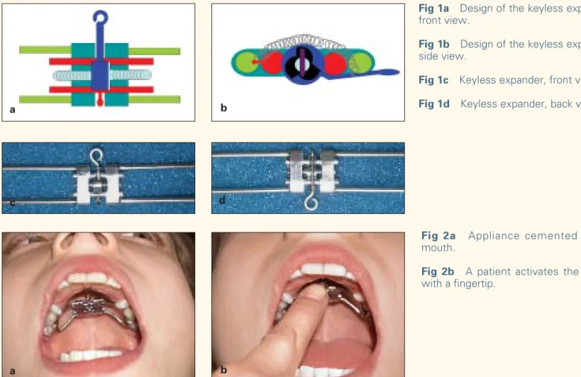
The appliance consists of a variety of components (US patent no. US 7,074,036 B1). Instead of using a key to turn the screw, the screw accommodates a wheel with a built-in, ratchet-like activation arm (Figs 1a to 1d). The arm automatically springs backward with the help of a nickel-titanium spring mechanism after a finger press. The wheel’s housing consists of 3 quartocircular indents and accommodates a pin that actively turns the screw in a uni-directional fashion and passively springs back. The patient
CASE 1
A female 14 years 6 months of age in the permanent denti-tion was diagnosed with a bilateral maxillary constricdenti-tion of 5 mm. She required an RPE appliance to resolve a skeletal and dental transverse discrepancy. We banded the maxillary first molars and premolars and took impressions for construction of a keyless expander. The appliance was cemented in place and the patient instructed to turn it once a day. She was seen 22 days later to monitor the expansion. The results are presented in Figs 3a and 3b and Figs 4a and 4b.
CASE 2
A female 13 years 5 months of age in permanent dentition was diagnosed with a bilateral maxillary constriction of 4.5 mm. She required an RPE appliance to resolve a skeletal and dental transverse discrepancy. We banded the maxil-lary first molars and took impressions for construction of a keyless expander. The appliance was cemented on the
Fig 1a Design of the keyless expander,
front view.
Fig 1b Design of the keyless expander,
side view.
Fig 1c Keyless expander, front view.
Fig 1d Keyless expander, back view.
Fig 2a Appliance cemented in the
mouth.
Fig 2b A patient activates the screw
with a fingertip.
a b
c d
Fig 3a Case 1, initial frontal view.
Fig 3b Case 1, initial occlusal view.
Fig 4a Case 1, midtreatment frontal
view (note the central diastema).
Fig 4b Case 1, midtreatment
occlusal view.
Fig 5a Case 2, initial frontal view.
Fig 5b Case 2, initial occlusal view.
Fig 6a Case 2, midtreatment frontal
view (note the correction of the poste-rior crossbite in the molar region).
Fig 6b Case 2, midtreament occlusal
view. a b a b a b a b
Fig 7a Case 3, initial frontal view.
Fig 7b Case 3, initial occlusal view.
Fig 8a Case 3, midtreatment frontal
view (note the midline diastema and correction of the crossbite in the right molar region).
Fig 8b Case 3, midtreatment
occlusal view (note the improvement of the arch symmetry).
Fig 9a Case 4, initial frontal view.
Fig 9b Case 4, initial occlusal view.
Fig 10a Case 4, midtreatment
frontal view (note the midline diastema and correction of the cross-bite in the molar region).
Fig 10b Case 4, midtreatment
occlusal view. a b a b a b a b
CASE 3
A female 14 years 1 month of age in permanent dentition was diagnosed with unilateral maxillary constriction of 7 mm. She required an RPE appliance to resolve a skeletal and dental transverse discrepancy. We banded her maxillary first molars and took impressions for construction of a key-less expander. The appliance is cemented on the first molar and bonded to the first premolar. The patient was instructed to turn the screw once a day and was seen 24 days later to monitor the expansion. The results are presented in Figs 7a and 7b and Figs 8a and 8b.
CASE 4
A female 12 years 6 months old in permanent dentition was diagnosed with a bilateral maxillary constriction of 5 mm. She required an RPE appliance to resolve a skeletal and dental transverse discrepancy. We banded the maxil-lary first molars and took impressions for the construction of the newly developed expander. The appliance is cemented on the first premolars and bonded to the first premolars. The patient was instructed to turn the screw once a day and was seen 14 days later to monitor the expansion. The results are presented in Figs 9a and 9b and Figs 10a and 10b.
CONCLUSION
The results of the above cases show that the keyless expander is an effective means to achieve maxillary expan-sion with minimum patient cooperation. The conventional expanders require a key for activation; if you do not com-plete the one-quarter turn, the next hole on the screw does not appear, thereby making it impossible for the patient to proceed activation. The keyless expander does not require a key, and the patient needs only turn the arm once a day with a finger tip. The other advantage is that after comple-tion, the screw does not unwind because of its design. The built-in arm allows for easy activation of the screw without the risk of dropping the key down a patient’s throat or activating the patient’s gag reflex. The risks of aspiration, swallowing, and injuring the palate have been eliminated. In this case report, the first phase (expansion) treatment was completed in an average of 3 weeks, enabling the second phase to begin without delay.
ACKNOWLEDGMENTS
The author would like to thank Dr Edward Seldin, Dr Donald Nelson, Dr Doil Kim, and Dr Ping-Lin Ben Chung for their contributions.
REFERENCES
1. Angell EC. Irregularities of teeth and their treatment. San Francisco Medical Press 1860;1:181–185.
2. Debbanne EF. A cephalometric and histologic study of the effect of orthodontic expansion of the midpalatal suture of the cat. Am J Orthod 1958;44:187–219.
3. Haas AJ. Gross reaction to the widening of the maxillary dental arch of the pig by splitting of the midpalatal suture. Am J Orthod 1959;45: 868–869.
4. Starnbach H, Bayne D, Cleall J, Subtelny JD. Facioskeletal and dental changes resulting from rapid maxillary expansion. Angle Orthod 1966;36:152–164.
5. Biederman W. Rapid correction of Class 3 malocclusion by mid-palatal expansion. Am J Orthod 1973;63:47–55.
6. Brossman RE, Bennett CG, Merow WW. Facioskeletal remodelling resulting from rapid palatal expansion in the monkey (Macaca cynomolgus). Arch Oral Biol 1973;18:987–994.
7. Chaconas SJ, Caputo AA. Observation of orthopedic force distribution produced by maxillary orthodontic appliances. Am J Orthod 1982;82: 492–501.
8. Tanne K, Sachdeva R, Miyasaka J, Yamagata Y, Sakuda M. A study of strain and stress levels in the circummaxillary sutural systems during rapid maxillary expansion: An approach using both the strain gauge technique and the theoretical stress analysis. J Osaka Univ Dent Sch 1986;26:151–165.
9. Melsen B. Palatal growth studied on human autopsy material. A histo-logic microradiographic study. Am J Orthod 1975;68:42–54. 10. Melsen B. A histological study of the influence of sutural morphology
and skeletal maturation on rapid palatal expansion in children. Trans Eur Orthod Soc 1972:499–507.
11. Murray JM, Cleall JF. Early tissue response to rapid maxillary expan-sion in the midpalatal suture of the rhesus monkey. J Dent Res 1971;50:1654–1660.
12. McNamara JA. Brudon WL. Orthodontics and Dentofacial Orthopedics, ed 2. Ann Arbor: Needham Press, 2001.
13. Nazif MM, Ready MA. Accidental swallowing of orthodontic expansion appliance keys: Report of two cases. ASDC J Dent Child 1983;50: 126–127.
14. Sfondrini MF, Cacciafesta V, Lena A. Accidental ingestion of a rapid palatal expander. J Clin Orthod 2003;37:201–202.