Zurich Open Repository and
Archive
University of Zurich
Main Library
Strickhofstrasse 39
CH-8057 Zurich
www.zora.uzh.ch
Year: 2019
No evidence of differences in cognitive control in children with autism
spectrum disorder or obsessive-compulsive disorder: An fMRI study.
Gooskens, Bram ; Bos, Dienke J ; Mensen, Vincent T ; Shook, Devon A ; Bruchhage, Muriel M K ;
Naaijen, Jilly ; Wolf, Isabella ; Brandeis, Daniel ; Williams, Steven C R ; Buitelaar, Jan K ; Oranje,
Bob ; Durston, Sarah
Abstract: Repetitive behaviors are among the core symptoms of both Autism Spectrum Disorder (ASD)
and Obsessive-Compulsive Disorder (OCD) and are thought to be associated with impairments in
cogni-tive control. However, it is still unknown how deficits in cognicogni-tive control and associated neural circuitry
relate to the quality or severity of repetitive behavior in children with these disorders. Therefore, we
investigated the behavioral and neural correlates of cognitive control using a modified stop-signal task in
a multicenter study of children (aged 8-12 years) with ASD, OCD and typically developing (TD) children
(N = 95). As both ASD and OCD have high levels of comorbidity with Attention Deficit/Hyperactivity
Disorder (ADHD), we did an exploratory analysis addressing ADHD-symptoms. We found that children
with ASD and OCD did not show deficits in cognitive control or changes in brain activity in task-relevant
neural networks when compared to TD children. However, increased activity in prefrontal brain areas was
associated with increased symptoms of comorbid ADHD. As such, this study does not support differences
in cognitive control or associated neural circuitry in children with ASD and OCD, but rather suggests
that changes in cognitive control in these disorders may be related to symptoms of comorbid ADHD.
DOI: https://doi.org/10.1016/j.dcn.2018.11.004
Posted at the Zurich Open Repository and Archive, University of Zurich
ZORA URL: https://doi.org/10.5167/uzh-172664
Journal Article
Published Version
The following work is licensed under a Creative Commons: Attribution-NonCommercial-NoDerivatives
4.0 International (CC BY-NC-ND 4.0) License.
Originally published at:
Gooskens, Bram; Bos, Dienke J; Mensen, Vincent T; Shook, Devon A; Bruchhage, Muriel M K; Naaijen,
Jilly; Wolf, Isabella; Brandeis, Daniel; Williams, Steven C R; Buitelaar, Jan K; Oranje, Bob; Durston,
Sarah (2019). No evidence of differences in cognitive control in children with autism spectrum disorder
or obsessive-compulsive disorder: An fMRI study. Developmental Cognitive Neuroscience, 36:100602.
DOI: https://doi.org/10.1016/j.dcn.2018.11.004
Contents lists available atScienceDirect
Developmental Cognitive Neuroscience
journal homepage:www.elsevier.com/locate/dcnNo evidence of differences in cognitive control in children with autism
spectrum disorder or obsessive-compulsive disorder: An fMRI study
Bram Gooskens
a,⁎, Dienke J. Bos
a, Vincent T. Mensen
a, Devon A. Shook
a,
Muriel M.K. Bruchhage
b, Jilly Naaijen
c, Isabella Wolf
d, Daniel Brandeis
d,e,f,g,h,
Steven C.R. Williams
b, Jan K. Buitelaar
c,i, Bob Oranje
a, Sarah Durston
a, the TACTICS consortium
aDepartment of Psychiatry, Brain Center Rudolf Magnus, University Medical Center Utrecht, Utrecht University, Utrecht, the NetherlandsbDepartment of Neuroimaging, King’s College London, Institute of Psychiatry, Psychology and Neuroscience, London, UK
cDepartment of Cognitive Neuroscience, Donders Institute of Brain, Cognition and Behaviour, Radboud University Medical Center, Nijmegen, the Netherlands dDepartment of Child and Adolescent Psychiatry and Psychotherapy, Central Institute of Mental Health, Medical Faculty, Mannheim/Heidelberg University, Mannheim, Germany
eDepartment of Child and Adolescent Psychiatry and Psychotherapy, Psychiatric Hospital, University of Zurich, Zurich, Switzerland fCenter for Integrative Human Physiology, University of Zurich, Zurich, Switzerland
gNeuroscience Center Zurich, University of Zurich, Zurich, Switzerland hETH Zurich, Zurich, Switzerland
iKarakter Child and Adolescent Psychiatry University Center, Nijmegen, the Netherlands
A R T I C L E I N F O
Keywords:
Autism spectrum disorder Obsessive-compulsive disorder Compulsive behavior Cognitive control
Attention deficit/hyperactivity disorder fMRI
A B S T R A C T
Repetitive behaviors are among the core symptoms of both Autism Spectrum Disorder (ASD) and Obsessive-Compulsive Disorder (OCD) and are thought to be associated with impairments in cognitive control. However, it is still unknown how deficits in cognitive control and associated neural circuitry relate to the quality or severity of repetitive behavior in children with these disorders. Therefore, we investigated the behavioral and neural correlates of cognitive control using a modified stop-signal task in a multicenter study of children (aged 8–12 years) with ASD, OCD and typically developing (TD) children (N= 95). As both ASD and OCD have high levels of comorbidity with Attention Deficit/Hyperactivity Disorder (ADHD), we did an exploratory analysis addressing ADHD-symptoms. We found that children with ASD and OCD did not show deficits in cognitive control or changes in brain activity in task-relevant neural networks when compared to TD children. However, increased activity in prefrontal brain areas was associated with increased symptoms of comorbid ADHD. As such, this study does not support differences in cognitive control or associated neural circuitry in children with ASD and OCD, but rather suggests that changes in cognitive control in these disorders may be related to symptoms of comorbid ADHD.
1. Introduction
Repetitive behaviors are among the core symptoms of neurodeve-lopmental disorders such as Autism Spectrum Disorder (ASD) and Obsessive-Compulsive Disorder (OCD) (American Psychiatric Association, 2013). ASD has an estimated prevalence of 1%, OCD has prevalence rates estimated between 1 and 3% in children and young adolescents (Baxter et al., 2015;Flament et al., 1988; Valleni-Basile et al., 1994). Although ASD and OCD are distinct disorders, it has been noted that children with ASD show increased rates of obsessive-com-pulsive symptoms (Leyfer et al., 2006) and vice versa, individuals with
OCD often show symptoms of autism (Ivarsson and Melin, 2008). As such, it has been suggested that obsessive-compulsive symptoms in ASD and OCD may have common neurobiological characteristics (Jiujias et al., 2017).
Yet, while there appear to be quantitative similarities, there are simultaneous qualitative differences in the repetitive behaviors of in-dividuals with ASD and OCD (Zandt et al., 2007,2009). For instance, individuals with ASD show more stereotyped repetitive behaviors, such as hoarding, touching and tapping behaviors, and these seem to sometimes have a positively reinforcing function. Conversely, in-dividuals with OCD often have recurrent intrusive and distressing
https://doi.org/10.1016/j.dcn.2018.11.004
Received 30 May 2018; Received in revised form 5 October 2018; Accepted 26 November 2018
⁎Corresponding author at: Department of Psychiatry, Brain Center Rudolf Magnus, University Medical Center Utrecht, Heidelberglaan 100, 3584 CX, Utrecht, The
Netherlands.
E-mail address:[email protected](B. Gooskens).
Available online 29 November 2018
1878-9293/ © 2018 Published by Elsevier Ltd. This is an open access article under the CC BY-NC-ND license (http://creativecommons.org/licenses/BY-NC-ND/4.0/).
thoughts (obsessions) and repetitive mental and behavioral rituals (compulsions) such as checking, repeating and counting behaviors, that may serve to reduce anxiety (McDougle et al., 1995;Zandt et al., 2007). This suggests that in addition to common neural circuits, there may also be distinct, disorder-specific pathways involved in the repetitive be-haviors seen in ASD and OCD.
Cognitive control is a broad concept including many behaviors re-lated to the ability to regulate one’s behavior, for example by stopping or suppressing ongoing behavior when it is no longer appropriate or required. This ability is crucial for successfully navigating the demands of daily life. Previous work has suggested that repetitive behavior in ASD and OCD may be related to impairments in cognitive control (Chamberlain et al., 2005; Hill, 2004; Moritz et al., 2002; Mosconi et al., 2009;Snyder et al., 2015), yet it has been challenging to find a consistent relation between repetitive behavior and cognitive control in ASD and OCD. Findings from studies investigating cognitive control in ASD using varying tasks have been inconsistent, with many reporting no deficits in ASD (Geurts et al., 2009; Yerys, 2015). To complicate matters further, both ASD and OCD have high levels of comorbidity with Attention Deficit/Hyperactivity Disorder (ADHD) (Abramovitch et al., 2015;Masi et al. (2006);Stevens et al., 2016;Grzadzinski et al., 2016;Mayes et al., 2012) and the neural circuits involved have been suggested to overlap (e.g.Ameis et al., 2016;Norman et al., 2016). As such, one hypothesis may be that differences in cognitive control in OCD and ASD may in part be driven by elevated symptoms of comorbid ADHD.
The stop-signal task has proven successful in assessing a specific aspect of cognitive control, the ability to withhold an already initiated motor response (Logan and Cowan, 1984; Verbruggen and Logan, 2008), especially in ADHD (Lipszyc and Schachar, 2010). This task can be used to estimate the stop-signal reaction time (SSRT), an indicator of the speed of the stopping process. Findings in OCD and ASD using this task have been mixed: increased SSRTs have been reported for in-dividuals with OCD (Chamberlain et al., 2007; Kang et al., 2013;
Mancini et al., 2018; Penadés et al., 2007; de Wit et al., 2012) and children with ASD (Geurts et al., 2004;Lemon et al., 2011), while other studies have found no differences (Adams and Jarrold, 2012;
Chantiluke et al., 2015;Fan et al., 2017;Ozonoff and Strayer, 1997;
Schmitt et al., 2017). Furthermore, associations between SSRT and symptom severity in OCD have been reported (Berlin and Lee, 2018; Mancini et al., 2018).
Similarities and differences in the neural signatures of OCD and ASD were recently reviewed byCarlisi et al. (2017). Both OCD and ASD were associated with reduced activation and gray matter volume of the dorsal anterior cingulate cortex (dACC) and medial prefrontal cortex (mPFC). Disorder-specific effects were found in basal ganglia and in-sula. These findings suggest that broadly, changes in frontostriatal and frontoinsular circuitry may underlie phenotypic overlap and distinc-tions between OCD and ASD. Yet, in children with ASD and OCD the neural correlates of changes in SSRT are unclear. To date, only one neuroimaging study has used the stop-signal task in ASD, and reported greater activation in left and right inferior frontal cortex compared to controls (Chantiluke et al., 2015). In OCD, there is some evidence for decreased activation of the cortico-striatal-thalamo-cortical (CSTC) loop, including the dorsolateral prefrontal cortex (DLPFC), inferior frontal gyrus (IFG), striatum and thalamus in children (Rubia et al., 2010;Woolley et al., 2008) and adults (Kang et al., 2013;de Wit et al., 2012) with the disorder.
In the current study, we set out to investigate shared and distinct changes in cognitive control and associated frontostriatal neural cir-cuitry in relation to the severity of repetitive behaviors in a sample of children with a primary diagnosis of ASD or OCD, and a group of age-and gender-matched typically developing children. We operationalized cognitive control as the ability to stop an ongoing response in the context of the stop-signal task, which was performed by all participants during fMRI. As it has proven challenging to recruit a sizeable sample of
children with OCD, this study was performed within a multi-center collaborative initiative, the Translational Adolescent and Childhood Therapeutic Interventions in Compulsive Syndromes (TACTICS) con-sortium. We hypothesized that (1) children with ASD and OCD would show prolonged SSRTs compared to typically developing children, in-dicating reduced cognitive control; (2) children with ASD and OCD would show a pattern of shared and distinct changes in associated neural circuitry during performance of the stop-signal task; and (3) the severity of compulsive behavior in children with OCD and ASD would be related to increased SSRTs and reduced activation in frontostriatal cognitive control circuitry.
2. Methods
The study was approved by local ethics committees for each site (Nijmegen and Utrecht: Commissie Mensgebonden Onderzoek Regio Arnhem-Nijmegen, 2013, NL nr: 42004.091.12; Mannheim: Ethics committee of the Medical Faculty Mannheim, Heidelberg University, 2013, nr: 213616 NMA; London: NRES Committee London -Camberwell St Giles, 2013, nr: 14/LO/1413).
2.1. Participants
We prescreened 212 participants and succeeded in including a total of 205 participants between 8 and 12 years of age in the study (ASD N = 62, OCD N = 42, TD = 101). We recruited participants at four different sites across Europe (King’s College London, London, United Kingdom (N = 34); Central Institute of Mental Health, Medical Faculty Mannheim, University of Heidelberg Mannheim; Germany (N = 33); Radboud University Medical Center and the Donders Institute for Brain, Cognition and Behaviour, Nijmegen, The Netherlands (N = 101); Brain Center Rudolf Magnus, University Medical Center Utrecht, Utrecht, The Netherlands (N = 37)) that were commissioned by a multicenter study (COMPULS:Naaijen et al., 2016) as part of the overarching TACTICS collaborative initiative (http://www.tactics-project.eu).
Parents or legal representatives of all children provided signed in-formed consent and children provided verbal assent. Participants with ASD or OCD were diagnosed according toThe Diagnostic and Statistical Manual of Mental Disorders, 4thedition, Text Revision (APA, 2000) or 5th
edition criteria (APA, 2013). For children with ASD, the clinical diag-nosis was confirmed by a trained psychologist at each site using the Autism Diagnostic Interview-Revised (ADI-R; Lord et al., 1994). For children with OCD, the Children’s Yale-Brown Obsessive Compulsive Scale (CY-BOCS; Scahill et al., 1997) were collected to assess OCD symptom severity. This interview was also performed in participants with ASD if screening questions confirmed the presence of clinically significant obsessions or compulsions. In addition, all parents were in-terviewed using the structured Diagnostic Interview Schedule for Children (DISC-IV, parent version;Shaffer et al., 2000), the Develop-ment and Well-being AssessDevelop-ment (DAWBA;Goodman et al., 2000) or the Kiddie Schedule for Affective Disorders and Schizophrenia (K-SADS;
Kaufman et al., 1997) to assess the presence of possible comorbidities. Total Intellectual Quotient (IQ) was estimated using a shortened ver-sion of the Wechsler Intelligence Scale for Children (WISC-III;Wechsler, 2003). Repetitive behavior was assessed using the Repetitive Behavior Scale – Revised questionnaire (RBS-R;Bodfish et al., 2000). In addition, ADHD symptomatology was assessed using the Conners’ Parent Rating Scale questionnaire (CPRS-R:L;Conners et al., 1998).
For both diagnostic groups, a concurrent diagnosis of the other disorder was an exclusion criterion (i.e. a comorbid OCD for a child with ASD, or vice versa). For the TD group, any psychiatric diagnosis for themselves or any first-degree relatives was an exclusion criterion. All included participants had a total IQ > 70 as well as sufficient comprehension and speaking abilities of the native language of the country in which the assessment took place. Finally, the presence of metal objects in the body (i.e. pacemaker, dental braces), neurological
B. Gooskens, et al. Developmental Cognitive Neuroscience 36 (2019) 100602
illness or other contra-indications for MRI-assessment were exclusion criteria for all groups. Six children (ASD N = 3, OCD N = 1, TD N = 2) dropped out after inclusion because of feeling anxious or claustrophobic when entering the MRI-scanner.
Four children with ASD were being treated with psychostimulants, two with antipsychotics, one with a combination of both and one child used low-dose naltrexone. Within the OCD group, seven children were being treated with antidepressants, one with antipsychotics and one with both. Participants were asked to abstain from stimulant medica-tion 24 h before scanning. Seven children with ASD had a current co-morbid diagnosis of ADHD, another two children had both coco-morbid ADHD and oppositional defiant disorder (ODD). In the OCD group, two participants had comorbid ADHD.
2.2. Stop-signal task
Participants completed a nine minute modified version of the stop-signal task adapted fromRubia et al. (2003), using Presentation soft-ware (Neurobehavioral Systems, Albany, California) during an fMRI session. Each trial started with a fixation cross (500 ms) displayed on a computer screen. During go-trials (80% of a total number of 294 trials), participants were instructed to hit the right button on a response pad as fast as possible with their right middle finger whenever they were presented with an arrow pointing to the right, and to hit the left button as fast as possible with their right index finger whenever they were presented with an arrow pointing to the left. During stop-trials (20% of a total number of 294 trials), a go-signal was followed by an arrow pointing upwards (stop-signal) and participants were instructed to withhold (stop) their button press. The mean inter-trial interval (ITI) was randomly jittered between 1.6 and 2.0 s to optimize statistical ef-ficiency. The delay between a go- and stop-signal (stop-signal delay: SSD) was dynamically changed (start: 250 ms) using an adaptive staircase algorithm, where whenever the participant stopped success-fully on a stop-trial, the SSD latency of the following stop-trial increased by 50 ms (max. 900 ms), thereby making it more difficult to stop. If the participant did not inhibit his/her response during the previous stop-trial, SSD latency decreased by 50 ms (min. 50 ms). This procedure ensured that the sessions concluded with an approximately equal number of successful and failed stop-trials.
Before participating in the MR session, children at each site were prepared for scanning using a mock scanner. In this session, children were familiarized with MR sounds, the button box needed for task completion, and lying still in the scanner environment. In addition, participants performed a brief practice session of the task. If a child (or his/her parent) reported anxiety to enter the MR scanner, the session was ended. This procedure has proven succesful in reducing anxiety for the MR session (Durston et al., 2009).
Our measures of interest for task performance were mean reaction time (MRT) on correct go-trials, stop-signal reaction time (SSRT), mean stop-signal delay (SSD), number of non-responses to go-trials (omis-sions) and number of incorrect responses to go-trials (commis(omis-sions). The SSRT was estimated using the integration method fromVerbruggen et al. (2013):first, reaction times (RT) to correct go-trials were rank ordered. Subsequently, thenth go-RT was selected, wherenwas derived by multiplying the number of correct go-trials by the probability that one respond to a stop-signal (P(respond | stop-signal)). The SSRT then was estimated by subtracting the mean SSD from thenth go-RT.
2.3. fMRI image acquisition
At the four different sites, comparable 3-Tesla MRI scanners were used (Siemens Trio and Siemens Prisma, Siemens, Erlangen, Germany; General Electric MR750, GE Medical Systems, Milwaukee, WI, USA; Philips 3 T Achieva, Philips Medical Systems, Best, The Netherlands). Scanner information across sites is available in Table S1 (Supplemental material).
2.4. Behavioral data analysis
Data from sixteen participants (ASD N = 11, OCD N = 3, TD N = 2) could not be analyzed due to incomplete task performance. We cleaned the data to optimize data quality for statistical analysis: we excluded participants with SSRT values below 50 ms (ASD N = 9, OCD N = 3, TD = 4), accuracy below 25% successful stop-trials (OCD N = 1), or accuracy below 60% of the total number of correct go-trials (ASD N = 1, OCD N = 3) (Congdon et al., 2012). Data from 162 participants (ASD = 39, OCD = 31, TD = 92) were available for behavioral ana-lysis.
2.4.1. Matching procedure
As there were differences in mean age for the children in the be-havioral sample (children with OCD where older than other groups [F(2, 159)= 7.424,p= .001]), propensity score matching (PSM) was
per-formed to create an age-matched sample of children with ASD, OCD and TD. PSM, implemented in SPSS 22.0, is a statistical method to pair individuals with similar values on a propensity score from a pool of participants. First, a multivariate logistic regression analysis was per-formed to obtain propensity scores, with age as a covariate. In order to minimize the number of exclusions, children with OCD and ASD were merged together in one group, and then matched 1:1 with a TD parti-cipant using a caliper set at .05. This procedure yielded three final di-agnostic groups that were matched for age (ASD = 38, OCD = 23, TD = 61) (Supplemental Table S2).
2.4.2. Statistical analyses
Behavioral data analysis was performed in SPSS version 22.0 (IBM). First, group differences in demographic and clinical measures were tested using the appropriate Pearson’s χ2-tests or one-way analyses of
variance (ANOVA). Levene’s test for homogeneity of variance and Shapiro-Wilk normality tests were used to check if assumptions of homogeneity of variance and normality were met. If data did not meet these assumptions, non-parametric tests were used (Kruskal Wallis rank sum test or Mann–Whitney U-test). Group differences on all task-per-formance measures (MRT, SSRT, SSD, number of omission and com-mission errors) were initially tested within a General Linear Model (GLM) framework using analyses of covariance (ANCOVA), with the task-performance measures as the dependent variables, diagnostic group as independent variable, and gender and site as covariates. However, if both gender and site did not render any significant effects, they were removed from the design in the final analyses. For significant main effects, we ran post-hoc pairwise comparisons between the three diagnostic groups. Lastly, we ran correlation analyses between task performance measures and symptom severity as assessed using the compulsive subscale and the total score of the RBS-R, and the total score of the CPRS-R. Unfortunately, the number of administered CY-BOCS in the ASD group was too low to include in any further analyses.
2.5. fMRI data analysis 2.5.1. fMRI preprocessing
fMRI preprocessing was performed using standard procedure in SPM12, as implemented in MATLAB R2015b. Data from three partici-pants (ASD N = 1, TD N = 2) were excluded due to incomplete fMRI data or problems with angulation. fMRI data were realigned to the first volume to correct for in-scanner head motion. Next, using the ArtRepair toolbox in SPM12, all volumes with frame-to-frame movement > 1 mm or > 1.5% standard deviation from the mean signal were substituted using linear interpolation from neighboring frames. Consequently, 24 participants (ASD N = 11, OCD N = 7, TD N = 6) were excluded from the fMRI analysis due to excessive head motion (absolute movement more than one voxel, N = 13), or replacement of more than 20% of total volumes in the ArtRepair step (N = 11). This resulted in 26 ASD, 16 OCD and 53 TD datasets to carry forward to the fMRI analysis. After
realignment and motion-correction, the fMRI data and anatomical T1-image were co-registered, followed by normalization to Montreal Neurological Institute (MNI) standard atlas and finally spatially smoothed with a 6 mm3full width at half maximum (FWHM) Gaussian
kernel.
2.5.2. Statistical analyses
At the first level, onsets of three event types (correct go-trials, successful stop-trials, failed stop-trials) were modeled using delta functions convolved with the canonical haemodynamic response func-tion (HRF). Six mofunc-tion estimafunc-tion parameters were included in the model as regressors of no interest.
Second-level random effects analyses were run for two contrasts of interest: successful stopping was investigated by contrasting successful stop trials with correct go trials (successful stop activation > go acti-vation). Failed stopping was investigated by contrasting failed stop trials with correct go trials (failed stop activation > go activation). We assessed whole-brain group differences in activation for these two contrasts using F-tests (three-way comparison) and t-tests (planned di-rect comparisons) at a Family-Wise Error (FWE) cordi-rectedp-value of .05. Whole-brain analyses were followed up with a data-driven region of interest (ROI) analysis. These data-driven ROIs showed high agree-ment with regions reported in earlier studies including older samples of subjects (Chantiluke et al., 2015; Rubia et al., 2010; Woolley et al., 2008).
For both contrasts, we used MarsBar (http://marsbar.sourceforge. net), to create 6 mm spheres around the peak voxel coordinates of re-gions in the frontostriatal cognitive control network showing significant whole-brain corrected (pFWE= .05) activation in typically developing
controls (Supplemental Table S3). This resulted in seven ROIs for suc-cessful stop trials, and eight ROIs for failed stop trials. We extracted the timecourses from these ROIs for between-group comparisons. We tested for between-group differences in brain activation within the ROIs for both contrasts within a General Linear Model (GLM) framework using
ANCOVA, with the mean signal from the ROIs as dependent variables, and diagnostic group (ASD, OCD, TD) and site as independent variables. We then compared children with ASD directly to children with OCD in a two-samplet-test planned contrast. Additionally, we assessed possible correlations between whole brain activity and activity from ROIs with the compulsive subscale and the total score of the RBS-R, and the total score of the CPRS-R.P-values were adjusted for multiple testing, using the Benjamini-Hochberg procedure to control the False Discovery Rate (FDR) (Benjamini and Hochberg, 1995).
2.5.3. Exploratory analyses
In a first exploratory follow-up analysis, we pooled the children with OCD and ASD into a single group to increase statistical power to detect any shared differences from TD children. In a second exploratory analysis, we performed a median split to group participants based on their scores on the RBS-R (score < 14 or > 13). A third exploratory analysis addressed ADHD symptoms, using a median split to create two groups based on CPRS-R score (score < 64 or > 63).
2.5.4. Power analysis
Using G*Power 3.1 (Faul et al., 2009), we conducted a post-hoc power analysis to estimate the sample size that would be needed to detect between-group differences. Sample sizeNwas computed as a function of the required power level 1 - ß (.80), the prespecified sig-nificance level α (.05), and the population effect size to be detected with probability 1 - ß (.10).
3. Results
3.1. Group characteristics
After propensity score matching, groups did not differ in age [F2, 119= 0.408, p= .666] gender composition [χ2 (2) = 4.019, p=
.134], hand preference [χ2 (2) = 0.515,p= .773], or estimated IQ
Table 1
fMRI participants’ demographic and clinical information.
ASD (N= 26) Mean (SD) OCD (N= 16) Mean (SD) TD (N= 53) Mean (SD) Test statistic P value Demographic measures Age (years) 11.33 (1.07) 10.92 (1.47) 10.76 (1.15) F2, 92= 2.000 .141 Gender (f/m) 9/17 9/7 24/29 χ2(2) =1.936 .380 Righthanded (in %) 92.3% 86.7 % 90.6 % χ2(2) =0.352 .838 Estimated IQ’s 108.88 (16.67) 100.72 (13.26) 111.92 (10.33) K-W χ2(2) = 7.999 .018a Clinical measures ADI-Revised -Social interaction 18.54 (5.30) – – -Social communication 13.67 (3.39) – – -Repetitive behavior 3.30 (2.48) – – CY-BOCS -Obsession – 7.19 (5.23) – -Compulsion – 10.19 (3.51) – -Total score – 17.38 (7.70) – RBS-Revised -Compulsivity 2.04 (2.64) 4.69 (3.03) 0.06 (0.30) K-W χ2(2) = 56.919 < .001b -Total score 20.04 (16.04) 16.47 (11.36) 0.64 (1.30) K-W χ2(2) = 62.981 < .001c CPRS-Revised: Longd -Inattention 62.85 (11.93) 57.36 (10.62) 45.63 (6.24) K-W χ2(2) = 34.954 < .001c -Hyperactivity 62.90 (13.16) 61.29 (10.51) 45.92 (3.78) K-W χ2(2) = 36.921 < .001c -Total score 64.40 (12.78) 59.92 (10.77) 45.26 (4.76) K-W χ2(2) = 38.695 < .001c
Abbreviations: ASD, autism spectrum disorder; OCD, obsessive-compulsive disorder; TD, typically developing group; SD, standard deviation; m/f, male/female; ADI, Autism Diagnostic Interview; CY-BOCS, Children’s Yale-Brown Obsessive-Compulsive scale; RBS, Repetitive-Behavior scale; CPRS, Conners’ Parent Rating scale. Comorbidities: ASD + ADHD (n= 5), ASD + ADHD + ODD (n= 1), OCD + ADHD (n= 2).
a TD > OCD. b OCD > ASD > TD. c ASD, OCD > TD.
dDisplayed scores are based on T-scores.
B. Gooskens, et al. Developmental Cognitive Neuroscience 36 (2019) 100602
scores [F2, 119= 1.844,p= .163]. Both children with ASD and OCD
scored higher on the compulsivity subscale and total score of the RBS-R than typically developing children. The mean CY-BOCS score for the OCD group was just above the clinical cut-off score (> 16) (see Supplement Table S2).
The reduced fMRI-sample was also matched in terms of age, gender and hand preference. However, IQ differed between groups, with lower scores for children with OCD compared to controls (seeTable 1).
3.2. Task performance
ANOVA showed no main effects of diagnostic group on any of the behavioral measures of interest (MRT, SSRT, SSD, number of omission and commission errors) in either the whole sample (Supplement Table S4) or the smaller fMRI sample (Table 2). These results did not sig-nificantly change after adding IQ or CPRS-R as a covariate to the design, or when children on medication were excluded. Within the whole sample, there was a main effect of site on MRT [F3, 110= 3.207,p=
.026] and SSD [F3, 110= 4.458,p= .005], but this did not affect the
effect of diagnostic status. Furthermore, correlational analysis showed a positive correlation between the compulsive subscale of the RBS-R and SSD (r(22) = -0.517,p= .012) in children with OCD. However, this correlation was not present in the fMRI group, suggesting that this ef-fect may not be replicable over studies. There were no correlations between measures of task performance and symptom severity, as as-sessed using the total score of the RBS-R or total score of the CPRS-R, that survived Bonferroni-correction.
3.3. Between-group differences in brain activation
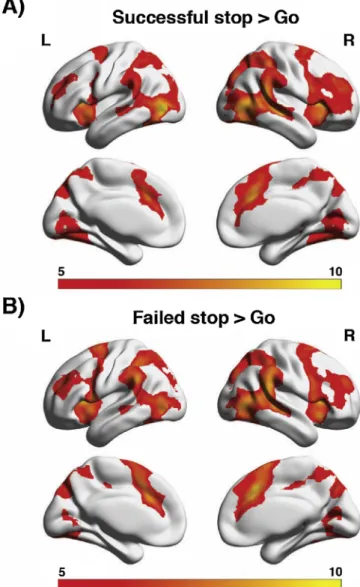
All groups showed the expected pattern of brain activation related to go-trials, including activation of left motor cortex. During stopping, participants showed predominantly right but also left middle frontal gyrus activity, extending into precentral gyrus / presupplementary motor area and the inner cortical structures of insula and cingulate gyrus (Fig. 1). There were no differences between groups in activation during successful or failed stop-trials that survived whole-brain cor-rection (pFWE< .05). Nor were there any between-group differences in
any of the ROIs that survived FDR-correction for multiple comparisons. These results did not significantly change after adding IQ or CPRS-R as a covariate to the design, or when children on medication were ex-cluded. The calculated effect sizes (ηp²) for non-significant findings did
not exceed .017. Correlational analyses yielded no significant associa-tions between whole brain activity and activity from ROIs with the compulsive subscale and the total score of the RBS-R, and the total
score of the CPRS-R, that survived Bonferroni-correction for multiple comparisons.
3.4. Between-group differences in brain activation based on symptom severity
As the three-group comparison yielded no significant differences in task-performance or brain activation between diagnostic groups, we merged the children with OCD and ASD into a single group to increase statistical power for an exploratory analysis. Again, we found no dif-ferences between children with a diagnosis and those without. When we divided the children with a diagnosis into two groups using a median-split analysis on their RBS-R scores, we similarly found no differences in task-performance or brain activity.
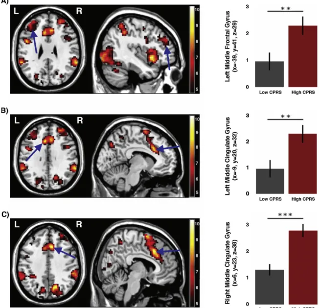
When we divided participants with ASD and OCD into two groups using a median split analysis on their CPRS-R scores, we found that children with higher ADHD symptom scores had more activity in left middle frontal gyrus (t = -2.782, df = 33,p= .009, d= 0.94), left middle cingulate gyrus (t = -3.316, df = 33,p= .002,d= 1.12) and right middle cingulate gyrus (t = -4.397, df = 33,p< .001,d= 1.48) during failed stop trials than children with lower scores (Fig. 2). During
Table 2
fMRI task performance for the diagnostic groups.
ASD (N= 26) Mean (SD) OCD (N= 16) Mean (SD) TD (N= 53) Mean (SD) Test statistic P value MRT 370.36 (87.36) 336.01 (111.96) 381.72 (144.27) F2, 92= 0.809 .448 SSRT 165.94 (71.68) 190.72 (79.99) 188.67 (69.32) F2, 92= 0.991 .375 SSD 561.27 (101.11) 516.08 (93.15) 516.41 (90.49) F2, 92= 2.160 .121 Omissionsa 2.02 % 2.43 % 2.24 % F 2, 92= 0.116 .891 Commissionsa 2.79 % 4.25 % 4.37 % F 2, 92= 2.304 .106 Successful stoppinga 52.4 % 51.2 % 51.4 % F2, 92= 1.093 .339
Abbreviations: ASD, autism spectrum disorder; OCD, obsessive-compulsive disorder; TD, typically developing group; SD, standard deviation; MRT = Mean reaction time, SSRT = Stop-signal reaction time, SSD = Stop-signal delay.
a Commissions, omissions and successful stopping are displayed in % of total
trials.
Fig. 1.A) Task activation across all groups for successful stop trials (successful
stop > correct go trials), thresholded atpFWE= 0.05, and B) failed stop trials
(failed stop > correct go trials), thresholded atpFWE= 0.05, showing robust
frontostriatal activation during cognitive control. The numbers above the col-orbars reflect t-values.
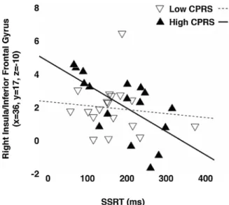
successful stop trials, the same group showed increased activity in the left middle cingulate gyrus (t = -2.067, df = 33, p= .047), left su-perior frontal gyrus (t = -2.145, df = 33,p= .039) and right middle frontal gyrus (t = -2.063, df = 33,p= .047), although these findings did not survive FDR-correction for multiple comparisons. Furthermore, activity in right insula/inferior frontal gyrus correlated negatively with SSRT for children with high ADHD scores (r= -.630,p= .007), but not for those with lower scores (r= -.114,p= .654) (Fig. 3). Groups did not differ in terms of task performance, despite the differences in brain activity.
3.5. Power analysis
The power analysis showed that with the effect-sizes found in the current study, a total of 923 participants would have been required to reach a power level of 0.80 and discriminate between children with ASD and OCD and typically developing volunteers, suggesting that any differences between children with and without diagnoses in terms of
brain activity and taks performance are in the range of noise and un-likely to be clinically meaningful.
4. Discussion
The aim of this study was to investigate the behavioral and neural correlates of cognitive control as assessed using a modified stop-signal task in a multicenter study of children with ASD, OCD and typically developing children. Children with ASD and OCD did not show the expected deficits in cognitive control, nor changes in brain activity in task-relevant neural networks. In fact, the only operationalization of behavioral symptoms that led to findings of changes in brain activity was for symptoms of ADHD. Here, children who had more symptoms showed increased activity in prefrontal regions during cognitive con-trol, compared to children with fewer ADHD symptoms. In addition, decreased cognitive control was associated with higher activation of the right insula/inferior frontal gyrus for the children with more parent-rated symptoms of ADHD.
Fig. 2.Results of the region of interest analysis during failed stopping, showing A) increased left middle frontal gyrus activation in the high CPRS-R group compared
to the low CPRS-R group, B) increased left middle cingulate gyrus activation in the high versus low CPRS-R group, and C) increased right middle cingulate gyrus activation in the high CPRS-R group. Y-axis reflects parameter estimates. The numbers next to the colorbars reflect t-values. Asterisks denote ** p < .01, *** p < .001. CPRS = Conners’ Parent Rating scale – Revised.
B. Gooskens, et al. Developmental Cognitive Neuroscience 36 (2019) 100602
Contrary to our expectations, we found no differences between children with OCD and TD children in stopping speed. Nevertheless, this finding converges with previous work showing similar SSRT in children with OCD and matched typically developing peers (Rubia et al., 2010; Wooley et al., 2008). In contrast, differences in SSRT have been found for adolescents and adults with OCD (Chamberlain et al., 2007; Kang et al., 2013; Penadés et al., 2007; de Wit et al., 2012), suggesting that for individuals with OCD, problems with cognitive control may emerge later in development.
Similarly, we found no differences in task performance between children with ASD and typically developing controls. Earlier findings in ASD have been mixed, with reports of both longer SSRT (Geurts et al., 2004; Lemon et al., 2011) and no difference in SSRT (Adams and Jarrold, 2012; Chantiluke et al., 2015; Ozonoff and Strayer, 1997;
Schmitt et al., 2017). The present findings contribute to a broader cognitive control literature in ASD that shows similar inconsistencies in findings (Ambrosino et al., 2014;Christ et al., 2007;Geurts et al., 2014;
Yerys, 2015), yet converges to suggest that children with ASD may show selective impairments in interference control (Adams and Jarrold, 2012) or proactive mechanisms (Schmitt et al., 2017) in the absence of problems with reactive stopping. Recent work has also suggested that the inconsistencies in findings may be driven by the type of stimulus used (Bos et al., 2018;Kuiper et al., 2016), where the ability to exert cognitive control may be selectively impaired in the context of stimuli that are highly salient to individuals with ASD or OCD (Bos et al., 2018;
Cascio et al., 2014;Kohls et al., 2018). Finally, it is an open question to which extent the existing studies in this area have been affected by co-morbid ADHD symptoms in participants, which are often not assessed. Consistent with our behavioral findings, we found no differences in brain activity during cognitive control in ASD and OCD. Previous stu-dies investigating the neural correlates of cognitive control in children with OCD using stop-signal tasks have reported decreased activation of frontal regions including the orbitofrontal gyrus, mesial/dorsolateral frontal gyrus, anterior cingulate, and insula, as well as subcortical re-gions such as thalamus, caudate, putamen and globus pallidus (Rubia et al., 2010; Wooley et al., 2008). In ASD, earlier studies have reported increased left-hemispheric inferior/middle frontal gyrus and orbito-frontal activation during stopping (Schmitz et al., 2006). Yet, our finding of no differences in frontostriatal brain activation is consistent with a growing body of literature using cognitive control paradigms and showing no differences in brain activation in children with ASD
(Ambrosino et al., 2014;Lee et al., 2009; extensively reviewed in Yerys et al., 2015).
Notably, we found that children with increased ADHD symptoms showed greater activation in left middle frontal gyrus and left/right middle cingulate gyrus during failed stop-trials compared to children with low ADHD symptoms (Fig. 2). These findings are partially in line with reports of increased activation in right dorsolateral prefrontal cortex (DLPFC) in children with ADHD (Pliszka et al., 2006). Yet, children with ADHD have also been reported to show reduced activa-tion in several prefrontal regions compared to typically developing children (Passarotti et al., 2010). Furthermore, at the behavioral level children with ADHD have been reported to have longer SSRTs (Lipszyc and Schachar, 2010), which we did not find in our children with ASD or OCD. Taken together, our findings suggest that children with ASD and OCD do not have a general deficit in cognitive control and associated frontostriatal neural circuits, but that changes in brain activation may rather be driven by the presence of ADHD symptoms.
A strength of our study is that it considers children with ASD and OCD with similar symptoms of compulsion in a single design. However, our findings should also be considered in light of some limitations: the sample size for our fMRI-analyses was relatively small (especially for the OCD-group). This was mostly due to loss of data due to subject motion during the fMRI session. Nevertheless, the remaining sample was well-matched for important demographic confounders. Furthermore, a post-hoc power analysis showed that a vast number of participants (N = 923) would have been necessary to detect between-group differences. Even though we cannot exclude that our finding of no differences between groups may have been caused by a lack of statistical power, it must be noted that the effect sizes for our primary between-group comparisons were extremely small (ηp² < .017),
sug-gesting that the clinical relevance of these effects would be minimal, even in an adequately powered study.
In conclusion, the findings from this study do not support differ-ences in cognitive control or associated neural circuitry in children with ASD and OCD. Instead, our results suggest that changes in cognitive control are more likely to be associated with symptoms of comorbid ADHD than compulsivity or repetitive behavior more generally.
Conflict of Interest
None.
Appendix A. Supplementary data
Supplementary material related to this article can be found, in the online version, at doi:https://doi.org/10.1016/j.dcn.2018.11.004.
References
Abramovitch, A., Dar, R., Mittelman, A., Wilhelm, S., 2015. Comorbidity between at-tention deficit/hyperactivity disorder and obsessive-compulsive disorder across the lifespan: a systematic and critical review. Harv. Rev. Psychiatry 23 (4), 245–262.
https://doi.org/10.1097/HRP.0000000000000050.
Adams, N.C., Jarrold, C., 2012. Inhibition in autism: children with autism have difficulty inhibiting irrelevant distractors but not prepotent responses. J. Autism Dev. Disord. 42 (6), 1052–1063.https://doi.org/10.1007/s10803-011-1345-3.
Ambrosino, S., Bos, D.J., van Raalten, T.R., Kobussen, N.A., van Belle, J., Oranje, B., & Durston, S., 2014. Functional connectivity during cognitive control in children with autism spectrum disorder: an independent component analysis. J. Neural Transm. 121 (9), 1145–1155.https://doi.org/10.1007/s00702-014-1237-8.
Ameis, S.H., Lerch, J.P., Taylor, M.J., Lee, W., Viviano, J.D., Pipitone, J., Nazeri, A., Croarkin, P.E., Voineskos, A.N., Lai, M.-C., Crosbie, J., Brian, J., Soreni, N., Schachar, R., Szatmari, P., Arnold, P.D., Anagnostou, E., 2016. A diffusion tensor imaging study in children with ADHD, autism spectrum disorder, OCD, and matched controls: dis-tinct and non-disdis-tinct white matter disruption and dimensional brain-behavior re-lationships. Am. J. Psychiatry 173 (12), 1213–1222.https://doi.org/10.1176/appi. ajp.2016.15111435.
American Psychiatric Association, 2000. Diagnostic and Statistical Manual of Mental Disorders. Author, Washington, DC (4th ed., text rev.).
American Psychiatric Association, 2013. Diagnostic and Statistical Manual of Mental Disorders, 5th ed. Author, Washington, DC.
Fig. 3.Negative correlation between stop-signal reaction time (SSRT) and right
insula/inferior frontal gyrus activity in children with ASD or OCD who show elevated symptoms of ADHD (solid line,r= -0.630,p= .007). There was no significant correlation in children with low symptoms of ADHD (dashed line,r
Baxter, A.J., Brugha, T.S., Erskine, H.E., Scheurer, R.W., Vos, T., Scott, J.G., 2015. The epidemiology and global burden of autism spectrum disorders. Psychol. Med. 45, 601–613.https://doi.org/10.1017/S003329171400172X.
Benjamini, Y., Hochberg, Y., 1995. Controlling the false discovery rate: a practical and powerful approach to multiple testing. J. R. Stat. Soc. Ser. B 55 (1), 289–300. Berlin, G.S., Lee, H.-J., 2018. Response inhibition and error-monitoring processes in
in-dividuals with obsessive-compulsive disorder. J. Obsessive. Compuls. Relat. Disord. 16, 21–27.https://doi.org/10.1016/J.JOCRD.2017.11.001.
Bodfish, J.W., Symons, F.J., Parker, D.E., Lewis, M.H., 2000. Varieties in repetitive be-havior in autism. J. Autism Dev. Disord. 30 (3), 237–243.https://doi.org/10.1023/ A:1005596502855.
Bos, D.J., Silverman, M.R., Ajodan, E.L., Martin, C., Silver, B., Brouwer, G., Jones, R.M., 2018. Rigidity Coincides With Reduced Cognitive Control to Affective Cues in Children With Autism.PsyArXivhttps://doi.org/10.31234/osf.io/faz45. Carlisi, C.O., Norman, L.J., Lukito, S.S., Radua, J., Mataix-Cols, D., Rubia, K., 2017.
Comparative multimodal meta-analysis of structural and functional brain abnorm-alities in autism spectrum disorder and obsessive-compulsive disorder. Biol. Psychiatry 82, 83–102.https://doi.org/10.1016/j.biopsych.2016.10.006. Cascio, C.J., Foss-Feig, J.H., Heacock, J., Schauder, K.B., Loring, W.A., Rogers, B.P.,
Bolton, S., 2014. Affective neural response to restricted interests in autism spectrum disorders. J. Child Psychol. Psychiatry Allied Discip. 55 (2), 162–171.https://doi. org/10.1111/jcpp.12147.
Chamberlain, S.R., Blackwell, A.D., Fineberg, N.A., Robbins, T.W., Sahakian, B.J., 2005. The neuropsychology of obsessive compulsive disorder: the importance of failures in cognitive and behavioural inhibition as candidate endophenotypic markers. Neurosci. Biobehav. Rev. 29 (3), 399–419.https://doi.org/10.1016/j.neubiorev. 2004.11.006.
Congdon, E., Mumford, J.A., Cohen, J.R., Galvan, A., Canli, T., Poldrack, R.A., 2012. Measurement and reliability of response inhibition. Front. Psychol. 3, 1–10.https:// doi.org/10.3389/fpsyg.2012.00037.
Chantiluke, K., Barret, N., Giampietro, V., Santosh, P., Brammer, M., Simmons, A., Rubia, K., 2015. Inverse fluoxetine effects on inhibitory brain activation in non-comorbid boys with ADHD and with ASD. Psychopharmacology 232 (12), 2071–2082.https:// doi.org/10.1007/s00213-014-3837-2.
Chamberlain, S.R., Fineberg, N.A., Menzies, L.A., Blackwell, A.D., Bullmore, E.T., Robbins, T.W., Sahakian, B.J., 2007. Impaired cognitive flexibility and motor in-hibition in unaffected first-degree relatives of patients with obsessive-compulsive disorder. Am. J. Psychiatry 164 (2), 335–338.https://doi.org/10.1176/ajp.2007. 164.2.335.
Conners, C.K., Sitarenios, G., Parker, J.D., Epstein, J.N., 1998. The revised Conners’ Parent Rating Scale (CPRS-R): factor structure, reliability, and criterion validity. J. Abnorm. Child Psychol. 26 (4), 257–268.https://doi.org/10.1023/
A:1022602400621.
Christ, S.E., Holt, D.D., White, D.A., Green, L., 2007. Inhibitory control in children with autism Spectrum disorder. J. Autism Dev. Disord. 37 (6), 1155–1165.https://doi. org/10.1007/s10803-006-0259-y.
Durston, S., Nederveen, H., van Dijk, S., van Belle, J., de Zeeuw, P., Langen, M., van Dijk, A., 2009. Magnetic resonance simulation is effective in reducing anxiety related to magnetic resonance scanning in children. J. Am. Acad. Child Adolesc. Psychiatry 48 (2), 206–207.https://doi.org/10.1097/CHI.0b013e3181930673.
Fan, S., Cath, D.C., van der Werf, Y.D., de Wit, S., Veltman, D.J., van den Heuvel, O.A., 2017. Trans-diagnostic comparison of response inhibition in Tourette’s disorder and obsessive-compulsive disorder. World J. Biol. Psychiatry 25, 1–11.https://doi.org/ 10.1080/15622975.2017.1347711.
Faul, F., Erdfelder, E., Buchner, A., Lang, A.E., 2009. Statistical power analyses using G*Power 3.1: tests for correlation and regression analyses. Behav. Res. Methods 41 (4), 1149–1160.https://doi.org/10.3758/BRM.41.4.1149.
Flament, M.F., Whitaker, A., Rapoport, J.L., Davies, M., Berg, C.Z., Kalikow, K., Shaffer, D., 1988. Obsessive compulsive disorder in adolescence: an epidemiological study. J. Am. Acad. Child Adolesc. Psychiatry 27 (6), 764–771.https://doi.org/10.1097/ 00004583-198811000-00018.
Geurts, H.M., Verte, S., Oosterlaan, J., Roeyers, H., Sergeant, J.A., 2004. How specific are executive functioning deficits in attention deficit hyperactivity disorder and autism? J. Child Psychol. Psychiatry 45 (4), 836–854.https://doi.org/10.1111/j.1469-7610. 2004.00276.x.
Geurts, H.M., van den Bergh, S.F.W.M., Ruzzano, L., 2014. Prepotent response inhibition and interference control in autism spectrum disorders: two meta-analyses. Autism Res. 7 (4), 407–420.https://doi.org/10.1002/aur.1369.
Geurts, H.M., Corbett, B., Solomon, M., 2009. The paradox of cognitive flexibility in autism. Trends Cognit. Sci. 13 (2), 74–82.https://doi.org/10.1016/j.tics.2008.11. 006.
Goodman, R., Ford, T., Richards, H., Gatward, R., Meltzer, H., 2000. The development and well-being assessment: description and initial validation of an integrated as-sessment of child and adolescent psychopathology. J. Child Psychol. Psychiatry 41 (5), 645–655.
Grzadzinski, R., Dick, C., Lord, C., Bishop, S., 2016. Parent-reported and clinician-ob-served autism spectrum disorder (ASD) symptoms in children with attention deficit/ hyperactivity disorder (ADHD): implications for practice under DSM-5. Mol. Autism 7 (7).https://doi.org/10.1186/s13229-016-0072-1.
Hill, E.L., 2004. Executive dysfunction in autism. Trends Cognit. Sci. 8 (1), 26–32.
https://doi.org/10.1016/j.tics.2003.11.003.
Ivarsson, T., Melin, K., 2008. Autism spectrum traits in children and adolescents with obsessive-compulsive disorder (OCD). J. Anxiety Disord. 22 (6), 969–978.https:// doi.org/10.1016/j.janxdis.2007.10.003.
Jiujias, M., Kelley, E., Hall, L., 2017. Restricted, repetitive behaviors in autism spectrum disorder and obsessive-compulsive disorder: a comparative review. Child Psychiatry
Hum. Dev. 48 (6), 944–959.https://doi.org/10.1007/s10578-017-0717-0. Kang, D.H., Jang, J.H., Han, J.Y., Kim, J.H., Jung, W.H., Choi, J.S., Kwon, J.S., 2013.
Neural correlates of altered response inhibition and dysfunctional connectivity at rest in obsessive-compulsive disorder. Prog. Neuro-Psychopharmacol. Biol. Psychiatry 40, 340–346.https://doi.org/10.1016/j.pnpbp.2012.11.001.
Kaufman, J., Birmaher, B., Brent, D., Rao, U., Flynn, C., Moreci, P., Ryan, M., 1997. J. Am. Acad. Child Adolesc. Psychiatry 36 (7), 980–988.https://doi.org/10.1097/ 00004583-199707000-00021.
Kohls, G., Antezana, L., Mosner, M.G., Schultz, R.T., Yerys, B.E., 2018. Altered reward system reactivity for personalized circumscribed interests in autism. Mol. Autism 9 (9), 1–12.https://doi.org/10.1186/s13229-018-0195-7.
Kuiper, M.W.M., Verhoeven, E.W.M., Geurts, H.M., 2016. The role of interstimulus in-terval and “Stimulus-type” in prepotent response inhibition abilities in people with ASD: a quantitative and qualitative review. Autism Res. 9 (11), 1124–1141.https:// doi.org/10.1002/aur.1631.
Lemon, J.M., Gargaro, B., Enticott, P.G., Rinehart, N.J., 2011. Executive functioning in autism spectrum disorders: a gender comparison of response inhibition. J. Autism Dev. Disord. 41 (3), 52–356.https://doi.org/10.1007/s10803-010-1039-2. Lee, P.S., Yerys, B.E., Della Rosa, A., Foss-Feig, J., Barnes, K.A., James, J.D., Kenworthy,
L.E., 2009. Functional connectivity of the inferior frontal cortex changes with age in children with autism spectrum disorders: a fcMRI study of response inhibition. Cereb. Cortex 19 (8), 1787–1794.https://doi.org/10.1093/cercor/bhn209.
Leyfer, O.T., Folstein, S.E., Bacalman, S., Davis, N.O., Dinh, E., Morgan, J., Lainhart, J.E., 2006. Comorbid psychiatric disorders in children with autism: interview develop-ment and rates of disorders. J. Autism Dev. Disord. 36 (7), 849–861.https://doi.org/ 10.1007/s10803-006-0123-0.
Lipszyc, J., Schachar, R., 2010. Inhibitory control and psychopathology: a meta-analysis of studies using the stop signal task. J. Int. Neuropsychol. Soc. 16, 1064–1076.
https://doi.org/10.1017/S1355617710000895.
Logan, G.D., Cowan, W.B., 1984. On the ability to inhibit thought and action: A theory of an act of control. Psychol. Rev. 91 (3), 295–327. https://doi.org/10.1037/0033-295X.91.3.295.
Lord, C., Rutter, M., Le Couteur, A., 1994. Autism Diagnostic Interview-Revised: a revised version of a diagnostic interview for caregivers of individuals with possible pervasive developmental disorders. J. Autism Dev. Disord. 24 (5), 659–685.
Mancini, C., Cardona, F., Baglioni, V., Panunzi, S., Pantano, P., Suppa, A., Mirabella, G., 2018. Inhibition is impaired in children with obsessive-compulsive symptoms but not in those with tics. Mov. Disord. 33 (6), 950–959.https://doi.org/10.1002/mds. 27406.
Masi, G., Millepiedi, S., Mucci, M., Bertini, N., Pfanner, C., Arcangeli, F., 2006. Comorbidity of obsessive-compulsive disorder and attention-deficit/hyperactivity disorder in referred children and adolescents. Compr. Psychiatry 47 (1), 42–47.
https://doi.org/10.1016/j.comppsych.2005.04.008.
Mayes, S.D., Calhoun, S.L., Mayes, R.D., Molitoris, S., 2012. Autism and ADHD: over-lapping and discriminating symptoms. Res. Autism Spect. Disord. 6 (1), 277–285.
https://doi.org/10.1016/j.rasd.2011.05.009.
McDougle, C.J., Kresch, L.E., Goodman, W.K., Naylor, S.T., Volkmar, F.R., Cohen, D.J., Price, L.H., 1995. A case-controlled study of repetitive thoughts and behavior in adults with autistic disorder and obsessive-compulsive disorder. Am. J. Psychiatry 152 (5), 772–777.https://doi.org/10.1176/ajp.152.5.772.
Moritz, S., Birkner, C., Kloss, M., Jahn, H., Hand, I., Haasen, C., Krausz, M., 2002. Executive functioning in obsessive–compulsive disorder, unipolar depression, and schizophrenia. Arch. Clin. Neuropsychol. 17 (5), 477–483.https://doi.org/10.1016/ S0887-6177(01)00130-5.
Mosconi, M.W., Kay, M., D’Cruz, A.-M., Seidenfeld, A., Guter, S., Stanford, L.D., Sweeney, J.A., 2009. Impaired inhibitory control is associated with higher-order repetitive behaviors in autism spectrum disorders. Psychol. Med. 39 (9), 1559–1566.https:// doi.org/10.1017/S0033291708004984.
Naaijen, J., de Ruiter, S., Zwiers, M.P., Glennon, J.C., Durston, S., Lythgoe, D.J., Buitelaar, J.K., 2016. COMPULS: design of a multicenter phenotypic, cognitive, genetic, and magnetic resonance imaging study in children with compulsive syndromes. BMC Psychiatry 16 (1), 361.https://doi.org/10.1186/s12888-016-1072-6.
Norman, L.J., Carlisi, C., Lukito, S., Hart, H., Mataix-Cols, D., Radua, J., Rubia, K., 2016. Structural and functional brain abnormalities in attention-deficit/hyperactivity dis-order and obsessive-compulsive disdis-order. JAMA Psychiatry 73 (8), 815.https://doi. org/10.1001/jamapsychiatry.2016.0700.
Ozonoff, S., Strayer, D.L., 1997. Inhibitory function in nonretarded children with autism. J. Autism Dev. Disord. 27 (1), 59–77.https://doi.org/10.1023/A:1025821222046. Passarotti, A.M., Sweeney, J.A., Pavuluri, M.N., 2010. Neural correlates of response in-hibition in pediatric bipolar disorder and attention deficit hyperactivity disorder. Psychiatry Res. 181 (1), 36–43.https://doi.org/10.1016/j.pscychresns.2009.07.002. Penadés, R., Catalán, R., Rubia, K., Andrés, S., Salamero, M., Gastó, C., 2007. Impaired
response inhibition in obsessive compulsive disorder. Eur. Psychiatry 22 (6), 404–410.https://doi.org/10.1016/j.eurpsy.2006.05.001.
Pliszka, S.R., Glahn, D.C., Semrud-Clikeman, M., Franklin, C., Perez III, R., Xiong, J., Liotti, M., 2006. Neuroimaging of inhibitory control areas in children with attention deficit hyperactivity disorder who were treatment naive or in long-term treatment. Am. J. Psychiatry 163 (6), 1052–1060.https://doi.org/10.1176/ajp.2006.163.6. 1052.
Rubia, K., Smith, A.B., Brammer, Michael J., Taylor, E., 2003. Right inferior prefrontal cortex mediates response inhibition while mesial prefrontal cortex is responsible for error detection. Neuroimage 20 (1), 351–358.https://doi.org/10.1016/S1053 8119(03)00275-1.
Rubia, K., Cubillo, A., Smith, A.B., Woolley, J., Heyman, I., Brammer, M.J., 2010. Disorder-specific dysfunction in right inferior prefrontal cortex during two inhibition tasks in boys with attention-deficit hyperactivity disorder compared to boys with
B. Gooskens, et al. Developmental Cognitive Neuroscience 36 (2019) 100602
obsessive-compulsive disorder. Hum. Brain Mapp. 31 (2), 287–299.https://doi.org/ 10.1002/hbm.20864.
Scahill, L., Riddle, M.A., McSwiggin-Hardin, M., Ort, S.I., King, R.A., Goodman, W.K., Leckman, J.F., 1997. Children’s yale-brown obsessive compulsive scale: reliability and validity. J. Am. Acad. Child Adolesc. Psychiatry 36 (6), 844–852.https://doi. org/10.1097/00004583-199706000-00023.
Wechsler, D., 2003. Wechsler Intelligence Scale for Children, 4th edn. PsychCorp, San Antonia, TX.
Schmitt, L.M., White, S.P., Cook, E.H., Sweeney, J.A., Mosconi, M.W., 2017. Cognitive mechanisms of inhibitory control deficits in autism spectrum disorder. J. Child Psychol. Psychiatry 59 (5), 586–595.https://doi.org/10.1111/jcpp.12837. Schmitz, N., Rubia, K., Daly, E., Smith, A., Williams, S., Murphy, D.G.M., 2006. Neural
correlates of executive function in autistic spectrum disorders. Biol. Psychiatry 59 (1), 7–16.https://doi.org/10.1016/j.biopsych.2005.06.007.
Shaffer, D., Fisher, P., Lucas, C.P., Dulcan, M.K., Schwab-Stone, M.E., 2000. NIMH di-agnostic interview schedule for children version IV (NIMH DISC-IV): description, differences from previous versions, and reliability of some common diagnoses. J. Am. Acad. Child Adolesc. Psychiatry 39 (1), 28–38. https://doi.org/10.1097/00004583-200001000-00014.
Snyder, H.R., Kaiser, R.H., Warren, S.L., Heller, W., 2015. Obsessive-compulsive disorder is associated with broad impairments in executive function: a meta-analysis. Clin. Psychol. Sci. 3 (2), 301–330.https://doi.org/10.1177/2167702614534210. Stevens, T., Peng, L., Barnard-Brak, L., 2016. The comorbidity of ADHD in children
di-agnosed with autism spectrum disorder. Res. Autism Spectr. Disord. 31, 11–18.
https://doi.org/10.1016/j.rasd.2016.07.003.
Valleni-Basile, L.A., Garrison, C.Z., Jackson, K.L., Waller, J.L., McKeown, R.E., Addy, C.L., Cuffe, S.P., 1994. Frequency of obsessive-compulsive disorder in a community sample
of young adolescents. J. Am. Acad. Child Adolesc. Psychiatry 33 (6), 782–791.
https://doi.org/10.1097/00004583-199407000-00002.
Verbruggen, F., Logan, G.D., 2008. Response inhibition in the stop-signal paradigm. Trends Cognit. Sci. 12 (11), 418–424.https://doi.org/10.1016/j.tics.2008.07.005. Verbruggen, F., Chambers, C.D., Logan, G.D., 2013. Fictitious inhibitory differences: how
skewness and slowing distort the estimation of stopping latencies. Psychol. Sci. 24 (3), 352–362.https://doi.org/10.1177/0956797612457390.
De Wit, S.J., De Vries, F.E., Van Der Werf, Y.D., Cath, D.C., Heslenfeld, D.J., Veltman, E.M., Van Den Heuvel, O.A., 2012. Presupplementary motor area hyperactivity during response inhibition: a candidate endophenotype of obsessive-compulsive disorder. Am. J. Psychiatry 169 (10), 1100–1108.https://doi.org/10.1176/appi.ajp. 2012.12010073.
Wooley, J., Heyman, I., Brammer, M., Frampton, I., McGuire, P.K., Rubia, K., 2008. Brain activation in paediatric obsessive-compulsive disorder during tasks of inhibitory control. Br. J. Psychiatry 192 (1), 25–31.https://doi.org/10.1192/bjp.bp.107. 036558.
Yerys, B.E., 2015. An update on the neurobiology of repetitive behaviors in autism. In: Hodapp, R.M., Fidler, D.J. (Eds.), International Review of Research in Developmental Disabilities, pp. 91–150.
Zandt, F., Prior, M., Kyrios, M., 2007. Repetitive behaviour in children with high func-tioning autism and obsessive compulsive disorder. J. Autism Dev. Disord. 37 (2), 251–259.https://doi.org/10.1007/s10803-006-0158-2.
Zandt, F., Prior, M., Kyrios, M., 2009. Similarities and differences between children and adolescents with autism spectrum disorder and those with obsessive compulsive disorder: executive functioning and repetitive behaviour. Autism 13 (1), 43–57.