61:2 (2013) 1–6 | www.jurnalteknologi.utm.my | eISSN 2180–3722 | ISSN 0127–9696
Full Paper
Jurnal
Teknologi
An Approach to Brain Tumor MR Image Detection and Classification
using Neuro Fuzzy
Mohd Ariffanan Mohd Basri a*, Mohd Fauzi Othmana, Abdul Rashid Husaina
aFaculty of Electrical Engineering, Universiti Teknologi Malaysia, 81310 UTM Johor Bahru, Johor, Malaysia *Corresponding author: [email protected]
Article history Received :31 May 2012
Received in revised form :10 October 2012
Accepted :5 January 2013 Graphical abstract
Abstract
Segmentation is an important step in many applications, being also important in those that deal with medical images. Thresholding is one of the most important and used techniques for image segmentation. Some segmentation techniques based on thresholding are performed in order to segment the tumors. The conventional method used in medicine for brain magnetic resonance (MR) images classification and tumors detection is by human inspection. The use of artificial intelligent techniques, for instance, neural networks, fuzzy logic and neuro fuzzy have shown great potential in this field. Hence, in this study, the neuro fuzzy system or ANFIS is applied for classification purposes. ANFIS is applied to classify the abnormal brain based on the location of the tumors. The performance of the ANFIS classifier is evaluated in terms of training performance and classification accuracy and the results confirmed that the proposed ANFIS classifier has potential in classifying the tumors.
Keywords: ANFIS; MRI; brain tumor; segmentation; classification Abstrak
Pensegmenan merupakan satu langkah yang penting dalam kebanyakan aplikasi, juga penting di dalam perkara yang berkaitan dengan imej perubatan. Pengambangan adalah salah satu teknik yang paling penting dan digunakan untuk pensegmenan imej. Beberapa teknik pensegmenan yang berdasarkan pengambangan dilakukan untuk mengsegmen barah. Kaedah konvensional di dalam bidang perubatan yang digunakan untuk mengklasifikasi dan mengesan barah pada imej resonan magnet (MR) otak adalah secara pemeriksaan manusia. Penggunaan teknik pintar tiruan, sebagai contoh, rangkaian neural, logik kabur dan neuro kabur telah menunjukkan potensi yang besar di dalam bidang ini. Oleh itu, dalam kajian ini, sistem neuro kabur atau ANFIS telah digunakan untuk tujuan pengkelasan. ANFIS telah digunakan untuk mengklasifikasikan otak yang tidak normal berdasarkan kedudukan barah. Prestasi pengkelas ANFIS dinilai dari segi prestasi latihan dan ketepatan pengkelasan dan keputusan mengesahkan bahawa pengkelas ANFIS yang dicadangkan mempunyai potensi dalam mengklasifikasikan barah.
Kata kunci: ANFIS; MRI; barah otak; pensegmenan; pengkelasan
© 2012 Penerbit UTM Press. All rights reserved.
1.0 INTRODUCTION
There are many brain tumors to classify and many different ways to classify them. For example, they are classified by the cell-type of origin, the degree of malignancy, their location, and so forth. Pathological classification means that the microscopic cell features of the tumor and its cell type of origin were used to classify it. Radiological classification means that the location of the tumor was used to classify it. The radiological classification was chosen to classify the brain tumors in this study.
Image processing techniques make it possible to extract
meaningful information from medical images.1 Brain
segmentation, which segments the brain region from medical brain images, is a part of medical image processing. Since MR
images can provide detailed information for various tissues (e.g.,
white matter, grey matter, skull, meninges, other organs, etc)2,3
numerous segmentation algorithms have been proposed for brain segmentation of MR brain images.
Automated segmentation and classification of tumors in different medical images is motivated by the necessity of high accuracy when dealing with human life. Also, computer assistance is demanded in medical institutions due to the fact that it could improve the results of humans in such a domain where false negative cases must be at a very low rate. It has been proven that double reading of medical images could lead to better tumor
detection. Since the cost involved in double reading is very high,good software to assist human in medical institutions is of great interest nowadays.
In this study, the classification of abnormal brain magnetic resonance images by using some prior knowledge like pixel intensity and some anatomical features is proposed. Currently, there is no method widely accepted. Therefore automatic and reliable methods for tumor detection are of great need and interest. The application of neuro fuzzy systems in the classification and detection of data for MR images problems are not fully utilized yet. This includes the clustering and classification techniques especially for MR images problems with huge scale of data and consuming times and energy if done manually. Thus, fully understanding the detection, classification or clustering techniques is essential to the development of neuro fuzzy systems, particularly in medical problems. Furthermore, fuzzy set theory plays an important role in dealing with
uncertainty when making decisions in medical applications.7
Neuro fuzzy systems are fuzzy systems which use ANNs theory in order to determine their properties (fuzzy sets and fuzzy rules) by processing data samples. A specific approach in neuro fuzzy development is the adaptive neuro fuzzy inference system (ANFIS), which has shown significant results in modeling nonlinear functions. The ANFIS learns features in the data set and adjusts the system parameters according to a given error
criterion.5 Successful implementations of ANFIS in biomedical
engineering have been reported, for classification6 and data
analysis.7
The pixel classification of biomedical image using neuro
fuzzy approach is presented in.10 A neuro fuzzy approach was
used to take advantage of neural network’s ability to learn, and membership degrees and functions of fuzzy logic, respectively. An approach for detection and specification of anomalies
present in medical images is proposed in.11 The idea is to combine
three metaphors: Neural Networks, Fuzzy Logic and Genetic Algorithms in a hybrid system. The Neural Network and Fuzzy Logic metaphors are coupled in one system called Fuzzy Neural Networks. The result shows that the suspect regions can be detected by using this technique.
Segmentation technique for magnetic resonance MR images of the brain based on the adaptive fuzzy leader clustering (AFLC)
algorithm has been evaluated in.12 This approach performs vector
quantization by updating the winning prototype of a competitive network through an unsupervised learning process. Segmentation of MR images is formulated as an unsupervised vector quantization process, where the value of a vigilance parameter restricts the number of prototypes representing the feature vectors. The choice of the misclassification rate as a quantitative measure shows that AFLC outperforms other existing segmentation methods.
A novel neuro fuzzy network which can efficiently reason fuzzy rules based on training data to solve the medical diagnosis
problems is proposed in.13 This study proposes a refined K-means
clustering algorithm and a gradient-based learning rule to logically determine and adaptively tuned the fuzzy membership functions for the employed neuro fuzzy network. Experimental results indicated that the proposed neuro fuzzy network with feature reduction can discover very simplified and easily interpretable fuzzy rules to support medical diagnosis.
The results of the studies in the literature have demonstrated that the neuro fuzzy is the promising method to classify the brain tumors. In this respect, in the present study the ANFIS is used for classifying the brain tumors.
2.0 BRAIN TUMOR DETECTION
In order to extract the tumors from the brain images a new approach has been done. The proposed technique involved brain tumor segmentation for the extraction of tumor from MR images. In this study, the proposed techniques have been implemented on a synthetic contrast enhanced MR images. The appearance of the brain tumor in MR images are simulated by synthesizing texture images from real MR images. All the synthetic MR images (total images are 40: 35 images are abnormal and 5 normal) consists of 256 x 256 pixels.
Thresholding is one of the most important and used techniques for image segmentation. In order to extract the interested objects from the image a suitable threshold value is needed in such a way that it separates those objects from the rest. Thus, the global image threshold using Otsu's method is used to computes the threshold value. Then, the computed threshold value is used to convert an intensity image to a binary image.
The morphological operations are well known techniques for the segmentation and enhancement of images and also for recognition purposes. In this study opening and dilation which is a morphological operation are used in order to extract the tumor. Opening and dilation are used to eliminate small details and smoothes the objects respectively.
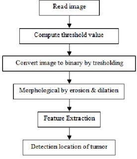
The fundamental objective in object recognition is to utilize a minimum number of features that can be used not only to identify a specific object, but also to distinguish between objects. In order to accomplish this objective, the right combination of distinguishing features needs to be obtained. In this study, it is found that the size of the objects can be used to distinguish tumor and the other objects. The location of the tumor can be found once the tumor is detected. Figure 1 shows the workflow of the proposed technique.
Figure 1 Detection of tumor workflow
An example of image processing in order to detect and find the location of tumor is illustrated in Figure 2.
b i i A a c x x 2 1 1 ) ( Figure 2 A sample of an abnormal Brain with detected tumor
3.0 DATA MODELING
Once the location of the tumor from the abnormal brain images is found, training data sets that contains the desired input/output data pairs of the targeted system is modeled. The coordinated of tumors which are in the matrix form are presented to ANFIS for training membership function parameters. Each row of the training data is a desired input/output pair of the target system to be modeled. Each row starts with an input vector and is followed by an output value. Therefore, the number of rows of the training data is equal to the number of training data pairs, and, since there is only one output, the number of columns training data is equal to the number of inputs plus one.
4.0 ADAPTIVE NEURO FUZZY INFERENCE SYSTEM (ANFIS)
ANFIS is an approach that integrates the interpretability of a fuzzy inference system with the adaptability of a neural
network.5,14,15,16,17 It is an attractive compromise between the
adaptability of a neural network and the interpretability of a fuzzy inference system. Moreover, because ANFIS has much less tunable parameters than traditional neural networks, it has the advantage of being significantly faster and more accurate than
many pure neural network-based methods.18
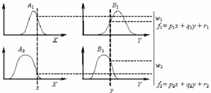
The ANFIS is a fuzzy Sugeno model put in the framework of adaptive systems to facilitate learning and adaptation. Such framework makes the ANFIS modeling more systematic and less reliant on expert knowledge. To present the ANFIS architecture, two fuzzy if-then rules based on a first order Sugeno model are considered:
Rule 1: If x is A1 and y is B1, then f1 = p1x + q1y + r1 Rule 2: If x is A2 and y is B2, then f2 = p2x + q2y + r2
where x and y are the inputs, Ai and Bi are the fuzzy sets, fi
are the outputs within the fuzzy region specified by the fuzzyrule,
pi, qi and ri are the design parameters that are determined during the training process. Figure 3 illustrates the reasoning mechanism for this Sugeno model where it is the basis of ANFIS model.
Figure 3 A two-input first-order Sugeno fuzzy model with two rules
The ANFIS architecture to implement these two rules is shown in Figure 4, in which a circle indicates a fixed node, whereas a square indicates an adaptive node. Adaptive neuro fuzzy inference system basically has 5 layer architectures and each of the function is explained in detail afterwards.
Figure 4 ANFIS architecture
In the first layer, all the nodes are adaptive nodes. The
outputs of layer 1 are the fuzzy membership grade of the inputs, which are given by:
O1,i = µAi(x) i = 1, 2 (1)
O1,i = µBi−2 (y) i = 3, 4 (2)
where µAi(x), µBi−2(y) can adopt any fuzzy membership
function. For example, if the bell shaped membership function is
employed, µAi (x) is given by:
(3)
where ai, bi and ci are the parameters of the membership
function, governing the bell shaped functions accordingly.
In the second layer, the nodes are fixed nodes. They are
labeled with π, indicating that they perform as a simple multiplier. The outputs of this layer can be represented as:
O2,i= wi= µAi(x)µBi(y) i = 1,2 (4)
2
,
1
,
2 , 3i
w
w
w
w
O
i i i i i i i i i i i i i w f w f w O5, 2 2 1 2 1 2 1 1f
w
w
w
f
w
w
w
f
2 2 1 1f w f w f ) ( ) ( 1 1 1 2 2 2 2 1 px q y r w p x q y r w f 2 2 2 2 2 2 1 1 1 1 1 1 ) ( ) ( ) ( ) ( ) ( ) (wx p wyq w r wx p w yq w r fIn the third layer, the nodes are also fixed nodes. They are labeled
with N, indicating that they play a normalization role to the firing strengths from the previous layer. The outputs of this layer can be represented as:
(5) which are the so-called normalized firing strengths.
In the fourth layer, the nodes are adaptive nodes. The output
of each node in this layer is simply the product of the normalized firing strength and a first order polynomial (for a first order Sugeno model). Thus, the outputs of this layer are given by:
O4,i wifi wi(pix qiy ri) i 1,2 (6)
In the fifth layer, there is only one single fixed node labeled
with ∑. This node performs the summation of all incoming signals. Hence, the overall output of the model is given by:
(7)
It can be observed that there are two adaptive layers in this ANFIS architecture, namely the first layer and the fourth layer. In the first layer, there are three modifiable parameters {ai, bi, ci},
which are related to the input membership functions. These parameters are the so-called premise parameters. In the fourth layer, there are also three modifiable parameters {pi, qi, ri},
pertaining to the first order polynomial. These parameters are
so-called consequent parameters.5
4.1 Learning Algorithm of ANFIS
The task of the learning algorithm for this architecture is to tune all the modifiable parameters, namely {ai, bi, ci} and {pi, qi, ri}, to
make the ANFIS output match the training data. When the
premise parameters ai, biand ciof the membership function are
fixed, the output of the ANFIS model can be written as:
(8)
Substituting Eq. (5) into Eq. (8) yields:
(9)
Substituting the fuzzy if-then rules into Eq. (9), it becomes: (10) After rearrangement, the output can be expressed as:
(11) which is a linear combination of the modifiable consequent parameters p1, q1, r1, p2, q2 and r2. The least squares method can
be used to identify the optimal values of these parameters easily. When the premise parameters are not fixed, the search space becomes larger and the convergence of the training becomes
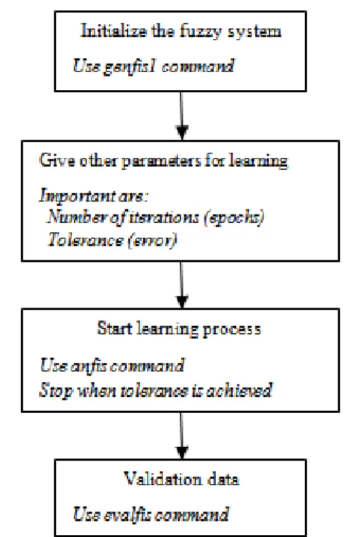
slower. A hybrid algorithm combining the least squares method and the gradient descent method is adopted to solve this problem. The hybrid algorithm is composed of a forward pass and a backward pass. The least squares method (forward pass) is used to optimize the consequent parameters with the premise parameters fixed. Once the optimal consequent parameters are found, the backward pass starts immediately. The gradient descent method (backward pass) is used to adjust optimally the premise parameters corresponding to the fuzzy sets in the input domain. The output of the ANFIS is calculated by employing the consequent parameters found in the forward pass. Figure 5 shows the basic flow diagram of computations in ANFIS.
Figure 5 Basic flow diagrams of computations in ANFIS
4.2 ANFIS Classifier
Both neural networks and fuzzy logic are universal estimators. They can approximate any function to any prescribed accuracy, provided that sufficient hidden neurons and fuzzy rules are available. Gradient descent and Backpropagation algorithms are used to adjust the parameters of membership functions (fuzzy sets) and the weights of defuzzification (neural networks) for fuzzy neural networks. ANFIS applies two techniques in updating parameters. The ANFIS is a FIS implemented in the framework of an adaptive fuzzy neural network. It combines the explicit knowledge representation of a FIS with the learning power of
ANNs.5 The objective of ANFIS is to integrate the best features of
fuzzy systems and neural networks. The advantage of fuzzy set is the representation of prior knowledge into a set of constraints to reduce the optimization research space is utilized. The adaptation of back propagation to structured network to automate fuzzy control parametric tuning is utilized from NN. For premise parameters that define membership functions, ANFIS employs gradient descent algorithm to fine-tune them. For consequent parameters that define the coefficients of each equation, ANFIS uses the least-squares method to identify them. This approach is thus called hybrid learning method since it combines gradient descent algorithm and least-squares method. To achieve good
generalization towards unseen data, the size of the training data set should be at least as big as the number of modifiable parameters in ANFIS. Functionally there are almost no constrains on the node functions of an adaptive network except for the requirement of piecewise differentiability. The neurons in ANFIS have different structures.
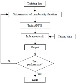
In this study, ANFIS is used as the classifier. ANFIS has to be trained before can be used as a classifier. The training data is a required argument to the ANFIS. To train the ANFIS, the input and output parameter of the membership functions have to be assigned. The task of training modifies all the modifiable parameters of adaptive layers. Once the ANFIS is trained, the testing data is used to validate the accuracy and the effectiveness of the trained ANFIS classifier. To improve classification accuracy the parameters of the membership function are modified to make the ANFIS output approach the expectation output. Figure 6 shows the flowchart of the ANFIS classifier.
Figure 6 Flowchart of ANFIS classification algorithms
5.0 EXPERIMENTS
Various experiments are performed and the sizes of the training and testing sets are determined by taking into consideration the classification accuracies. In this study, a total of 35 data sets (abnormal brain) have been used. The data sets are divided into two separate data sets – the training data sets (20 subjects) and the testing data sets (15 subjects). The training data sets are used to train the ANFIS, whereas the testing data sets are used to verify the accuracy and the effectiveness of the trained ANFIS model for the classification of brain tumors. There are a total of 27 fuzzy rules in the architecture of the ANFIS using a 3 types (generalized bell, triangular and pi) shaped membership function. The ANFIS is implemented by using MATLAB software package.
6.0 RESULTS AND DISCUSSION
The classifier is able to classify the abnormal brain. The ANFIS uses 20 training data in 40 training periods and the step size for parameter adaptation had an initial value of 0.01. At the end of 40
training periods, the final error convergence value is 2.5x10⁻³ as
shown in Figure 7.
Figure 7 ANFIS Errors
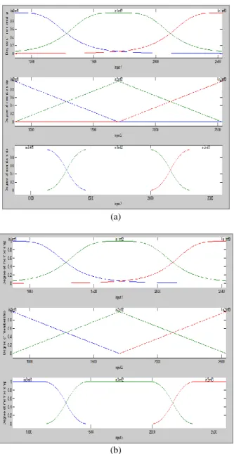
In a real world domain, just like the one used in the present study, all of the features used in the descriptions of instances may have different levels of relevancy. Therefore, in the present study, changes of the final (after training) membership functions with respect to the initial (before training) membership functions of the input parameters are examined. Membership function of each input parameter is divided into three regions, namely, small, medium, and large. The examination of initial and final membership functions indicates that there are considerable changes in the final membership functions but the change is very small. Figure 8 shows the initial and final membership function of the inputs 1, 2 and 3 using the generalized bell, triangular and pi shaped membership function.
Table 1 The ANFIS errors Type of errors Average Training Error 0.0038 Checking Error 0.1296 Global Error 0.0667
(a)
(b)
Figure 8 Membership function: (a) before training, (b) after training
After training, 15 testing data sets are used to validate the accuracy of the ANFIS classifier for the classification of brain tumors. The test results of the ANFIS are presented in Table 2. According to Table 2, 1 subject is classified incorrectly by the ANFIS. Iris data sets are used as the benchmark for this study in investigating the classifier accuracy. The test performance of the ANFIS is determined by the computation of the statistical parameters such as specificity, sensitivity and accuracy. The values of these statistical parameters are given in Table 3. As it is seen from Table 3, the ANFIS classified MRI and Iris subjects with the accuracy of 93.33% and 96%, respectively.
Table 2 The test results of ANFIS
Type of Data Number of subjects
Number of subjects classified correctly MRI 15 14
Iris 25 24
Table 3 The values of statistical parameters Statistical parameters Values
Specificity 93.33%
Sensitivity 96%
Accuracy 94.67%
7.0 CONCLUSION
This paper presents a new application of ANFIS classifier for the MR abnormal brain image classification. The system involves two major modules, one that performs classification and the other one that segment the tumors from the images. In the segmentation phase the system performed well and successfully detects the tumors in the images. Experimental result indicates that the classifier is workable with accuracy greater than 90%. Overall, the system performed well, but there is still a lot can be improved in the future works.
Acknowledgement
This paper is a part of a publication series on Research and Development in Signal, Image and Sensors in Biomedical Engineering Applications.
References
[1] C. Lee, S. Huh, T. A. Ketter, M. Unser. 1998. Comput. Biol. Med. 28: 309.
[2] M. S. Atkins, B. T. Mackiewich. 1998. IEEE T. Med. Imaging. 17: 98.
[3] D. W. Shattuck, S. R. Sandor-Leahy, K. A. Schaper, D. A. Rottenberg, R.
M. Leahy. 2001. Neuroimage. 13: 856.
[4] L. Kuncheva, F. Steimann. 1999. Artif. Intell. Med. 16: 121. [5] J. S. R. Jang. 1993. IEEE T. Syst. Man Cy. 23: 665.
[6] S. Y. Belal, A. F. G. Taktak, A. J. Nevill, S. A. Spencer, D. Roden, S. Bevan. 2002. Artif. Intell. Med. 24: 149.
[7] I. Virant-Klun, J. Virant. 1999. Comput. Biomed. Res. 32:305.
[8] A. Abraham, B. Nath. 2000. A Review of a Decade of Research,
Technical Report, School of Computing and Information Technology. Monash University. Australia.
[9] N. Kasabov. 1996. Foundations of Neural Networks, Fuzzy Systems and
Knowledge Engineering. MIT Press. Cambridge. [10] R. J. Oweis, M. J. Sunna. 2005. J. Electr. Eng. 56: 146.
[11] N. Benamrane, A. Aribi, L. Kraoula. 2006. IEEE Proc. Geom. Model. Imaging.
[12] R. Castellanos, S. Mitra. 2000. IEEE Symp. Comput.-Based Med. Syst.
[13] C. M. Hong, C. M. Chen, S. Y. Chen, C. Y. Huang. 2006. IEEE Int. Joint
Conf. Neural Networks.
[14] F. Frattale Mascioli, G. Martinelli. 1998. Signal Process. 64: 347. [15] J. S. R. Jang. 1996. Proc. Fifth IEEE Int. Conf. 12: 1493.
[16] M. Panella, A. Rizzi, F. M. F. Mascioli, G. Martinelli. 2001. IFSA World Congress and 20th NAFIPS Int. Conf. 1: 340.
[17] J. Jang, C. Sun, E. Mizutani. 1997. Neuro-Fuzzy and Soft Computing. New Jersey: Prentice Hall. USA.