Hamstring Strength and Morphology
Progression after Return to Sport from Injury
JENNIFER L. SANFILIPPO1, AMY SILDER2, MARC A. SHERRY3, MICHAEL J. TUITE4, andBRYAN C. HEIDERSCHEIT1,3,5 1
Departments of Biomedical Engineering and Athletics University of Wisconsin-Madison, Madison, WI;2Departments of Bioengineering and Orthopaedic Surgery Stanford University, Stanford, CA;3Sports Rehabilitation University of Wisconsin Health Sports Medicine, Madison, WI;4Department of Radiology University of Wisconsin-Madison, Madison, WI; and5Department of Orthopedics and Rehabilitation University of Wisconsin-Madison, Madison, WI
ABSTRACT
SANFILIPPO, J. L., A. SILDER, M. A. SHERRY, M. J. TUITE, and B. C. HEIDERSCHEIT. Hamstring Strength and Morphology Progression after Return to Sport from Injury.Med. Sci. Sports Exerc., Vol. 45, No. 3, pp. 448–454, 2013.Purpose: Hamstring strain reinjury rates can reach 30% within the initial 2 wk after return to sport (RTS). Incomplete recovery of strength may be a contributing factor. However, relative strength of the injured and unaffected limbs at RTS is currently unknown. The purpose was to characterize hamstring strength and morphology at the time of RTS and 6 months later.Methods: Twenty-five athletes who experienced an acute hamstring strain injury participated after completion of a controlled rehabilitation program. Bilateral isokinetic strength testing and magnetic resonance imaging (MRI) were performed at RTS and 6 months later. Strength (knee flexion peak torque, work, and angle of peak torque) and MRI (muscle and tendon volumes) measures were compared between limbs and over time using repeated-measures ANOVA.Results: The injured limb showed a peak torque deficit of 9.6% compared to the uninjured limb at RTS (60-Isj1,PG0.001) but not 6 months after. The knee flexion angle of peak torque decreased over time for both limbs (60-Isj1,PG0.001). MRI revealed that 20.4% of the muscle cross-sectional area showed signs of edema at RTS with full resolution by the 6-month follow-up. Tendon volume of the injured limb tended to increase over time (P= 0.108), whereas muscle volume decreased between 4% and 5% in both limbs (PG0.001).Conclusions: Residual edema and deficits in isokinetic knee flexion strength were present at RTS but resolved during the subsequent 6 months. This occurred despite MRI evidence of scar tissue formation (increased tendon volume) and muscle atrophy, suggesting that neuromuscular factors may contribute to the return of strength.Key Words:MAGNETIC RESONANCE IMAGING, KNEE FLEXION TORQUE, REHABILITATION, MUSCLE VOLUME
H
amstring reinjury rates have been reported as high as 30% within the same season for Australian Foot-ballers (19). Incomplete recovery and inadequate rehabilitation have been suggested as explanations for this high reinjury rate (7). In particular, a residual strength deficit in the injured limb is considered one of the primary causes (8,9,23,24,30); however, the extent of strength loss at the time of return to sport (RTS) has not yet been investigated. Persistent strength deficits have been observed in individ-uals with recurrent hamstring injuries, despite returning to athletic competition (9). For example, in comparison to the unaffected limb, a 10% reduction in concentric peak torquewas noted in subjects having experienced injury 2–12 months prior, with a 22% strength deficit during eccentric testing (9). In addition to reduced peak torque production, the knee flexion angle at which peak concentric torque occurs has been found to increase in previously injured limbs (4). This finding suggests that torque production at longer muscle lengths may be compromised. Considering that the susceptibil-ity for sustaining a muscle strain injury is greatest during ec-centric loading in a lengthened position (10,11,17,18,22), these observed strength deficits, particularly at longer muscle lengths, likely increase reinjury risk.
Atrophy of the previously injured muscle may also con-tribute to the persistent strength loss (9). A substantial re-duction in biceps femoris long head volume has been found in 950% of individuals with a prior injury despite having returned to athletic competition (25). Although the rehabili-tative process plays an important role in the return of muscle size after the acute injury (25), changes in the relative amount of connective tissue may also impede recovery (15). Scar tissue adjacent to the site of original injury has been ob-served as early as 6 wk (7) and as late as 23 months after injury (25). The presence of scarring has been shown to al-ter thein vivomuscle contraction mechanics, generating lo-calized regions of high tissue strains near the site of prior Address for correspondence: Bryan C. Heiderscheit, PT, Ph.D., Department
of Orthopedics and Rehabilitation, University of Wisconsin, 1300 Univer-sity Ave., MSC 4120, Madison, WI 53706-1532; E-mail: heiderscheit@ ortho.wisc.edu.
Submitted for publication June 2012. Accepted for publication October 2012. 0195-9131/13/4503-0448/0
MEDICINE & SCIENCE IN SPORTS & EXERCISEÒ CopyrightÓ2013 by the American College of Sports Medicine DOI: 10.1249/MSS.0b013e3182776eff
BASIC
injury of the biceps femoris (26). The presence of scar tis-sue may increase the overall stiffness of the musculoten-don unit (5,14), although this idea has not been scientifically tested. It is likely that, together, these changes in muscle mor-phology after a hamstring strain injury may compromise the return of normal muscle function.
The purpose of this study was to characterize isokinetic hamstring strength and morphology at the time of RTS after a controlled rehabilitation program for an acute strain in-jury. Tests were then repeated 6 months after RTS to provide additional insights into the recovery process over time. This study provides a unique understanding of the extent of heal-ing at the time of RTS and how this healheal-ing progresses over the following 6 months. We hypothesized that, at RTS, the injured muscle would display weakness and associated changes in hamstring muscle morphology based on magnetic reso-nance imaging (MRI).
METHODS
Participants.Twenty-five recreational athletes (20 male and 5 female; age = 24T9 yr, height = 1.7T0.5 m, weight = 73.8 T 25.8 kg) participated in the study. All subjects sustained an acute hamstring strain injury and completed a controlled rehabilitation program. Subjects were recruited from the University of Wisconsin Health clinics and Univer-sity of Wisconsin recreation facilities. To qualify, subjects needed to be 16–50 yr and involved in athletics a minimum of 3 dIwkj1. The subjects must have sustained an acute, sudden-onset hamstring injury within the prior 10 d and dis-play two or more of the following symptoms: palpable pain along any of the hamstring muscles, posterior thigh pain without radicular symptoms during a straight leg raise, weak-ness with resisted knee flexion, or pain with resisted knee flexion. Exclusion criteria included complete hamstring mus-cle disruption (grade 3) or avulsion, posterior thigh pain originating from another source (e.g., inguinal or femoral hernia, nerve entrapment, lumbosacral pathology), or any co-morbidity that might prevent participation in a rehabilitation program. Each subject or parent/guardian provided written informed consent before testing, in accordance with the Uni-versity of Wisconsin’s Health Sciences Institutional Re-view Board.
Protocol.After enrollment, subjects immediately began a controlled rehabilitation program under the supervision of the same physical therapist (M.A.S.). The exercises included in the rehabilitation program were the same as those pre-viously published (3,12). Each subject continued the rehabili-tation program until established RTS criteria were met, including no significant pain with straight leg raise, full iso-metric hamstring strength against manual resistance in prone at 90-and 15- of knee flexion, no tenderness to palpation, and no apprehension during full effort, sport-specific move-ments. Once cleared to RTS by the treating physical therapist, each subject underwent an MRI examination and isokinetic strength assessment; these measures were repeated 6 months
later. Subjects were encouraged to continue the rehabilita-tion exercises (three times per week for 8 weeks) on an inde-pendent basis; rehabilitation compliance after RTS was not monitored.
Strength testing.Subjects were positioned on an iso-kinetic dynamometer (Biodex Multi-Joint System 2; Biodex Medical Systems, Inc., Shirley, NY) such that the hip was flexed to 90-, and the dynamometer and knee joint axes were aligned. Strapping was used over the shank, thigh, and waist to minimize secondary joint movement. Each subject performed maximum effort knee flexion/extension testing through full range of motion during two conditions: con-centric at 60-Isj1(5 repetitions) and concentric at 240-Isj1 (15 repetitions). Eccentric knee flexion testing at 30-Isj1 (3 repetitions) was also performed with the final 13 subjects because this test was added after the study had begun. Be-fore each test, subjects received four submaximal practice trials. Joint angle and torque were recorded after being cor-rected for gravity. Full knee extension was defined as 0-.
Peak torque and angle-to-peak torque for each testing condition were calculated consistent with Brockett et al. (4). All analyses were limited to the repetitions at each speed containing the highest peak torque values (3 repetitions at 60-Isj1, 12 repetitions at 240-Isj1, and 2 repetitions at 30-Isj1). Torque–angle curves from the select repetitions at each speed were compiled and sorted in relation to move-ment direction (i.e., flexion/extension) and knee flexion angle. This resulted in one torque–angle curve for each sub-ject, speed, and direction. Next, every successive block of nine data points from the compiled torque angle curve was replaced with an average value. Finally, for each subject, speed, and direction, a second-order polynomial curve was fit to the torque data that were within 10% of the peak torque measurement for that particular condition. Peak torque and angle-of-peak torque were determined from the resulting poly-nomials. Work was calculated from 0-to 90- of knee flex-ion by integrating knee flexflex-ion torque with respect to time. In addition, the mixed hamstring-to-quadriceps ratio (H:Q) was calculated as hamstring eccentric peak torque at 30-Isj1 relative to quadriceps concentric peak torque at 240-Isj1(19). MRI.Images were obtained for each subject on a 1.5T Twin Speed magnetic resonance scanner (General Electric Healthcare, Milwaukee, WI) using a phased array torso coil. Each MRI examination included three scans: iterative de-composition of water and fat with echo asymmetry and least-squares estimation (IDEAL) combined with three-dimensional spoiled gradient echo imaging (20), T2-weighted fat-suppressed fast spin-echo coronal scan, and T2-weighted fat-suppressed fast spin-echo axial scan. T2 imaging details were as follows: IDEAL coronal three-dimensional slab, TR = 12.5 ms, three echoes (one echo per TR) with TE = 4.4, 5.0, and 6.6 ms, 15-flip angle; matrix,T41.7 kHz bandwidth, 384 256 matrix with 46 46 cm field of view with 84 slices, and 1.4 mm slice thickness for a true spatial resolution of 1.2 1.81.4 mm3(interpolated to 0.9 0.9 7 mm3). Water and fat images were created
BASIC
using homodyne reconstruction performed online (21,31). Coronal T2-weighted scan—4-mm slice thickness, 4.4-mm slice interval, 512512 matrix, 90-flip angle, and 2200/ 9.7 TR/TE. Axial T2-weighted scan—5-mm slice thickness, 5-mm slice interval, 256256 matrix, 90-flip angle, and 3200/89 TR/TE.
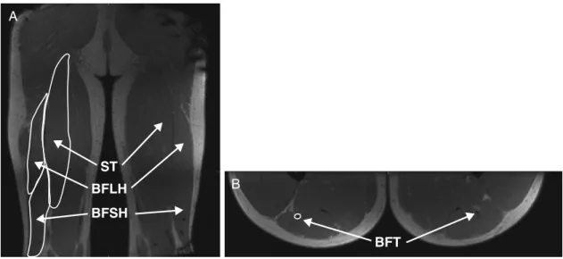
The two MRI examinations were analyzed by the same investigator (M.J.T.) at different time points to avoid biased measurements. The total injured area over all muscles was determined at the level where the injury had the largest abso-lute axial cross-sectional area. Specifically, the cross-sectional area of the injury was calculated from the mediolateral width (ML) and anteroposterior depth (AP) using the formula, 0.25PMLAP (2,7,24,27,30). In addition, muscle and tendon–scar volumes of the biceps femoris long head (BFLH), biceps femoris short head (BFSH), semitendinosus (ST), and proximal conjoint biceps femoris and semitendi-nosus tendon (BFT) were determined at both time points and for both limbs by the same investigator (J.L.S.) using manual segmentation (Mimics Software; Materialize Corp., Ann Arbor, MI) (Fig. 1). The structure boundaries were manually outlined on each coronal slice for muscles and axial slice for tendons in which the structure of interest was
pres-ent. Intraobserver variability for this technique has been reported at G5% for both muscle (13,29) and tendon (25). Volume was then calculated by summing the cross-sectional area of each slice and multiplying by the interslice distance. Absolute muscle volumes were analyzed for the BFLH, BFSH, and ST, whereas the percent difference between limbs at each time point was assessed for the BFT because of its smaller comparative size and potential for error.
Statistical analysis.Three subjects did not undergo testing at the 6-month follow-up because they experienced an injury after RTS during sports participation (two ham-string strains, one anterior cruciate ligament tear). Only the complete data sets from the 22 subjects were included in the analysis, with the exception of the H:Q peak torque ratio, which was based on only 13 subjects. The outcome mea-sures analyzed included peak torque, angle-of-peak torque, work (60-Isj1and 240-Isj1), H:Q peak torque ratio, muscle volumes (BFLH, BFSH, and ST), and tendon volumes (BFT). Two-factor repeated-measures ANOVA (limb-by-time) were performed to compare all outcome measures (PG0.05), ex-cept BFT volumes were compared over time using a depen-dentt-test.Post hoctesting was performed as needed using Tukey HSD (Statistica 6.0; StatSoft, Inc., Tulsa, OK). FIGURE 1—Manual segmentation was used to determine (A) the bilateral volumes of the biceps femoris long head (BFLH), short head (BFSH), and semitendinosus (ST), as well as (B) the tendon–scar tissue volumes of the proximal conjoint biceps femoris–semitendinosis tendon (BFT).
TABLE 1. Isokinetic strength testing measures (meanTSD) at 60-Isj1and 240-Isj1for each limb performed at return to sport (RTS) and 6 months after RTS.
60-Isj1
Limb
Peak Torque (NImIkgj1) Angle of Peak Torque (-)b Work (JIkgj1)
RTSa 6 months RTS 6 months RTSa 6 months
Injured 1.15T0.29 1.30T0.26 39.9T14.6 28.8T11.6 107.0T32.0 126.8T31.0
Uninjured 1.28T0.29 1.28T0.28 40.1T14.1 30.0T15.0 116.6T33.4 124.1T36.2
240-Isj1
Limb
Peak Torque (NImIkgj1)c Angle of Peak Torque (-) Work (JIkgj1)c
RTS 6 months RTS 6 months RTS 6 months
Injured 0.74T0.24 0.80T0.22 43.2T11.1 40.3T8.5 16.7T5.5 18.8T4.8
Uninjured 0.81T0.23 0.85T0.21 45.1T10.2 41.8T8.4 19.0T5.2 20.1T5.2
a
Peak torque (PG0.001) and work (P= 0.003) at 60-Isj1 were less in the injured limb than in the uninjured limb at RTS. bAngle of peak torque at 60-Isj1 for both limbs decreased over time (PG0.001), reflecting a longer hamstring length. cPeak torque (P= 0.021) and work (P= 0.006) at 240-Isj1 were less in the injured limb than in the uninjured limb.
BASIC
RESULTS
MRI examination at the time of injury revealed 16 sub-jects sustained an injury primarily to the biceps femoris; 4, to the semimembranosis; and 2, to the ST. The average time away from sport was 26 d (range = 17–49 d).
Strength.Isokinetic strength testing at 60-Isj1revealed significant limb-by-time interactions for peak torque (P G 0.001) and work (P= 0.002; Table 1). On average, the in-jured limb had a 9.6% deficit in peak torque (P G 0.001) and a 6.4% deficit in work (P= 0.003) at RTS compared to the uninjured limb. These differences resolved by the 6-month follow-up (Fig. 2). The knee flexion angle of peak torque decreased (PG0.001) for both limbs from RTS to the 6-month follow-up, reflecting a shift in peak torque devel-opment to a longer hamstring length (Table 1 and Fig. 2).
Testing at 240-Isj1showed a main effect for limb, with less peak torque (P= 0.021) and less work (P= 0.006) pro-duced by the injured limb compared to the uninjured limb. No significant difference in angle of peak torque was present between limbs or over time.
The H:Q ratio (30-Isj1eccentric : 240-Isj1 concentric) revealed a main effect for limb (P= 0.023), with the injured limb having a smaller ratio compared to the uninjured limb (RTS: injured 1.30T0.26, uninjured 1.62T0.31; 6 months: injured 1.39T0.26, uninjured 1.46T0.15).
Morphology.At the time of RTS, the percent of muscle area showing signs of injury (i.e., T2 hyperintensity) when considering all involved muscles was 20.4% T 19.4%. By the 6-month follow-up, no evidence of injury was visible on MRI (Fig. 3). The muscle volumes of the ST (4.1%,P= 0.024) and BFSH (6.1%,P= 0.010) for both limbs decreased from RTS to 6-month follow-up, whereas the BFLH volume (3.1%,P= 0.078) showed a similar trend. To obtain a
rep-resentation of the overall biceps femoris muscle volume, we summed the BFLH and BFSH volumes and found a signif-icant decrease (5.2%, P= 0.010) for both limbs from RTS to 6 months after RTS (Table 2). Only BFSH showed a significant difference between limbs (P = 0.036), with the muscle volume of the injured limb being larger than the un-injured limb over both time points.
At RTS, the BFT volume of the injured limb was 4.4%T 0.3% smaller than the uninjured limb. At the 6-month follow-up, the injured limb’s BFT volume was 29.9% T 82.9% larger; however, this change over time was not significant (P= 0.108).
DISCUSSION
The purpose of this study was to examine the effects of an acute hamstring strain injury on strength and morphology
FIGURE 2—Representative torque versus angle data for an injured limb, overlaid with a polynomial curve fitted to the top 10% of the torque measures. The increase in torque and decrease in angle of peak torque from time of return to sport (RTS) to the 6-month follow-up is shown.Error barsrepresent the nine averaged data points when the repeated cycles were compiled.
TABLE 2. Volumes (meanTSD) determined from MRI for the biceps femoris long head (BFLH), short head (BFSH), and semitendinosus (ST) muscles.
Limb Volume (mm3) Change over Time (%) RTS 6-month follow-up BFLH Injured 237.2T46.3 229.3T45.0 j3.0T9.2 Uninjured 244.9T49.6 236.9T47.6 j3.2T6.7 BFSHa,b Injured 113.1T35.3 103.5T30.7 j7.5T12.3 Uninjured 103.0T28.0 97.7T25.0 j4.7T10.4 STa Injured 260.2T61.6 250.5T63.1 j3.3T11.2 Uninjured 265.5T66.9 250.3T59.2 j4.9T8.2 Muscle and tendon volumes were estimated at RTS and 6 months after RTS. aMain effect over time (BFSH,P= 0.010; ST,P= 0.024) indicating that the muscle volume of both limbs decreased over time.
bMain effect between limbs (P= 0.036) indicating that muscle volume of the injured limb was larger than that of the uninjured limb.
FIGURE 3—T2-weighted magnetic resonance image shows (A) resid-ual edema in the injured limb (left side of image) at return to sport, with (B) no remaining edema present at the 6-month follow-up.
BASIC
at RTS and after the subsequent 6 months. The injured limb showed a strength deficit at RTS (60-Isj1: peak torque, 9.6%; work, 6.4%) compared to the uninjured limb, with 20% of the cross-sectional area still showing signs of injury on MRI. Six months after RTS, this strength deficit resolved, despite the ST and biceps femoris muscles of both limbs experiencing 4%– 5% atrophy.
This study is the first to assess hamstring strength at the time of RTS after a strain injury. The observed strength deficit of the injured limb at RTS is consistent with the 10% deficit reported in individuals having already returned to sport for periods ranging from 2 to 12 months (9). Unlike Croisier et al. (9), our results showed full strength recov-ery by 6 months after RTS. This difference may be reflec-tive of the greater number of subjects with prior hamstring strain injuries: 46% of the subjects in Croisier et al. (9) com-pared to only 13% (three subjects) of the subjects in the current study.
The strength deficit present at the time of RTS is likely related to the remaining muscle injury observed on MRI (T2 hyperintensity). On average, 20% of the muscles’ cross-sectional area showed signs of injury at RTS with the MRI performed an average of 26 d after injury (range = 13–49 d). These findings are consistent with those of Askling et al. (2); at 21 d after injury, 26% of the muscle area showed signs of injury, with a reduction to 17% by 42 d after injury. In the current study, there was no evidence of remaining injury on MRI performed at the 6-month follow-up. Thus, it appears that healing progressively continues after RTS and is com-pleted within the subsequent 6-month period.
Given the relationship between muscle size and strength, we anticipated muscle hypertrophy would accompany the strength gains. However, our results showed an average at-rophy of 4%–5% in the hamstring muscles of the injured limb from RTS to the 6-month follow-up. This atrophy was not limited to the most involved muscle but was observed in the BFLH, BFSH, and ST, despite the ST being the primary muscle injured in only two subjects and no subjects having primary involvement of the BFSH. Prior work has observed an apparent compensation among agonist muscles after a strain injury. Specifically, a 10% hypertrophy in the BFSH appeared to offset the corresponding 13% atrophy in the BFLH in individuals with a history of strain injury to the BFLH (25). This potential compensation was not observed during the 6-month period after RTS in the current study because both the BFLH and the BFSH showed a similar degree of atro-phy. Consequently, another explanation for the observed strength gains must exist, such as neuromuscular influences. Considering the degree of injury still evident on MRI at RTS, a protective neuromuscular inhibition may exist at that time to limit peak torque and minimize reinjury risk. At the 6-month follow-up, all indication of injury observed on MRI had resolved, and we reemphasize that muscle vol-ume had decreased. Therefore, we propose that part of the strength gains over time can be explained by removal of the neuromuscular inhibition during complete muscle healing,
regardless of muscle volume changes. Indeed, reduced ac-tivation of the hamstring muscles with a corresponding re-duction in peak torque has been observed in individuals after a hamstring strain injury (28). However, because we did not collect electromyography data in the current study, we are unable to confirm this relationship.
It has been suggested that the time of RTS after injury is, in part, influenced by psychosocial factors such as fear and apprehension (1,6). For example, increased fear of movement and reinjury has been associated with decreased perceived function in individuals nearing completion of re-habilitation after anterior cruciate ligament reconstruction (6). Specific to athletes with a recent hamstring strain injury, insecurity when performing a ballistic hip motion has been observed at the time of RTS testing, despite having passed common clinical strength and flexibility tests (1). As such, fear or apprehension within our subjects at the time of RTS may partially explain the corresponding reduction in strength. However, we do not believe these psychosocial factors played a primary role because all subjects in our study were re-quired to complete a variety of sport-specific movements at full effort without apprehension before being cleared to RTS and the isokinetic strength testing.
One likely explanation for the observed hamstring atro-phy may be that the subjects reported a decrease in ath-letic participation from RTS to the 6-month follow-up. On average, our subjects reported participating in sports 5.6T 1.2 dIwkj1before injury and only 3.7T2.1 dIwkj1at the 6-month follow-up. A reduction in activity could also ex-plain the decreased muscle volumes observed in the unin-jured limb. Why there was a reduction in activity level is not fully understood because it could suggest residual symp-toms or be related to a change in athletic season. Many of the subjects in this study were injured during their in-season athletic play. When the 6-month follow-up testing was per-formed, the subjects may have been out-of-season and there-fore less active.
Earlier research has shown that individuals with a history of multiple hamstring strain injuries display a greater knee flexion angle of peak torque (41-) compared to their unin-jured limb (30-) (9). Surprisingly, we did not observe a side-to-side difference in the angle of peak torque at either RTS or the 6-month follow-up. However, two interesting obser-vations were present. First, both limbs displayed an average knee flexion angle of peak torque of approximately 40-at RTS, 10-more flexed than at the 6-month follow-up. Sec-ond, the angle of peak torque for both limbs at the 6-month follow-up (È30-) was consistent with values previously reported among individuals who never incurred a hamstring strain injury (4).
Hamstring-to-quadriceps (H:Q) ratio (30-Isj1 eccentric: 240-Isj1concentric) values less than 1.05 have been pro-posed as a predictor of reinjury (8). We observed H:Q values above this criterion in both limbs at RTS and the 6-month follow-up. Although we did not observe a significant limb-by-time interaction (P= 0.120), we did find the H:Q of the
BASIC
injured limb to be consistently smaller than the uninjured limb. It is important to note that eccentric isokinetic testing at 30-Isj1was added at the midpoint of the study, and only the final 13 subjects completed this testing; thus, further investigation is warranted.
It is widely recognized that scarring accompanies heal-ing after a strain injury. Scar tissue has been observed as early as 6 wk after an initial injury (9) and found to persist on a long-term basis (25). Considering the biceps femoris or ST were the primary muscles injured in 83% of our sub-jects (n= 19/23), we anticipated the BFT volume would be larger in the injured limb and increase across time between measurement sessions. However, the 35% increase in BFT volume between RTS and the 6-month follow-up was not significant (P= 0.108), primarily because of the large vari-ability in volumes changes observed between subjects.
It has been proposed that the increased knee flexion angle of peak torque in injured muscles is a result of scar tissue formation and the effective shortening of the adjacent mus-cle fibers (4). Interestingly, we only observed changes in BFT volume in the injured limb, whereas the shift in angle of peak torque was bilateral. Thus, injury-induced scar tis-sue formation is likely not the sole cause of changes in angle of peak torque. Again, we suggest that neuromuscular in-fluences may be present. Reduced or delayed muscle acti-vation could produce a similar effect, causing peak torque to occur at a greater knee flexion angle as we observed here. Despite being cleared to RTS, the presence of continued edema and strength deficits suggest that an extended con-valescent period may be necessary to achieve full recovery. The required amount of time needed for this to occur is currently unknown. Our findings indicate that recovery occurs by 6 months; however, it is unlikely that this full period is necessary. Future work is needed to more closely identify the time point of recovery. Further, it appears that the battery of clinical tests used in the current study for de-termining clearance of RTS was inadequate because all sub-jects in this study were cleared for RTS despite the presence
of these residual deficits. Inclusion of objective measures such as isokinetic strength seems warranted in determining readiness of RTS. Although a limited number of reinjuries were observed during the 6-month period of this study, we performed a preliminary comparison of the measures at RTS between those who did and did not sustain reinjury. No sig-nificant differences between these subject groups were ob-served for any of our outcome measures. Thus, the influence that the strength deficits and edema present at RTS have on reinjury risk remains an area of future study.
Certain limitations within the study should be consid-ered when interpreting its findings. We opted to report ab-solute muscle volumes rather than normalizing the volume of the injured limb to the uninjured limb. Although nor-malizing would likely reduce the potential error associated with the segmentation process, muscle volume can quickly change with associated changes in physical activity; thus, the uninjured limb would not serve as a proper reference over time. However, because tendon properties are more dif-ficult to change as a result of physical activity (16), we did report normalized tendon volumes.
Our findings indicate that, at the time of RTS after a hamstring strain injury, the injured limb displays a strength deficit compared to the uninjured limb and shows MRI ev-idence of muscle injury. By 6 months after RTS, the strength deficit and signs of injury are fully resolved while scar tissue exists. Bilateral changes in angle of peak torque appear to occur independent of scar tissue formation, suggesting a neuromuscular influence. The influence of these combined factors on reinjury risk is uncertain at this time.
This work was funded by the National Football League Med-ical Charities, the National Institutes of Health (1UL2RR025012) and the University of Wisconsin Sports Medicine Classic Fund.
The authors would like to acknowledge Scott Hetzel for his as-sistance with the statistical analyses.
The authors have no conflicts of interest.
The results of the present study do not constitute endorsement by the American College of Sports Medicine.
REFERENCES
1. Askling CM, Nilsson J, Thorstensson A. A new hamstring test to complement the common clinical examination before return to sport after injury. Knee Surg Sports Traumatol Arthrosc. 2010; 18(12):1798–803.
2. Askling CM, Tengvar M, Saartok T, Thorstensson A. Acute first-time hamstring strains during high-speed running: a longitudinal study including clinical and magnetic resonance imaging findings.
Am J Sports Med. 2007;35(2):197–206.
3. Baquie P, Reid G. Management of hamstring pain. Aust Fam Physician. 1999;28(12):1269–70.
4. Brockett CL, Morgan DL, Porske U. Predicting hamstring strain injury in elite athletes.Med Sci Sports Exerc. 2004;36(3):379–87. 5. Butler DL, Juncosa N. Functional efficacy of tendon repair
pro-cesses.Ann Rev Biomed Eng. 2004;6:303–29.
6. Chmielewski TL, Jones D, Day T, Tillman SM, Lentz TA, George SZ. The association of pain and fear of movement/reinjury with function during anterior cruciate ligament reconstruction rehabili-tation.J Orthop Sports Phys Ther. 2008;38(12):746–53.
7. Connell DA, Schneider-Kolsky ME, Hoving JL, et al. Longitudi-nal study comparing sonographic and MRI assessments of acute and healing hamstring injuries.AJR Am J Roentgenol. 2004;183(4): 975–84.
8. Croisier JL, Ganteaume S, Binet J, Genty M, Ferret JM. Strength imbalances and prevention of hamstring injury in professional soccer players: a prospective study.Am J Sports Med. 2008;36(8): 1469–75.
9. Croisier J, Forthomme B, Namurois M, Vanderthommen M, Crielaard J. Hamstring muscle strain recurrence and strength per-formance disorders.Am J Sports Med. 2002;30(2):199–203. 10. Garrett WE, Safran MR, Seaber AV, Glisson RR, Ribbeck BM.
Biomechanical comparison of simulated and nonstimulated skele-tal muscle pulled to failure.Am J Sports Med. 1987;15(6):448–54. 11. Heiderscheit BC, Hoerth DM, Chumanov ES, Swanson SC, Thelen BJ, Thelen DG. Identifying the time of occurrence of a hamstring strain injury during treadmill running: a case study.Clin Biomech (Bristol, Avon). 2005;20(10):1072–8.
BASIC
12. Heiderscheit BC, Sherry MA, Silder A, Chumanov ES, Thelen DG. Hamstring strain injuries: recommendations for diagnosis, rehabilitation, and injury prevention.J Orthop Sports Phys Ther. 2010;40(2):67–81.
13. Holzbaur KR, Murray WM, Gold GE, Delp SL. Upper limb muscle volumes in adult subjects.J Biomech. 2007;40(4):742–9. 14. Huijing P, Baan G. Myofascial force transmission: muscle
rela-tive position and length determine agonist and synergist muscle force.J Appl Physiol. 2003;94(3):1092–107.
15. Jarvinen T, Jarvinen T, Kaariainen M, Kalimo H, Jarvinen M. Muscle injuries: biology and treatment.Am J Sports Med. 2005;33: 745–64.
16. Kubo K, Kanehisa H, Ito M, Fukunaga T. Effects of isometric training on the elasticity of human tendon structuresin vivo.J Appl Physiol. 2001;91(1):26–32.
17. Lieber RL, Friden J. Mechanisms of muscle injury gleaned from animal models.Am J Phys Med Rehabil. 2002;81(11):S70–9. 18. Lieber R, Friden JJ. Muscle damage is not a function of
mus-cle force but active musmus-cle strain.J Appl Physiol. 1993;74(2): 520–6.
19. Orchard J, Best T. The management of muscle strain injuries: an early return versus the risk of recurrence.Clin J Sports Med. 2002;21(1):3–5.
20. Reeder SB, McKenzie CA, Pineda AR, et al. Water–fat separation with IDEAL gradient-echo imaging.J Magn Reson Imaging. 2007; 25:644–52.
21. Reeder SB, Pineda AR, Wen Z, et al. Iterative decomposition of water and fat with echo asymmetry and least-squares estimation (IDEAL): application with fast spin-echo imaging.Magnet Reson Med. 2005;54:636–44.
22. Schache AG, Koulouris G, Kofoed W, Morris HG, Pandy MG. Rupture of the conjoint tendon at the proximal musculotendinous
junction of the biceps femoris long head: a case report.Knee Surg Sports Traumatol Arthrosc. 2008;16(8):797–802.
23. Schiltz M, Lehance C, Maquet D, Bury T, Crielaard J, Croisier J. Explosive strength imbalances in professional basketball players.
J Athl Training. 2009;44(1):39–47.
24. Schneider-Kolsky ME, Hoving JL, Warren P, Connell DA. A comparison between clinical assessment and magnetic resonance imaging of acute hamstring injuries. Am J Sports Med. 2006; 34(6):1008–15.
25. Silder A, Heiderscheit B, Thelen D. MR observations of long-term musculotendon remodeling following a hamstring strain in-jury.Skeletal Radiol. 2008;37(1):1101–9.
26. Silder A, Thelen DG, Heiderscheit BC. Effects of prior hamstring strain injury on strength, flexibility, and running mechanics.Clin Biomech. 2010;25:681–6.
27. Slavotinek JP, Verrall GM, Fon GT. Hamstring injury in ath-letes: using MR imaging measurements to compare extent of muscle injury with amount of time lost from competition.AJR Am J Roentgenol. 2002;179:1621–8.
28. Sole G, Milosavljevic S, Nicholson H, Sullivan SJ. Selective strength loss and decreased muscle activity in hamstring injury.
J Orthop Sports Phys Ther. 2011;41(5):354–63.
29. Tingart MJ, Apreleva M, Lehtinen JT, Capell B, Palmer WE, Warner JJ. Magnetic resonance imaging in quantitative analysis of rotator cuff muscle volume.Clin Orthop Relat Res. 2003;415: 104–10.
30. Verrall GM, Kalairajah Y, Slavotinek JP, Spriggins AJ. Assess-ment of player performance following return to sport after ham-string muscle strain injury.J Sci Med Sport. 2006;9:87–90. 31. Yu H, Reeder SB, Shimakawa A, Brittain JH, Pelc NJ. Field map
estimation with a region growing scheme for iterative 3-point water– fat decomposition.Magnet Reson Med. 2005;54:1032–9.