Acute Myeloid Leukemia
Frederick R. Appelbaum, Jacob M. Rowe, Jerald Radich, and John E. Dick
Through the hard work of a large number of investi-gators, the biology of acute myeloid leukemia (AML) is becoming increasingly well understood, and as a consequence, new therapeutic targets have been identified and new model systems have been developed for testing novel therapies. How these new therapies can be most effectively studied in the clinic and whether they will ultimately im-prove cure rates are questions of enormous
importance. In this article, Dr. Jacob Rowe presents a summary of the current state-of-the-art therapy for adult AML. His contribution emphasizes the fact that AML is not a single disease, but a number of related diseases each distinguished by unique cytogenetic markers which in turn help determine the most appropriate treatment. Dr. Jerald Radich continues on this theme, emphasizing how these cytogenetic abnormalities, as well as other muta-tions, give rise to abnormal signal transduction and
how these abnormal pathways may represent ideal targets for the development of new therapeutics. A third contribution by Dr. Frederick Appelbaum describes how AML might be made the target of immunologic attack. Specifically, strategies using antibody-based or cell-based immunotherapies are described including the use of unmodified antibod-ies, drug conjugates, radioimmunoconjugates, non-ablative allogeneic transplantation, T cell adoptive immunotherapy and AML vaccines. Finally, Dr. John Dick provides a review of the development of the NOD/SCID mouse model of human AML emphasiz-ing both what it has taught us about the biology of the disease as well as how it can be used to test new therapies. Taken together, these reviews are meant to help us understand more about where we are in the treatment of AML, where we can go and how we might get there.
I. CURRENT STANDARD THERAPYOF ADULT ACUTE MYELOID LEUKEMIA
Jacob M. Rowe, MD*
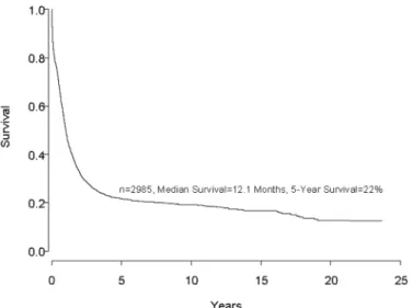
Despite important advances in the therapy of acute my-eloid leukemia (AML) the majority of patients will die from their disease (Figure 1). Progress in therapy and supportive care over the past three decades has led to gradual improvement in the overall results, especially in adults up to age 55-60 years (Figure 2). However, very little progress has been made in the long-term survival of older adults with AML (Figure 3). Since the median age of patients with AML is 64 years, this older group of patients represents the majority with this disease, and the outcome of therapy remains frustratingly disappoint-ing. This section will review current strategies for in-duction and post-remission therapy focusing on newly diagnosed younger adults. Possible strategies for older adults will then be discussed briefly.
It is no longer appropriate to consider all subgroups of AML as a single entity. The most important prognos-tic factors determining the outcome of therapy are the acquired genetic changes in the leukemic cells; deter-mined by conventional cytogenetic techniques, fluores-cence in situ hybridization (FISH) analysis or polymerase chain reaction (PCR). Thus, the following discussion will focus on acute promyelocytic leukemia (APL) and all other AMLs divided into three recognized prognostic groups: those exhibiting favorable, intermediate or un-favorable cytogenetics at presentation (Table 1). Acute Promyelocytic Leukemia—t(15;17)—
PML-RARααααα
APL is the subtype of acute leukemia where the greatest progress has been made over the past decade. It is the most curable subtype of AML and the most important development leading to the dramatic improvement in sur-vival has been the introduction of all-trans retinoic acid (ATRA). While the incorporation of ATRA has led to these remarkable results, differentiation therapy with ATRA is associated with unique toxicities not previously observed with conventional cytotoxic therapy.
* Department of Hematology and Bone Marrow Transplant, Rambam Medical Center, Haifa, 31096, Israel
Induction therapy
Historically, induction therapy for APL included an anthracycline and cytarabine. APL was known to be particularly sensitive to anthra-cyclines, in part due to significantly lower Pgp expression and other resistance markers in APL cells compared to other subtypes of AML.1 It
may also be that for this reason the long-term results in APL with conventional chemotherapy have historically been significantly better than in other forms of AML.2
Several studies have confirmed that both daunorubicin and idarubicin, used as single agents, induce a CR in 60%-80% of patients.3,4
Importantly, a retrospective analysis by the Southwest Oncology Group (SWOG) showed an improved survival in patients with APL when the dose of anthracycline was increased (70 mg/ m2 ) but without a change in cytarabine dose.5 It
has now been established that ATRA is an im-portant part of induction therapy for APL. The benefit of ATRA in induction may not necessar-ily be a dramatic change in the initial CR rate;6
rather, incorporation of ATRA has a major im-pact on the number of patients that may be cured in this disease.7-9
Several studies have shown that when ATRA is combined with single-agent anthracycline the results are at least as good as when cytarabine is also added.4,10 It is likely that the standard form
of induction therapy for APL will rely on ATRA with an anthracycline without the addition of cytarabine (Table 2). Despite theoretic consid-erations, there is no evidence for the superiority of any anthracycline in APL. Using ATRA and an anthracycline, a CR rate of greater than 80% may be reasonably expected. However, despite the remarkable impact of ATRA in the treatment of AML, the induction mortality remains ap-proximately 10%, and acquired retinoid resis-tance contributes to relapse in approximately 20-30% of patients.7-9,11
While the incidence of coagulopathy and bleeding have diminished significantly with ATRA therapy, the retinoic acid syndrome (RAS) is now the major toxicity associated with ATRA. Among patients treated with ATRA alone the incidence is approximately 15%. The mortality from this syndrome has declined over time, likely reflecting earlier recognition and in-stitution of dexamethasone. Furthermore, the concurrent administration of chemotherapy with ATRA may decrease the incidence of RAS.13
Figure 1. Survival of almost 3,000 consecutive patients treated on ECOG protocols for newly diagnosed acute myeloid leukemia (AML) since 1973. The only exclusion from this curve is acute promyelocytic leukemia (APL) patients treated on all-trans retinoic acid (ATRA).
Figure 2. Patients ≤≤≤≤≤ 55 years with newly diagnosed acute myeloid leukemia (AML) treated on Eastern Cooperative Oncology Group (ECOG) protocols since 1973.
Figure 3. Patients > 55 years with newly diagnosed acute myeloid leukemia (AML) treated on Eastern Cooperative Oncology Group (ECOG) protocols since 1973.
Post-remission therapy
Although CR may be achieved using ATRA alone, most patients relapse with this therapy. Consolidation chemo-therapy after CR is mandatory, although the best form of such therapy is unknown. In most studies consolida-tion chemotherapy has been anthracycline-based, and several studies have included high-dose cytarabine.7,9,12
However, the administration of lower doses of cytarabine appears to be just as efficacious,13 and a recent
prospec-tive study has suggested that patients do as well without cytarabine in either induction or consolidation.10 Thus,
it is likely that, just as in induction, there is probably little role for cytarabine in consolidation, although this remains a subject of study in current clinical trials. Most centers administer at least two courses of
post-remis-sion therapy following induction with ATRA and anthra-cyclines, although, as in all types of AML, there are no prospective data establishing the number of courses of intensive post-remission consolidation.14 Clearly, the
ob-jective of post-remission therapy is complete eradica-tion of the leukemic clone with lack of deteceradica-tion of PML-RARα by PCR. This remains critical as the persistence of such minimal residual disease predicts for relapse.9
Maintenance therapy
Randomized trials have suggested that maintenance therapy with ATRA is a critical component of therapy for APL.7,8 There has also been a suggestion that when
ATRA maintenance is combined with low-dose chemo-therapy this may further improve the long-term survival.8
Thus, it appears that patients with APL may benefit from maintenance ATRA, with or without continuous low-dose chemotherapy, particularly those patients who present with higher risk for recurrent disease.8
Investigational approaches
While the outlook for patients with APL has improved dramatically over the past decade, complacency is to be discouraged. Over 20% of patients presenting with APL will die from this disease. This includes some
pa-Table 1. Cytogenetic classification.
SWOG Criteria27 MRC criteria2: As for SWOG, except:
-Favorable t(15;17) – with any other abnormality
inv(16)/t(16;16)/del(16q) – with any other abnormality
t(8;21) – without del(9q) or complex karyotype t(8;21) – with any other abnormality
Intermediate +8, -Y, +6, del(12p) abn 11q23
normal karyotype del(9q), del(7q) – without other abnormalities
Complex karyotypes (≥ 3 abnormalities, but < 5 abnormalities) All abnormalities of unknown prognostic significance
Unfavorable -5/del(5q), -7/del(7q),
t(8;21) with del(9q) or complex karyotype inv(3q), abn 11q23, 20q,
21q, del(9q), t(6;9) t(9;22), abn 17p,
Complex karyotypes (≥ 3 abnormalities) Complex karyotypes (≥ 5 abnormalities)
Unknown All other clonal chromosomal aberrations with fewer than 3 abnormalities
Abbreviations: SWOG, Southwestern Oncology Group; MRC, Medical Research Council; abn, abnormalities of
Table 2. Treatment of acute promyelocytic leukemia [t(15;17) or PML-RARα α α α α postitive]... INDUCTION ATRA + anthracycline-based chemotherapy
(i) anthracycline alone if cannot give ATRA (ii) probably, no need for cytarabline
CONSOLIDATION Anthracycline-based chemotherapy x 1 – 2 cycles role of cytarabine unproven
MAINTENANCE ATRA ± low-dose chemotherapy
Current investigation Role of arsenic in patients who have not relapsed
Possible future investigation Role of autologous PBSC transplants in patients who have become negative for PML-RARα
tients who relapse even after achieving PML-RARα mo-lecular negativity after induction and consolidation therapy.9 Arsenic trioxide has been established as an
im-portant agent for the treatment of ATRA-resistant APL. Whether this should be incorporated into the therapy of newly diagnosed APL is being currently investigated in clinical trials.
Allogeneic stem cell transplant has no role in APL in first remission. However, impressive results have been shown using autologous transplant for relapsed APL using cells that are molecularly negative.15 This has never
been prospectively studied in patients in first remission who have been successfully treated with ATRA-anthra-cyclines. It is speculative whether such therapy, using peripheral blood with its attendant low mortality, can further improve the long-term cure rate.
Therapy for Acute Myeloid Leukemia Other Than Acute Promyelocytic Leukemia
Induction therapy
Classic studies by the CALGB two decades ago resulted in the development of the standard induction regimen, which consists of daunorubicin, 45mg i.v. for three days, and cytarabine, 100mg i.v. by continuous infusion for seven days.16 For patients less than 55 to 60 years old,
an initial CR of 60-75% can be expected. However, mul-tiple randomized studies compared daunorubicin at a dose of 45mg/m2—while keeping the dose of cytarabine
constant—with idarubicin,17 amsacrine,18 aclacinomycin
A19 and mitoxantrone.20 Virtually all of these agents have
been shown to be either unequivocally superior, or at least with a strong trend towards an improvement, when compared with 45mg/m2 of daunorubicin. Thus, for
pa-tients not treated on a clinical trial it is no longer appro-priate to use 45mg/m2 of daunorubicin; rather, a higher
dose of daunorubicin should be used or an alternative anthracycline or anthraquinone, such as idarubicin or mitoxantrone.
Over the years many variations of the stan-dard 3+7 regimen have been developed and all yield approximately similar results. Intensify-ing induction therapy through the use of higher doses of cytarabine or the addition of etoposide, while not affecting the initial CR, clearly may have an effect on the disease-free survival.21
However, although intensifying induction therapy may affect the duration of remission in AML, it is not clear that the increased toxicity through more profound myelosuppression is ad-vantageous given the possibility that a similar intensification might be safely added during postremission therapy.22
Currently, the 3+7 induction regimen is
rec-ommended for all newly diagnosed patients with AML, including those who present with unfavorable cytoge-netics. The latter group also includes most patients with therapy-related or other secondary leukemias. Although multiple publications have advocated alternative regi-mens,23 there are no data that any form of therapy
pro-vides a better outcome than standard induction therapy consisting of an anthracycline and cytarabine. Three large cooperative groups that evaluated this reported CR rates of 55-58% in adult patients presenting with unfa-vorable cytogenetics (Table 3).2,24-27
Postremission Therapy
While induction therapy may be identical, the choice of postremission therapy must be determined by the prog-nostic group, most importantly, the cytogenetics at pre-sentation (Table 4).
AML with favorable cytogenetics
Although over the past decade there have been several large prospective studies24-26,28 of postremission therapy
in AML, only two of the large studies have rigorously analyzed post-remission data by cytogenetic prognostic groups.2,27 The initial response rate to induction in
pa-tients with favorable cytogenetics was approximately 85%. With intensive postremission therapy the overall survival at 5 years exceeds 50%.2,27 There are many
che-motherapeutic strategies for postremission therapy, al-though it is al-thought by many that high-dose cytarabine is a critical element for the success of postremission therapy.29 However, while the data confirm its
effective-ness as postremission therapy, it is doubtful that we need to subscribe to a dogma that one cannot do the same with other regimens. A recent publication, based on CALGB data, suggested that it may be inappropriate not to administer 3-4 cycles of high-dose cytarabine to pa-tients with the t(8;21) abnormality, in which the disease-free survival was about 60%.29 However, the MRC
re-Table 3. Results of induction therapy in adults with acute myeloid leukemia according to cytogenetic prognostic groups.
Favorable Intermediate Unfavorable
n CR n CR n CR
MRC2 (excluding children) 289 90% 853 84% 130 57%
ECOG/SWOG27 121 84% 278 76% 184 55%
GOELAM24 48 87% 226 76% 36 58%
Results of induction therapy from 3 cooperative groups. Identical standard induction therapy was used in all cytogenetic subtypes. A remarkable concor-dance among the 3 groups is demonstrated, although a rigorous comparison cannot be made due to minor differences among the studies in the classifica-tion of the cytogenetic prognostic groups.
Abbreviations: MRC, Medical Research Council; ECOG, Eastern Cooperative Oncology Group; SWOG, Southwestern Oncology Group; GOELAM, Groupe Ouest-Est Leukemies Aigues Myeloblastiques
ported an identical disease-free survival in a much larger cohort of patients without using high-dose cytarabine.2,25
Whether autologous stem cell transplantation should be offered as a part of postremission strategy to patients with favorable cytogenetics remains controversial. Data from the CALGB have suggested that intensive chemo-therapy yields results that are unlikely to be improved by the substitution of autologous transplantation.29 In
contrast, the US Intergroup Study26,27 suggested that
au-tologous transplantation may be particularly useful in this group of patients. In the MRC AML 10 study, pa-tients were randomized to receive autologous stem cell transplant after four cycles of therapy and this was com-pared with an observation arm.25 The patients with
fa-vorable cytogenetics had a markedly lower relapse rate than patients who did not receive an autologous trans-plant, although a high procedural mortality rate in adults (18%) resulted in being ultimately no difference in the overall survival. The reported data need to be cautiously interpreted and may be influenced by small cohorts of patients, as in the CALGB data29 or the US Intergroup
Study.26 Except for young patients in whom fertility
re-mains a consideration, it is probably reasonable to use pe-ripheral autologous stem cell transplantation in experienced centers that have demonstrated a consistently low morbid-ity and a therapy-related mortalmorbid-ity of less than 3-5%.
None of the randomized studies demonstrated an advantage for allogeneic transplant for this group of pa-tients, and given the relatively high transplant-related mortality, this procedure cannot be recommended as stan-dard therapy for such patients. Whether newer methods using less severely myeloablative regimens and relying on the immunological effect of GVL may yield improved results remains to be determined in prospective studies.30
AML with intermediate risk cytogenetics
If an HLA-matched family donor is available, it seems likely that this should be the recommended therapy for patients up to age 55-65 years. Data have consistently shown that this form of therapy provides the best anti-leukemic effect as judged by the relapse rate.31,32 The
study with the largest cohort of prospectively evaluated patients with this subgroup of AML have reported a 3-year survival rate of 65% with a relapse risk at 3 3-years of 18%.2,25 It should be noted, however, that this
advan-tage for allogeneic transplant was not demonstrated in the US Intergroup Study,26,27 albeit in a smaller cohort of
patients.
Timing of allogeneic transplantation in first remis-sion has never been prospectively established. For lo-gistic reasons it may often be necessary to administer chemotherapy after achievement of CR until the avail-ability of a donor and transplant center is established. However, retrospective analysis from the IBMTR sug-gests that for patients proceeding to allogeneic trans-plantation in AML there is no added benefit in receiving additional postremission therapy, and if an HLA-matched donor is available, based on the current available data, patients should be referred for this procedure as soon as possible.33
Patients who do not have an HLA-matched sibling should receive intensive postremission chemotherapy using high-dose cytarabine or similar regimen. The op-timal dose of high-dose cytarabine—anywhere from 1.5 g/m2 to 3 g/m2—the optimal duration and the total
num-ber of doses have never been prospectively established. Only one retrospective analysis, combining several his-toric studies, suggested that three cycles of high-dose cytarabine are better than a single course.29 While these
are important practical questions affecting the
manage-Table 4. Suggested schema for the post-remission therapy of adults with newly diagnosed acute myeloid leukemia (except APL) according to cytogenetic prognostic groups.
Favorable Intermediate Unfavorable Induction
Standard anthracycline + cytarabine (3 + 7) or similar
Post Remission Therapy
(i) HLA-matched family High-dose cytarabine or Allogeneic transplant as soon as possible
donor available similar x 2-3 cycles
(ii) No donor ± autologous peripheral High-dose cytarabine or High-dose cytarabine or
stem cell transplant similar x 2-3 cycles similar x 2-3 cycles autologous transplant ± autologous transplant
Future Investigations Addition of gemtuzumab ozogamicin either to Allogeneic transplant from intensive chemotherapy or pre-autologous transplant alternative donors –
matched unrelated (MUD) or haploidentical
Because of the extremely high rate of relapse in this group of patients and the poor long-term outcome, it is important to note that select reports of alternative do-nors in this group of patients, which used either matched unrelated donors or haploidentically matched family donors, have shown long-term survival rates of 40-50% in patients undergoing such procedures in first remis-sion.36-39 Whether such information on select patients can
be applied to the group as a whole remains to be deter-mined in clinical trials.
Bone Marrow Transplantation
Over the past decade a major effort was made to deter-mine the role of bone marrow transplantation (BMT) in adult AML, especially autologous transplantation, com-pared to intensive chemotherapy. Several large prospec-tive studies were designed and much effort was expended on these trials.9,24,26,29 The results have been confusing
and the data difficult to interpret. It seems that a decade later this issue has still not been resolved. However, cer-tain points need to be emphasized.
1. The preponderance of the data demonstrate that autologous transplants provide better antileukemic ac-tivity than intensive chemotherapy, as judged by the re-lapse rate (Table 5).
2. Historically, the superior antileukemic benefit of autotransplants was negated by the high procedural mor-tality of autologous transplants (14% in the US Inter-group study and 18% in the MRC AML 10 study). Were this mortality to be reduced by about 10% then the con-clusions from these studies may be quite different. Cur-rent use of peripheral blood as a source of stem cells for autotransplants is associated with an extremely low pro-cedural mortality.35
3. All the major reported trials used bone marrow as the source of the stem cells and may not be relevant to current clinical practice in many centers. Aside from the higher procedural mortality, the morbidity when bone ment of many patients with AML, there is little current
enthusiasm among cooperative groups to study this pro-spectively in clinical trials.
There are multiple reports of autologous transplants for AML including many patients with intermediate cyto-genetics. However, it is difficult to identify and analyze large cohorts of patients who have received this therapy. The MRC study reported a relapse rate of 35% for pa-tients who have also received an autotransplant com-pared with 55% among patients receiving intensive che-motherapy only. The 5-year survival was 56% versus 48%.2 It is generally assumed that those patients going
on to an autologous transplant should receive prior in-tensive chemotherapy as the best method of in vivo purg-ing. For such patients the intensity of postremission therapy as well as the number of cycles required are unknown and have also never been prospectively stud-ied. Although some of the best results from phase II data of autologous transplants in AML have been reported when patients received no postremission therapy prior to the transplant,34 the preponderance of the data
sug-gest that using several cycles of postremission therapy should be given prior to transplant. Preliminary data have also reported that peripheral blood stem cells can be re-liably collected after two cycles of intensive chemo-therapy such as high-dose cytarabine with a subsequent very low transplant-related mortality rate.35 Several
coop-erative groups are currently evaluating the role of gemtu-zumab ozogamicin, a humanized monoclonal antibody against CD33 linked to calicheamicin, given in addition to high-dose cytarabine or prior to autologous transplant. AML with unfavorable cytogenetics
This group has long been recognized to have the poor-est outcome among patients with AML. While the ini-tial response rate may exceed 50% (Table 3), the overall long-term survival remains poor, whatever mode of postremission therapy is employed. If a family-matched HLA donor is available, patients should be referred for this procedure as soon as possible after induction therapy. Although this probably represents current practice there aren’t abundant prospective data that support this. The MRC AML 10 study reported that among the group with unfavorable cytogenetics allogeneic transplant did not offer any advantage.2 However, there were only 13
pa-tients in this group. In contrast, the US Intergroup study reported a clear advantage for patients undergoing an allogeneic transplant27 with a 5-year survival of 44%
compared to 15% for patients undergoing chemotherapy. However, once again, this was based on a very small subgroup of 18 patients using an intent-to-treat analysis and only 11 patients actually received this form of therapy. Nevertheless, this appears to represent a therapy with the greatest potential for prevention of relapse.
Table 5. Relapse following allogeneic transplant, autologous transplant and chemotherapy.
Allogeneic Autologous Chemo-Study transplant transplant therapy GIMEMA, 199528 24% 40% 57%
GOELAM, 199724 28% 45% 55%
*MRC, 199825 19% 35% 53%
ECOG/SWOG, 199826 29% 48% 61% * Data for children excluded. In the MRC study, BMT was compared with an observation arm after 4 cycles of chemotherapy, rather than a direct comparison with high-dose chemotherapy as in the other studies.
Abbreviations: See Table 3; GIMEMA, Gruppo Italiano Malattie Ematologiche Maligne dell’Adulto
marrow is used is considerable with a median time to neutrophil recovery of approximately 25 days and over 7 weeks for platelet recovery. With such morbidity and mortality, using bone marrow, there had to be a vastly superior advantage to transplants for this to be consid-ered a better option than chemotherapy.
4. However, most importantly, data from these stud-ies are impossible to interpret due to the small number of patients analyzed per subgroup. The first disappoint-ment was the very high dropout rate of patients, typical in all transplant studies. This dropout occurs at several stages. Only a proportion of patients eligible for ran-domization after induction actually go on to randomiza-tion. Also, a significant number of randomized patients never go on to receive the transplant. In the US Inter-group study approximately half of the patients who were randomized to an autotransplant never received one.26
The validity of intent-to-treat analyses is uncertain when half of the patients do not receive their intended therapy. Furthermore, when these studies were designed over a decade ago AML was considered as a single entity and the number of patients for these studies was determined based on this consideration. With identification of the importance of prognostic groups, it became crucial to consider each of the prognostic subgroups. Once the data for these subgroups are broken down, the number in each patient group is extraordinarily small making any analy-sis difficult and potentially misleading. As an example, the US Intergroup study26 accrued over 800 patients, and
116 were assigned to autologous transplant. However, only 63 (54%) of these 116 patients actually completed this therapy. These 63 patients were then divided into 3 subgroups: favorable, intermediate and unfavorable cy-togenetics, leading to an analysis based on numbers that statisticians would never have agreed to had this been defined as the primary endpoint at the time of design of the study.27 In fact, it has been estimated that in order to
conduct such a study in AML patients and obtain mean-ingful results with data that could be reliably analyzed, an excess of 7,000 patients would be required. Thus, it is unlikely that such a study will ever be carried out, which is the main reason why conflicting results from these transplant studies have been reported, especially when analyzed by subgroups. The most telling example of this may be gleaned from the analysis of the value of high-dose cytarabine in AML patients with favorable cy-togenetics. The results from the CALGB studies report a disease-free survival in excess of 60%.29 In contrast,
the US Intergroup study reported that for this group of patients the 5-year survival was only 35%. This same study reported that when the identical therapy was ap-plied to a group with less favorable cytogenetics —the intermediate group—the 5-year survival was 55%. These data, while providing some biologic information,
emphasize the pitfalls of drawing conclusions when small cohorts are involved and the inability to compare data between different studies.
Acute Myeloid Leukemia in Older Adults
Older adults have a dismal long-term prognosis that has not improved much over the past two decades (Figure 3). They have more unfavorable prognostic factors at presentation and their treatment is made more difficult by their inability to withstand intensive chemotherapy.40
Further, the type of postremission therapy that they re-ceive is generally considered to be sub-optimal even for the more favorable prognostic groups seen in younger adults. The critical factor is that a biologically unfavor-able disease is treated sub-optimally.
Older adults who do not have significant co-mor-bidities should be treated with standard induction therapy. With such therapy 50% of such patients can achieve a CR.41,42 The major difficulty relates to the selection of
postremission therapy. Though patients can generally tolerate one cycle of cytarabine given at somewhat lower doses than is usually given for younger adults,41 it has
never been shown that this improves the long-term out-come. This population represents the majority of indi-viduals with AML, and major efforts are needed to de-termine the best therapy in this group of patients with the aim of achieving a possible cure in some patients and a prolongation of the disease-free survival in many others. Maintenance therapy has been studied in the past with some evidence that this can prolong the disease-free survival.43 Current strategies aim to emphasize
non-myeloablative immune modulation following induction and limited intensive post-remission therapy and include phase III studies evaluating the role of IL-2/histamine, IL-2 (CALGB) and Flt 3 ligand (ECOG/SWOG/ CALGB). Future strategies to prolong the disease-free survival in older adults include proposed studies of gemtuzumab ozogamicin, farnesyltransferase inhibitors and bcl-2 antisense oligonucleotides. Clearly, this is the area with the greatest challenge in AML: applying a less severely myeloablative form of postremission therapy that may, nevertheless, cure the most unfavorable prog-nostic type of the AML. Achieving this aim has so far been elusive. Such breakthroughs will, likely, also ben-efit younger adults.
II. MOLECULAR TARGETSIN ACUTE MYELOID LEUKEMIA
Jerald Radich, MD*
Acute myeloid leukemia (AML) is a heterogeneous dis-ease characterized by a myriad of genetic defects. These include translocations involving oncogenes and
transcrip-tion factors, activatranscrip-tion of signal transductranscrip-tion pathways, and alterations of growth factor receptors. If each type of genetic lesion found in AML involved a distinct pro-cess to cause leukemia, then “targeted” therapy directed at specific genetic lesions would be futile—quite sim-ply, there would be too many potential targets. How-ever, it appears that many of the pathways perturbed in leukemogenesis interact, so that a limited number of specific targets may be useful in a wide variety of AML cases. Indeed, if multiple genetic abnormalities are needed to cause and sustain AML, then the blunting of a single aberrant pathway may be enough to eliminate the proliferative advantage and curb the disease.
In this short review we will examine the signaling pathways involved in leukemogenesis, with specific em-phasis as to how these pathways can be utilized as tar-gets for novel therapy.
Normal Signal Transduction
Signal transduction pathways are designed to translate extracelluar signals (e.g., stimulation to respond to cytokine ligands, interferons) into intracellular action (proliferation, differentiation, survival). Perhaps the best understood pathway involves signaling utilizing the ras family of guanosine nucleotidases (GTPases). A highly simplified cartoon of the ras signal transduction path-way is shown in Figure 4 (see color page 541). The most important components include the following:
Receptor tyrosine kinase (RTK): These ligand binding receptors include PDGF, Fms, c-kit, and Flt 3.1-4 In general their structure includes an
extracel-lular ligand binding region consisting of 5 immuno-globulin-like domains, transmembrane and juxta-membrane domains, and an intracellular domain with kinase activity (Figure 5; see color page 541). Grb-2 and SOS: Grb-2 is an adaptor protein that functionally bridges the association of the RTK with ras. SOS is a guaninine nucleotide exchange pro-tein that facilitates ras-GDP→ras-GTP exchange.
Ras.Harvey (H), Kirsten (K), and N-ras are 21 kd GDP/GTP-binding proteins that serve as the hub of signal transduction.5,6 All three ras proteins are
ex-pressed in most tissues, but the constellation of
muta-tions (i.e., N-ras vs. K-ras) tend to be disease spe-cific.
GAP, NF-1: These are GTPase activating proteins, that catalyze the inherently slow GTPase activity of ras, converting active GTP to an inactive ras-GDP form.7
Briefly, extracellular ligand (L) interacts with the RTK that causes receptor dimerization (Figure 4). This prompts activation of the RTK and subsequent receptor autophosphorylation. This phosphorylated RTK can in turn phosphylate and activate Grb-2, the adaptor pro-tein, which when coupled to SOS, causes activation of the ras. Inactive ras remains bound with GDP, and the interaction with Grb2-SOS causes ras to become acti-vated by GTP binding. This produces a conformational change in ras, and promotes interaction with downstream effector proteins. Since ras has an intrinsically slow GTPase activity, the switch back to the inactive ras-GDP state necessitates the activity of the GTPase-activating proteins (p210 GAP and NF-1).
Activation of ras causes activation of several down-stream pathways, which may effect cell proliferation, differentiation, and apoptosis.8-12 The serine/threonine
kinase Raf is activated by direct association with ras, and in turn activates the MAP/ERK kinase (MEK), which activates downstream mitogen-activating protein kinases (MAPK), such as extracellular signal-regulated kinases (ERK 1 and 2).13 These in turn phosphylate cytoplasmic
targets (Rsk, Mnk) that translocate to the nucleus, caus-ing activation of transcription involved in proliferation. Ras activation also may influence cytoskeleton organi-zation through activation of Rac and Rho. In addition, ras may promote cell cycling through the activation of Cyclin D dependent kinases (CDKs), by interacting alone, with Raf, or with Myc. Lastly, ras may play a part in inhibiting apoptosis. The activation of PI-3 kinase by ras activates c-Akt, which has been demonstrated to pro-tect against apoptosis. Thus, ras pathways may take part in an extraordinary range of pathways regulating cell proliferation and death.
The initiation of the signal cascade takes place at the intracellular membrane, and thus ras must move from the cytoplastic space to that site of action. For mem-brane association, ras proteins must undergo a post-trans-lation modification called prenypost-trans-lation, which adds an isoprenoid moiety to the cytoplasmic ras.14 This
prenylation is accomplished by the enzymes farnesyl and geranylgeranyl transferases, which add 15 and 17-mer isoprenoids, respectively, to the ras protein. Prevention of prenylation keeps ras in the cytoplasmic space and is the underlying rationale for therapy directed at the inhi-bition of farnesyl transferase.
The Janus kinase-signal transducer and activator of transcription (Jak/Stat) pathway is utilized by many
* Fred Hutchinson Cancer Research Center, 1100 Fairview Ave N, D4-100, P.O. Box 19024, Seattle WA 98109-1024
Acknowledgements: I thank Drs. Derek Stirewalt and Soheil Meshinchi for their helpful comments. In addition, because of size constraints of this review, important work from many colleagues had to be omitted. I apologize that I could not include all the contributions they have made to this field.
members of the cytokine receptor superfamily (erythro-poietin, interferons, granulocyte colony stimulating fac-tor [G-CSF]).15,16 These receptors, unlike the RTK noted
above, lack their own intrinsic tyrosine kinase activity. Binding of ligand to receptor causes autophosphorylation of Jaks, and the activated Jaks in turn phosphorylate the receptor. These phosphorlyated receptors are docking sites for signaling proteins, including Stats, which are in turn activated by phosphorylation. The activated Stats form dimers (either homodimers, or heterodimers with other Stats), translocate into the nucleus, and bind to specific DNA sequences, regulating gene transcription (Figure 4). There are at least 4 Jaks and 7 Stats, and the complex interaction between these regulate gene tran-scription in an elaborate fashion that is both gene and tissue specific. Further complexity arises in that Jaks and Stats may play a role in pathways not strictly in the Jaks/ Stat pathway (e.g., Jak/STAT activation may occur through activation from the RTK/ras/MAPK pathway). Abnormal Signal Transduction in AML
Perturbations in the signal transduction pathway are com-mon in AML and occur through a variety of mechanisms. The precise cellular consequences of such inappropri-ate activation are unknown, but functionally it can be thought of as an uncontrolled activation of downstream targets causing inappropriate signals for proliferation and survival.
Tyrosine kinase receptor mutations
Mutations in the Fms, Kit, and Flt3 RTKs have been de-scribed frequently in AML (Table 6). Activating point mutations in the kinase domains of Fms have been de-scribed in 5-10% of selected AML cases.17-19 Deletions,
insertions, and point mutations have been found occa-sionally in the Kit receptor, often in cases with a mast-cell phenotype.20,21 However, mutations in the Flt3 RTK
appear quite common and may be the most common mutation so far discovered in AML.22-29
The Flt3 receptor is preferentially expressed on he-matopoietic stem cells and mediates stem cell differen-tiation and proliferation. Flt3 receptor activation causes proliferation of AML cells in vitro, as it appears to both stimulate proliferation and inhibit apoptosis of the AML cells. Recently a unique mutation has been described in the Flt3 gene, whereby a fragment of the JM domain-coding sequence (exons 11 and 12) is duplicated in di-rect head-to-tail orientation. This creates a so-called in-ternal tandem duplication (ITD) mutation (Figure 5). The length of the ITD varies from approximately 20-200 base pairs and the duplicated sequence is always in-frame. In vitro studies have shown that mutant Flt3/ITD receptors are dimerized in a ligand-independent manner, leading to autophosphorylation of the receptor through
consti-tutive activation of the tyrosine kinase moieties, and leads to autonomous, cytokine independent growth in the mutant cells. Activation of signaling proceeds through the ras/MAPK and Stat 5 pathways.30,31
Several studies have explored the prevalence and significance of the Flt3/ITD mutation. Common themes in these studies are that the mutation is associated with a pronounced leukocytosis, that the prevalence appears to increase with age, and that the presence of the Flt3/ITD may be associated with a poor prognosis, particularly in the pediatric population. The prevalence of the Flt3/ITD in two Japanese pediatric studies ranged from 5-11%, and was associated with a poor clinical outcome.23,32
More recently, analysis of 91 pediatric AML patients treated on a single Children’s Cancer Group (CCG) study was performed and showed that 15 of 91 samples (16.5%) were positive for the Flt3/ITD.29 None of the
patients with the Flt3/ITD had unfavorable cytogenetic markers. Despite this, the remission induction rate was 40% in patients with the Flt3/ITD compared to 74% in patients without the Flt3/ITD, and event-free survival at 8 years was only 7% for those with the Flt3/ITD com-pared to 44% for patients without a mutation. The re-sults in adult AML are not as conclusive in regards to Flt3 and outcome. Two retrospective adult studies dem-onstrated that the presence of the Flt3/ITD was associ-ated with poor outcome. Rombouts et al found Flt3/ITDs in 18/81 patients (22%), and found that the complete response rate (47% vs. 80%), relapse rates (75% vs. 26%), and leukemia-free survival rates (< 10% vs. ~40%) were significantly poorer in patients with Flt3 mutations compared to patients without mutations.33 Kiyoi found
the Flt3 mutation in 43/201 (22%) newly diagnosed AML cases but found no effect of the mutation on CR rates. However, survival among those with the Flt3 mutation was inferior to those without the mutation (~20% vs. 50%).24 On the other hand, a large study (N = 143) of
“older” (> 55 years) AML cases from SWOG revealed that the presence of Flt3 /ITD did not have an adverse effect on outcomes.27 However, in this elderly AML
group, response and outcome was universally (and pre-dictably) poor in all genetic subgroups. This study found a very high rate of Flt3/ITD in 34% of cases, thus forti-fying the association of increasing Flt3 with increasing age.
Table 6. RTK mutations in acute myeloid leukemia.
Gene Mutation Prevalence Refs.
Flt 3 Internal tandem duplication 15-30% 39-49 Point mutation 5-10% 51 Fms Point mutation 10-20% 35, 36 Kit Point mutation, deletion, insertion <10% 37, 38
Point mutations in the Flt3 activation loop have re-cently been described in 7% of adult AML patients.34
Curiously these point mutations were not associated with leukocytosis, unlike Flt3/ITDs, although those having a point mutation shared an equally poor event-free sur-vival as those with the Flt3/ITD compared to patients without the mutation. While Flt3/ITD activation seems to work through ERK and Stat5 pathways, it is unknown if the point mutations behave in the same fashion. ras mutations
Mutations in N-, K-, or H- ras occur in approximately 10-30% of AML cases (Table 7).35-40 They also occur in
myelodysplastic syndrome (MDS) (~5-20%),17,41
juve-nile CML (20-30%),42,43 and CMML (30-50%).17,44 Ras
mutations occur rarely in blast phase CML.44,45 In
leuke-mia (as opposed to solid tumors), mutations are predomi-nately in N-ras, less commonly in K-ras, and quite in-frequently in H-ras. These mutations are point muta-tions in codons 12, 13, and 61, with rare excepmuta-tions,46
and act to prevent the hydrolysis of ras-GTP; effectively, this causes the activated ras to be constitutively stuck in the “on” position. Intuitively this activation would likely lead to constitutive activation of downstream pathways normally controlled by ras, but this may not entirely be the case. For example, in a study of primary AML cases, fully one-half had constitutive activation of ERK, but none of these cases had ras mutations. In addition, mu-tated ras has been found in vitro to bypass some of the normal signal transduction intermediaries and bind di-rectly with the transcription activator, JunB.
NF-1 mutations
Children with neurofibromatosis have an increased in-cidence of juvenile CML (JCML), often associated with a mutation in the NF-1 gene. The structure and function of NF-1 is similar to GAP, so that a decrease in its activ-ity promotes the maintenance of the ras-GTP state, pre-sumably inappropriately activating the transduction path-way.47,48 N-ras mutations also occur often in patients with
JCML, but only in those with normal NF-1.49,50 GAP
mu-tations in other myeloid leukemias are rare.51
Aberrant activation of the Jak/Stat pathway.
The most direct example of aberrant activation of Jak/ Stat are from translocations that either directly involve genes or directly activate the pathway. For example, the t(9;12) translocation involving Tel-Jak2 has been found in both lymphocytic and myeloid leukemia.52,53 This
translocation contains the helix-loop-helix oligomeriza-tion domain of Tel and the catalytic domain of Jak2. Dimerization promotes the activation of Jak, which in turn leads to constitutive activation of the downstream Stat 5. Similar activation of Stat 5 occurs from the propriate kinase activity of Bcr-Abl. In addition, inap-propriate activation of Stats 1 and 3 have been found in primary acute leukemia cells. For example, mutations in the RTK Flt3 appear to activate Stat 5, whereas acti-vating mutations in c-kit appear to activate Stat 3 and (to a lesser degree) Stat 1.54
Translocations
Translocations have been described that activate the ras pathway directly. For example, in CML specific domains on the BCR moiety of the chimeric BCR-ABL protein interact with the docking protein Grb-2, which then couples with SOS to activate ras. In CMML, the t(5;12) translocation involves the Tel gene and the β chain of PDGFR.55 The Tel dimerization causes
autophosphoryla-tion of PDGFR, which activates ras via an interacautophosphoryla-tion with Grb-2/SOS. In addition, the central role of ras activation in CMML is underlined by the frequent mutation in N-ras in those patients without the t(5;12) translocation. Summary
Taken together, aberrations in signal transduction path-ways appear to be quite common in AML. In fact, two studies have measured the frequency of ras and Flt3 mutations in a single population and found that just these two genes account for 30-50% of patients with muta-tions involving the RTK/ras pathway. Analysis of Kit and Fms receptors might increase the prevalence even further. Thus, drugs designed to target this pathway, par-ticularly at downstream “choke points,” might be effec-tive in a surprisingly large number of AML cases. New Drugs Aimed at Molecular Targets
To reiterate, mutations in the ras-mediated signal trans-duction pathway are present in 30-50% of AML by di-rect mutational analysis. Indeed, approximately 50% of AML cases at diagnosis have abnormal phosphorylation of ERK, indicative of inappropriate pathway activation. Targeted therapy could be leveraged at many fronts. Rationale for RTK inhibition
As noted above, mutations in RTKs are a common ab-normality in AML. The exciting success of the tyrosine
Table 7. Ras mutations in myeloid leukemia.
Disease Prevalence Refs.
Acute myeloid leukemia 15-30% 22-27 Myelodysplastic syndrome 5-30% * 28, 29 Juvenile chronic myeloid leukemia 20-30% ** 30, 31 Chronic myeloid leukemia Rare 32, 33
*Perhaps depending on phase of disease. ** If wild type NF-1.
kinase inhibitor (TKI) STI571 in CML has caused a flurry of activity toward developing TKIs directed at aberrant RTK function.56,57 Unfortunately, while STI571
inhibits Bcr-Abl, Abl, and PDGF, it has limited activity on Flt3 or Fms;58 however, it has substantial activity
against c-kit and may be effective in the small subset of leukemia patients harboring that mutation. In c-kit mu-tant cell lines, the addition of STI571 inhibits Kit autophosphorylation and effectively blocks activation of ERK and Akt.59 Inhibition of Flt3 in mouse and human
leukemia cells can be accomplished with the TKIs herbimycin A and AG1296, which inhibit mutant Flt3 autophosphorylation as well as abrogate in vitro growth independence of Flt3/ITD cell lines.59
Treatment with the novel tyrosine kinase inhibitor SU5416 has recently been described in an AML patient in refractory second relapse.60 SU5416 blocks the
activ-ity both vascular endothelial growth factor receptor 2 (VEGFR-2) and the stem cell factor (SCF) receptor c-kit. The patient treated had evidence by flow cytometry of blasts that expressed both VEGFR-2 and c-kit. SU5416 monotherapy was instituted and by 12 weeks a CR was achieved. This CR was durable for an additional 6 months on maintenance SU5416 therapy alone. Analy-sis of the bone marrow microenvironment revealed a decrease in microvessel density suggesting a decline in angiogenesis caused by VEGFR-2 activity. The report is the first documented durable remission induced with specific RTK inhibition in AML.
Ras inhibition
Farnesyltransferase inhibitors (FI) target the post-trans-lational modification of ras to prevent subcellular local-ization necessary for participation in signal transduction. The first phase I trial of a FT inhibitor in hematological malignancies has recently been completed in 35 adults with refractory and relapsed acute leukemias.61 The
non-peptidomimetic FT inhibitor R115777 was given at doses from 100 mg b.i.d. to 1200 mg b.i.d. for up to 21 days. Dose-limiting neurotoxicity was encountered at 1200 mg b.i.d. The overall response rate in 25 AML patients was 32% (8/25, with 6 partial and 2 complete responses). Biochemical assays showed that the FT activity was in-hibited by a dose of 600 mg b.i.d., and at this level clini-cal responses were seen in 2/7 (29%) AML patients, sug-gesting that this may be a reasonable drug level for fu-ture phase 2 trials. Activation of the MAPK pathway was found in 8 patients, and in 4 this activity was curbed after R115777 treatment. It is unclear if the patients with ERK response were the same that revealed clinical re-sponses. Curiously, none of the 25 AML patients had evidence of N-ras mutations; RTK mutations were not evaluated. However, 3/5 patients with monosomy 7, a defect that may be associated with aberrant ras
expres-sion, had a clinical response, implicating some activity against ras activity. How, then, is R115777 working in the bulk of these patients? Other effectors of ras activa-tion, such as RhoB, and members of the PI3/AKT-2 path-way, need farnesylation for activity, and perhaps these downstream effectors represent more crucial targets for FT inhibition.62,63
There may be other targets for ras inhibition, based on its necessary physical interactions with downstream effectors. X-ray crystal structures of normal and onco-genic ras have been determined, and crucial binding sites of SOS, GAP, and Raf have been defined.56,57 These are
rational targets for small molecules designed to block these areas of protein-protein interaction. Moreover, elu-cidation of the conformational changes that occur in the mutant ras protein structure may provide structural tar-gets for therapy. Lastly, the finding that mutated ras binds Jun, perhaps bypassing normal ras signaling pathways, offers a potential target that would maintain normal ras function.
Rationale for Jak/Stat inhibitors
Activation of the Jak/Stat pathway appears common in AML, especially involving Stats 3 and 5.Inhibiting ac-tivation of Stat could be accomplished at the receptor level (by blocking ligand binding or inactivating RTK activity), by blocking Stat phosphorylation and subse-quent dimerization, by small molecule interactions with the SH2 domain, or by small molecules targeting Stat consensus DNA binding domains.64 In addition,
oligo-nucleotide therapy has been directed at inhibiting Stat expression.65 These novel approaches are currently in
the pre-clinical phase of development. The Discovery of New Molecular Targets
The study of the molecular biology of leukemia has been limited by the painstaking process of gene identification and the difficulty of unraveling the complicated networks that drive normal (and abnormal) cellular function. How-ever, the payoff of the Human Genome Project and the advances in micro-engineering and informatics has ush-ered in an area of genetic research where it is possible to study > 10,000 genes simultaneously by use of mRNA expression arrays. While this technology is in its infancy, it has already been demonstrated that it may be a power-ful tool for finding new biological classification meth-ods in leukemia and lymphoma. For example, the work of Golub et al suggests that gene arrays can be used to determine a set of genes that distinguish ALL from AML, and holds considerable promise towards a molecular clas-sification of cancer.66 As a model approach, the study
demonstrated the feasibility of molecular classification and described a general strategy for discovering new classification schemes independent of previous
knowl-edge or biases. Such technology may also be used to study pathways as well. Experimental studies have been successfully performed in yeast, where pathways have been mapped by using controlled manipulations of vari-ables followed by mRNA expression analysis.67 This has
elucidated considerable, unanticipated “cross-talk” be-tween several MAPK mediated pathways, such as those regulating filamentous growth and mating responses. Similar approaches can be imagined in human cancers. Unanticipated and unique pathways might be uncovered in leukemia cells that are quite different than normal pathways. The effects of potential drugs on various path-ways can be assessed, and interactions discovered that would likely never be apparent with conventional meth-odology.
Conclusion
Leukemia cells bypass normal control of growth, differ-entiation, and apoptosis. However, in skirting these nor-mal checks and balances, they place themselves in the tenuous position of relying on aberrant cellular mecha-nisms for survival. For example, if the activation of the ras pathway causes both inappropriate proliferation and a block in apoptosis, the inactivation of the pathway may both decrease the proliferation drive, as well as relieve the check of programmed death. The results with STI571 have shown how ungoverned signal transduction can be used as “pharmaceutical judo” to control disease when the aberrant signal is suddenly blocked. Further charac-terization of signaling pathways in AML, partnered with novel drug development, promises a new approach to the treatment of leukemia.
III. IMMUNOLOGIC APPROACHESTOTHE TREATMENTOF ACUTE MYELOID LEUKEMIA
Frederick R. Appelbaum, MD*
The creation of an effective immunologic approach to the treatment of acute myeloid leukemia (AML) has been a goal of many researchers over the past two decades. Finally, with the development of gemtuzumab ozog-amicin (Mylotarg), there is one example of a therapy based at least in part on an immunologic approach that has won FDA approval for the treatment of AML. Re-cent advances in immunology give us hope that this ex-ample will be neither the last nor the best. This brief article offers a review of preclinical and clinical work currently underway in the field.
Antibody-Based Approaches to AML Unconjugated monoclonal antibodies
Unconjugated monoclonal antibodies can kill tumor cells in one of three general ways. They can induce fatal im-munologic injury via complement-dependent cytotox-icity (CDC) or antibody-dependent cell-mediated cyto-toxicity (ADCC). They can react with cell receptors re-sulting in signal transduction events that directly lead to apoptosis without a significant contribution from CDC or ADCC. And finally, they can, at least theoretically, block the binding of other factors necessary for cell survival.
The only unconjugated antibodies systematically studied in AML target the CD33 antigen. CD33 is a gly-coprotein found on blasts from more than 90% of cases of AML. It is expressed on almost all normal early my-eloid and erythroid progenitors but is not on normal he-matopoietic stem cells or nonhehe-matopoietic tissue. Al-though the normal function of CD33 is not known, it is thought to be a member of the Siglec family with immunoreceptor tyrosine-based inhibitory motifs in its cytoplasmic domain. There is no evidence that ligation of CD33 induces apoptosis; thus, use of unconjugated antibody therapy in AML directed at CD33 is thought to depend on the antibody’s ability to inflict immunologic injury on the tumor cell.
Initial studies of murine anti-CD33 antibodies showed that the antibody could be administered with relatively little toxicity other than fever and chills, that there was rapid uptake of antibody in marrow and spleen, and that antigenic sites on leukemic and normal cells were saturated with antibody doses of 5-10 mg/m2.1,2
While transient drops in circulating blast counts occurred in some patients, no sustained responses were seen, dem-onstrating that the murine antibody was incapable of initiating an effective immunologic response.
In an effort to increase immunologic potency, the Sloan-Kettering group developed a chimeric humanized form of one anti-CD33 antibody, termed HuM195. In a phase II trial of this agent in 35 patients, transient drops in peripheral blast counts were seen, and one patient with less than 30% blasts at the start of treatment achieved a complete remission.3 Attempts to augment the
immuno-logic reactivity of HuM195 by combining it with IL-2 did not lead to a marked increase in clinical activity.4
Given the relative lack of activity in overt AML, subsequent trials of unconjugated HuM195 have been conducted in patients in clinical remission. In one study, patients with acute promyelocytic leukemia (APL) in remission following treatment with chemotherapy and retinoic acid were given 3 mg/m2 HuM195 twice weekly
for six doses. Of 27 patients in first remission, 22 had evidence of minimal residual disease by reverse tran-scription PCR assay for PML/RARα rearrangements. Of
* Fred Hutchinson Cancer Research Center, 1100 Fairview Ave N, D5-310, P.O. Box 19024, Seattle WA 98109-1024
these 22, 11 became PCR negative after antibody therapy.5 Current trials are examining HuM195 in
com-bination with retinoic acid and arsenic trioxide in APL and in combination with conventional chemotherapy for patients with AML in first relapse.
Drug conjugates and immunotoxins
Although unconjugated antibodies to CD33 do not have major clinical activity in overt AML, the antibodies are capable of reaching sites of leukemia, saturating the an-tigenic sites on leukemic cells and internalizing after cell surface binding. These observations together with the fact that CD33 is absent from the surface of the normal hematopoietic stem cell and all non-hematopoietic sites suggested that antibodies to CD33 might serve as an ef-fective vehicle to target potent drug conjugates to leuke-mic cells while sparing the normal hematopoietic stem cell and normal organs.
Working with investigators from Lederle Laborato-ries, the Seattle group developed the drug immuno-conjugate Mylotarg, which joins a humanized anti-CD33 IgG4 antibody to the potent antitumor antibiotic calicheamicin. Mylotarg was initially evaluated in a phase I dose escalation trial involving 40 patients with refractory or relapsed AML.6 Patients received
0.25-9 mg/m2/dose q14 days for up to 3 doses. Dose
escala-tion was stopped at 9 mg/m2 because at that dose more
than 90% of antigenic sites on leukemic cells were saturated. The most common side effects were fever, chills, transient transaminasemia and the expected myelosuppression. Peripheral blast counts dropped in virtually all patients receiving doses of 6 mg/m2 or
more of drug. When all dose levels were considered, 8 of 40 patients (20%) had complete clearance of bone marrow blasts, 3 of whom had full hematopoietic re-covery.
The combined results of three multicenter phase II trials of Mylotarg have recently been reported.7 In these
studies, 142 patients with AML in first relapse were treated with 9 mg/m2 of Mylotarg on days 1 and 14. Most
patients experienced the post-infusion syndrome of fe-ver, chills and hypotension, but this usually cleared by 8 hours. Grade 3-4 mucositis (4%), nausea and vomiting (11%) and serious documented infections (28%) were less frequent than seen with most aggressive reinduction regimens. Grade 3-4 elevation in transaminases was seen in 17% of patients and was usually transient, although one of the 142 patients died of apparent veno-occlusive disease of the liver. Overall, 30% of patients achieved remission, defined as less than 5% blasts in marrow and recovery of red cell and neutrophil counts to normal and platelet transfusion independence. Of these 30, 11 had not recovered to 100,000 platelets prior to receiving sub-sequent post-reinduction therapy. Based on its activity
and favorable safety profile, Mylotarg was approved by the FDA for the treatment of CD33 positive AML in first relapse in patients 60 years and older who are not good candidates for aggressive reinduction regimens.
An analysis of leukemic blasts from 126 of the 142 patients treated on the phase II Mylotarg studies de-scribed above found that increased surface expression of P-glycoprotein (Pgp) and increased Pgp function, as demonstrated by cyclosporine (CSA) inhibitable dye efflux, correlated with treatment failure.8 Specifically,
52% of samples from patients who failed to achieve re-mission exhibited dye efflux compared to only 24% of those from patients who achieved remission. Similarly, apoptosis induced by in vitro exposure to Mylotarg was reduced in blasts with high levels of Pgp expression and in those from patients who failed to achieve remission. Addition of cyclosporine to cultures containing Mylotarg significantly increased apoptosis in approximately 1/3 of such cases.
Current trials of Mylotarg include the use of the agent as de novo treatment in older individuals, in combina-tion with cytarabine or cytarabine plus an anthracycline, as a debulking agent prior to nonablative transplanta-tion, as maintenance therapy in younger patients in re-mission, and in combination with cyclosporine.
As an alternative to targeting CD33, Frankel et al have developed a fusion protein consisting of granulo-cyte-macrophage colony-stimulating factor (GM-CSF) combined with diphtheria toxin as a means to specifi-cally target myeloid cells. An early report of a phase I dose escalation trial reported on 26 patients with relapsed or refractory AML.9 Frequently seen toxicities were
gen-erally limited to fever and chills, and in 3 of the 26 patients greater than 90% reduction in marrow blasts was seen. Radioimmunoconjugates
Radioimmunoconjugates have been investigated as therapy for AML both as a stand-alone treatment and in the context of hematopoietic cell transplantation. Be-cause leukemia cells are adjacent to normal hematopoi-etic stem cells, the only real hope of delivering adequate doses of radiotherapy to the tumor cell without also ir-radiating normal stem cells would be with a radionu-clide possessing an extremely short path length, such as an alpha emitter like 213Bi. The Sloan-Kettering group
has explored a 213Bi-HuM195 conjugate in a dose
esca-lation trial.10 Seventeen patients with recurrent AML
were treated with HuM195 conjugated to 0.28-1 mCi/ kg 213Bi and, although blasts decreased in numbers in 12
of 17, no complete responses occurred and pancytope-nia was significant at the higher dose levels.
Given the obvious concern that radionuclides tar-geted to leukemic cells would irradiate adjacent normal stem cells, most trials of antibody targeted radiotherapy
for AML have now been conducted in the context of hematopoietic cell transplantation. To date, three differ-ent targets have been explored: CD33, CD45 and CD66. The rationale for these studies comes, in part, from prior randomized trials attempting to identify an optimal dose of total body radiation (TBI) included in transplant pre-parative regimens. In these studies, it was found that in-creasing the dose of TBI from 12 to 15.75 Gy signifi-cantly reduced the risk of post-transplant relapse but was associated with an increase in non-relapse mortality.11,12
If, by using radioimmunoconjugates, the dose of radia-tion to sites of leukemia could be similarly increased without affecting normal tissues such as the liver, lung and gastrointestinal mucosa, then cure rates might be significantly improved.
An initial trial in Seattle involved the use of 131
I-labeled anti-CD33 in 9 patients with recurrent AML.2
The trial design involved first administering a trace-labeled dose of the antibody to determine biodistribution and, if more radiation was predicted to be delivered to the marrow, spleen and other known sites of leukemia than to normal organs (a finding termed “favorable biodistribution”), treating patients on a dose escalation trial by adding increasing doses of 131I-labeled to
anti-CD33 to a standard 120 mg/kg cyclophosphamide (CY) plus 12 Gy TBI regimen. Although rapid uptake of the trace-labeled antibody in the marrow and spleen was seen, the residence time of the radionuclide was very short, due to antigen-antibody internalization and dehalogenation, and in only 4 of 9 patients was “favor-able” biodistribution found.
Jurcic et al from Sloan-Kettering have explored a similar approach, adding 131I-M195 (120-230 mCi/m2)
in 2-4 divided doses to a standard busulfan (BU) 16 mg/ kg plus CY 120 mg/kg preparative regimen.13 They
re-port that among 19 patients all engrafted, and no unex-pected toxicities were seen. However, because of the short retention of the radionuclide in the marrow, this approach required multiple infusions of the radionuclide to achieve the desired marrow dose, which led to pro-longation of the preparative regimen and the peri-trans-plant neutropenic phase. Given this experience, the Sloan-Kettering group is now exploring the use of 90
Y-HuM195, a radioimmunoconjugate that they hypothesize should reside longer in marrow and leukemic sites. An initial phase I trial appears to support this hypothesis.14
Their group is now exploring 90Y-HuM195 plus etoposide
as a stem cell transplant preparative regimen.
CD45 has been explored as an alternative target for radioimmunotherapy in AML. It is expressed by most hematopoietic cells, save mature red cells and platelets, and is not expressed by non-hematopoietic cells. Com-pared to CD33, it is found in far higher copy numbers per cell and does not internalize upon antibody binding.
Preclinical studies in mice and macaques showed that
131I-labeled anti-CD45 antibodies could deliver higher
doses of radiation to spleen (13-fold) and marrow (3- to 4-fold) than to any normal organ.15,16
Given these data, Matthews and the Seattle group conducted a phase I trial in which patients with recur-rent acute leukemia were given a trace-labeled dose of an 131I-labeled murine anti-CD45 antibody (BC8)
fol-lowing which biodistribution studies were conducted.17
If favorable biodistribution was found, patients went on to receive increasing doses of 131I conjugated to BC8
combined with CY (120 mg/kg) plus 12 Gy TBI fol-lowed by marrow transplantation. Among 44 patients, favorable biodistribution was found in 84%, with mar-row and spleen receiving 6-13 cGy/mCi as opposed to 2.8, 1.8 and 0.6 delivered to liver, lungs and total body. Mucositis became dose limiting at doses above 10.5 Gy delivered to normal organs. This, in turn, meant that it was possible to administer approximately 20 Gy of mar-row via the radioimmunoconjugate in addition to the stan-dard CY plus TBI regimen. Approximately 30% of pa-tients treated on this study became long-term survivors, but given patient heterogeneity, no conclusions about possible impact of the added radioimmunotherapy on outcome are possible.
A phase II trial of 131I-BC8 combined with standard
dose BU (16 mg/kg) plus CY (120 mg/kg) as a prepara-tive regimen for patients with AML with first remission is ongoing.18 The dose of 131I-BC8 is calculated to
de-liver 5.25 Gy to de-liver, or approximately 12 Gy and 29 Gy to marrow and spleen. Among 40 patients so far treated, relapse rates have been low (15%), grade 3-4 toxicity acceptable, and 70% of patients are calculated to be alive, disease-free, at 5 years.
CD66 is present on maturing hematopoietic cells but not on leukemic blasts. However, by targeting CD66, it should be possible to deliver radiation to leukemic cells as innocent bystanders. Bunjes et al have been explor-ing the use of 88Re-anti-CD66 as an adjunct to a
stan-dard preparative regimen and report that they can de-liver approximately 15 Gy to marrow in addition to a standard preparative regimen.19 Among 36 patients with
myeloid malignancies, 58% are alive in remission 17 months after treatment with this approach.
Cellular-Based Approaches to AML Allogeneic hematopoietic cell transplantation
Although the existence of a graft-versus-leukemia (GVL) effect has been recognized since it was first described by Barnes et al in 1956,20 only in the last few years have
investigators begun to focus on allogeneic HSCT as a potential immunotherapeutic approach rather than pri-marily as a vehicle for delivering high dose therapy.21