Abstract
Introduction: Benign lymphocytosis is one of the most common causes of leucocytosis, there is a heterogenous group of diseases could cause benign lymphocytosis. The spectrum of lymphocytes vary among these diseases. The immune response will include innate immune activation as well as adaptive immune stimulation, however there is no definite specification for different disease category. Aim of the study: To assess the frequency of Natural Killer [NK] and t natural Killer cells [TNK], as well as immune response responsible for benign lymphocytosis. Also asses their relation to each other in cases of benign lymphocytosis. Subject and Methods: Seventy four cases showing reactive lymphocytosis were investigated using flowcytometry technique for determination of percentage of NK and TNK cells, in parallel with 74 apparently healthy controls. Results: A highly significant increase in NK and TNK were found in cases with lymphocytosis compared to control group with p value < 0.001. Also a significant negative correlation were found between NK cells percentage and lymphocytes count with P value < 0.05. Conclusion: The levels of NK and TNK cells percentage are increased in cases of benign lymphocytosis in relation to normal controls which is an indicator of early immune response.Keywords
Benign lymphocytosis, NK cells, TNK cells, Innate immune response1. Introduction
Lymphocytosis is a heterogeneous group of disorders characterized by increased lymphocytes above the normal threshold in the peripheral blood. It includes benign and malignant lymphocytosis. Discrimination between them is built on many aspects as morphology, age, clinical picture and certain laboratory tests [1]. Although there is no clear cut off between benign and malignant lymphocyte count’s, a predictive points as age and lymphocytes count have been published to be a considerable points [2]. Also Modification of the standard cut off for investigating the lymphocytosis (5.0x10⁹)/L is suggested to be correlated with age than being fixed for all age groups [3].
NK cells are phenotypically identified by expression of CD56 with lack of surface CD3 [4]. There is evidence that NK progenitor commitment take place in the bone marrow then migrate to lymph node and then appear in the peripheral blood and bone marrow in last maturation stages. Five maturation stages have been identified [5]. The phenotypic features are the discriminating points, as very early stage is positive for CD34, CD45RA and CD10, then it loses CD10
* Corresponding author:
nagwamostafa2006@yahoo.com (Nagwa Hassanein) Published online at http://journal.sapub.org/cmd
Copyright©2019The Author(s).PublishedbyScientific&AcademicPublishing This work is licensed under the Creative Commons Attribution International License (CC BY). http://creativecommons.org/licenses/by/4.0/
and gain CD117 and CD161, After that it loses CD34 and gain CD94 followed by gaining killer inhibitory receptor (KIR) and NKP46 with bright CD56. In the last stage it gains CD16 with dim expression of CD56 [5]. Figure 1.
NK cells function is categorized into two major action, first it directly kills certain infected or transformed cells via perforin/granzyme or death receptor (eg. FAS, TRAIL) receptor. the second role is secreting certain cytokines and chemokines that influence the host immune response, for example interferon –gamma (INF-γ), it modulate immune response through shaping T helper -1 response, activation of antigen presenting cells to up regulate MHC class 1 expression, activate macrophage Killing of intracellular Pathogens and have anti-proliferative effect on viral and malignant transformed cells [6].
The function of NK cells differ according to maturation step, while bright CD56 cells cannot kill cells, CD56 dim cells are effector cytolytic cells helpful in the innate immune response [7]. The bright CD56 cells are mainly cytokine producer for INF-γ which modulate the immune response making it as a connector between innate and adaptive immune response [8].
Figure (1). Model of human NK-cell development. (1) Bone marrow–derived CD34+CD45RA+ HPCs circulate in the blood and (2) extravasate across lymph node high endothelial venules to enter the parafollicular space. There, (3) pro-NK cells are activated to progress through distinct stages of maturation (far right) to create both CD56bright and CD56dim NK cells. Maturing CD56dim NK cells return to the circulation via the efferent lymph (4), whereas some CD56bright NK cells remain within the secondary lymphoid tissue to interact with DCs (5). [5]
Different subtype secretes different lymphokines [11]. It bears the NK markers CD56, CD16, CD161, CD122, CD38, also produce NK characteristic cytokines as IL-4 and INFˠ [12]. Until now there is no definite evidence about the ontogeny of this cells however evidence have been documented about origin of committed progenitors. Bone Marrow is the location of TNK cell committed progenitors and pathway of maturation is Notch independent [13]. TNK cell T cell receptor [TCRs] recognize glycolipid antigen presented by CD1d instead of the peptide antigen presented by MHC class I and II used to stimulate regular T cells [14]. The function of NKT cells is mainly the production of a large quantities of interferon- gamma, IL-4 and granulocyte-colony –stimulating factor, as well as multiple other cytokines and chemokine’s such as IL-2, Interleukin-13, Interleukin -17, interleukin -21 and tumor necrosis factor-alpha (TNF-α). When NKT cells recognize a microbial lipid agent which is presented by CD1d expressing antigen presenting cells, this initiate a pathway to fight infection and enhance hum oral immunity, thus NKT cells support and help B cells which produce antibody against pathogens [15].
Although many studies discussed about the role of NK and TNK cells in different disease spectrum, no study discuss values of TNK cells in patient with benign lymphocytosis and its relation to NK cell level.
2. Subjects and Methods
2.1. Subjects
Seventy four cases of adult patient presented to Al Zahraa hospital between Feb 2018 through August 2018. Patient samples were collected from outpatient clinics as well as from inpatient departments. Cases were selected based on lymphocytosis with no neutrophilia. Informed consent was obtained from every individual shared in this study. Complete blood picture followed by immunophenotyping to determine benign cases versus malignant cases were done.
Then NK and TNK cells percentage were determined based on phenotypic signature by flow cytometery. Seventy four apparently healthy persons samples with normal CBC were processed in parallel to patient for CBC and NK, TNK and lymphocytes count.
2.2. Method
2.2.1. Technique for Immunophenotyping Using Flow Cytometry
EDTA peripheral blood from both cases and control was performed by EDTA whole blood lyse wash technique, starting with counting the WBC, then incubation of 50 ul blood with monoclonal antibodies (MOAb) for 15 minute in a dark cold place, followed by add of 2ml of working lyse solution for every tube, then incubated for 10 minutes followed by centrifugation and wash of the pellet twice using phosphate buffer saline (PBS) solution, then a fixative were added before acquisition. Acquisition was done using cell Quest soft ware on FACS-Calibur four color instrument Becton Dicknson (BD) San Jose California. Antibodies used were labeled in the following fluroscent dyes: CD3 FITC, CD19 PE, CD56 APC.
Analysis was done using the same soft ware by gating lymphocytes and quantifying NK and TNK out of that. CD3/CD56 were the phenotype of TNK cells were NK were CD56 positive and CD3 negative.
Confirmation of B cell polyclonality were concluded using the combination of CD19, Kabba and Lambda markers.
2.2.2. Statistical Method
while range in controls (11.0-16.3x10³\ul)) with mean 13.4x10³\ul) Platelet [PLT]: count range in patient was (139-595x103/ul) with mean 298), while range in control (143-419x103\ul) with mean 250 x103\ul).
There is a highly significant increase in NK and TNK cell percentage in patients compared to control group regard
relation to controls. p<0.001. (figure 2). There was a significant negative correlation between percentage of NK cells and lymphocytes count. Figure (5), table (2). While there was no significant correlation between TNK cells percentage and lymphocytes count as well as between NK and TNK cells percentages.
Table (1). Comparison between cases and controls regard, lymphocytes count, NK and TNK cells count There is a highly significant increase of NK and TNK cells percentage in cases in relation to controls. p<0.001
Patient group N (74) Control group N (74)
Mean SD Mean SD P value
Lymphocytes count x 103 \UL 4.97 .77 2.0 1.0 < 0.001
NK cells % 9.44 6.34 1.02 0.83 < 0.001
TNK cells % 6.19 4.06 1.19 1.67 < 0.001
Table (2). Show the correlation between NK cells, TNK cells percentages and lymphocytes count
Correlation Coefficient NK cells % T/NK cells%
Lymphocyte count R Value -.235 -.080-
P value .044 .497
Figure (3). Demonstrate the difference between cases and control regard NK cells %
Figure (5). Demonstrate the correlation between NK and lymphocytes count
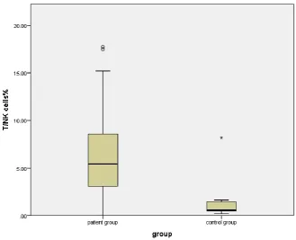
Figure (6). Shows on the left side forward scatter versus side scatter dot plot with the upper one represent a case while the lower one represent a control. The gate (P1) was localized to lymphocyte population. On the right side, it shows CD3 versus CD56, the upper one represent a case while the lower one represent the control same as left side. The population in Magenta represent the TNK cells with double positive CD3 and CD56 while the yellow population represent the NK cells with expression of CD56 and lack of CD3 expression
SSC
FSC
CD3
4. Discussion
Reactive lymphocytosis is attributed to many reasons as viral infection, bacterial infection, autoimmune disease, vaccination, drug hypersensitivity, endocrine disorder, stress, smoking and malignancy [18]. Reactive lymphocytosis is observed in type 2 diabetes mellitus significantly than the healthy people [19]. Altered lymphocyte profiles were reported in obesity, a major risk factor for type 2 diabetes [20].
The threshold of lymphocytosis is established to be equal to or increased than 4.5x10³ /UL. Benign causes could include also stress, trauma, vigorous exercise, post splenectomy and some cases of thalassemia intermedia, [21]. Typically malignant lymphocytes will be monomorphic, however reactive will show a pleomorphic shapes. Confirmation is based on phenotyping of the cells [22].
Defense mechanism against viral infection is attributed to both innate and acquired immune response, while the adaptive immune response rely mainly on T and B cells, the innate immune response is mastered by NK cells [23]. NK cells are dominant cells in the innate immune response. It has many strategy to control viral infection, like secreting cytokines as interferon gamma to control the spread of viruses [24]. Also direct cytolytic effect as well as use of death receptor mediated lyses [25]. Recently it was proven that NK cells also participate in adaptive immune response by keeping memory related to infection by certain viruses [26].
Our results show a highly significant increase of NK cells in cases of lymphocytosis in relation to control group. This result in suppression and control of Viruses. This is supported by the existence of certain genetic mutation which is associated with deficiency of NK cells in cases with recurrent viral infection as herpes virus, varicella virus, papilloma virus infection [26]. also suppression to the autoimmune process as proven by flaring up of autoimmune disease in NK deficient cases. To our knowledge, there was no published data about the frequency of NK or TNK cells in cases of benign lymphocytosis also no published data about relation between NK and TNK cells in such cases.
This study uncover an increase of TNK cells in cases of reactive lymphocytosis group compared with healthy control group. This is supported by the finding that person with congenital deficient TNK cells are susceptible to certain infection as Pseudomonas aeruginosa, Streptococcus pneumoniae, Salmonella typhi murium, Mycobacterium tuberculosis, Listeria monocytogenes, and Borrelia burgdorferi. Also viruses including influenza, cytomegalovirus, and coxsackie B3 viral diseases [27].
Also this study show a negative correlation between NK cells count and lymphocytes count, the explanation for that is the increase of NK could be the beginning of immune response before augmentation which will be followed by increase in the T and B lymphocytes which will predominate and lead to underestimation of the NK cells count and then lymphocytosis will be mainly T and B cells.
The results show that there is no correlation between NK and TNK cells count in cases of benign lymphocytosis, this could be explained by that these cells plays different role based on the cause of lymphocytosis with different mechanisms, although both cells could play a role in the innate immune response [28, 29].
The limitation of this study was lack of subgrouping of different causes of benign lymphocytosis, since different causes carry different pathogenesis and immune modulation based on the pathogen involved or the suspected inflammatory condition as in autoimmune disease and other metabolic inflammatory condition.
5. Conclusions
The levels of NK and TNK cells percentage are increased in cases of benign lymphocytosis in relation to normal controls which is an indicator of early immune response.
6. Recommendations
Our recommendation is to investigate different disease category separately to define the signature of both NK and TNK cells count clearly and to uncover any relation between both of these cells clearly.
REFERENCES
[1] Macintyre EA, and Linch DC. Lymphocytosis: is it Leukemia and when to treat. (1988). Postgraduate Medical Journal. 64; 42-47.
[2] Sun P, Kowalski EM, Cheng CK, Shawwa A, Liwski RS, Juskevicius R. (2014). Predictive significance of absolute lymphocyte count and morphology in adults with a new onset peripheral blood lymphocytosis. J Clin Pathol. Dec; 67(12): 1062-6.
[3] Tseng V, Morgan AS, Leith CP, Yang DT. (2014). Efficient assessment of peripheral blood lymphocytosis in adults: developing new thersholds for blood smear review by pathologists. Clin Chem Lab.
[4] Ritz J, Schmidt RE, Michon J, Hercend T, Schlossman SF. (1988). Characterization of functional surface structures on human natural killer cells. Adv Immunol.; 42: 181-211. Review.
[5] Caligiuri MA, Human Natural Killer cells, (2008). Blood, August, volume 112, Number 3; 461-469.
[6] Bryceson YT, March ME, Ljunggren HG, Long EO. (2006). Activation, co-activation, and costimulation of resting human natural killer cells. Immunol Rev; 214: 73-91.
[11] Vicari AP, Herbelin A, Leite-de-Moraes MC, Von Freeden-Jeffry U, Murray R, Zlotnik A. (1996). NK1.1+ T cells from IL-7-deficient mice have a normal distribution and selection but exhibit impaired cytokine production. Int Immunol. Nov; 8(11): 1759-66.
[12] Bean, A.G.D., Godfrey, D.I., Ferlin, W.G., Santos-Argumedo, L., Parkhouse, M. E., Howard, M. C. and Zlotnik, A., (1995). CD38 expression on mouse T cells: CD38 Defines functionally distinct subsets of αβTCR+CD4–CD8–thymocytes. Int. Immunol. 7:213–221. [13] Charoudeh HN, Tang Y, Cheng M. Cilio C M, Jacobsen
S.E.W and Sitnicka E. Identification of an NK/T cell–restricted progenitor in adult bone marrow contributing to bone marrow– and thymic-dependent NK cells. 2010 Blood 116:183-192.
[14] Shissler SC, Webb TJ. (2018). The ins and outs of type I iNKT cell development. Mol Immunol. Nov 28; 105: 116-130.
[15] Van Kaer, L., Parekh, V. V., & Wu, L. (2012). Invariant natural killer T cells as sensors and managers of inflammation. Trends in immunology, 34(2), 50-8.
[16] Chan YH. (2003a). Biostatistics 102: Quantitative Data – Parametric & Non-parametric Tests. Singapore Med J.; 44(8): 391-396.
[17] Chan YH (2003b). Biostatistics 104: Correlational Analysis. Singapore Med J.; 44(12): 614-619.
[18] George, TI: Diagnostic approach to lymphocytosis, (2015). The Hematologist: November-December. Volume 12, Issue 6.1-6.
Press, Great clarendon street, oxford, OX26DP, UK, page 116.
[22] Andrews JM, Cruser DL, Myers JB, Fernelius CA, Holm MT, Waldner DL. (2008). Using peripheral smear review, age and absolute lymphocyte count as predictors of abnormal peripheral blood lymphocytoses diagnosed by flow cytometry. Leuk Lymphoma. Sep; 49(9): 1731-7.
[23] Mandal A and Viswanathan C, Natural Killer cells: in health and disease, (2015). Hematol Oncol stem cell Ther 2015; 8(2); 47-55.
[24] Lee SH, Miyagi T, Biron CA: (2007). Keeping NK cells in highly regulated antiviral warfare. Trends Immunol; 28: 252–259.
[25] Lee SH, Biron CA. (2010). Here today – not gone tomorrow: roles for activating receptors in sustaining .NK cells during viral infections. Eur J Immunol; 40: 923–932.
[26] O’Sullivan TE, Sun JC, Lanier LL. (2015). Natural Killer Cell Memory. Immunity.; 43: 634–645.
[27] Mace EM, Hsu AP, Monaco-Shawver L, Makedonas G, Rosen JB, Dropulic L, Cohen JI, Frenkel EP, Bagwell JC, Sullivan JL, et al. (2013). Mutations in GATA2 cause human NK cell deficiency with specific loss of the CD56 (bright) subset. Blood.; 121:2669–2677.
[28] Gao Y, Williams AP. (2015). Role of Innate T Cells in Anti-Bacterial Immunity. Front Immunol. Jun 11; 6: 302. [29] Crome SQ, Ohashi PS, (2018). Immunoregulatory functions