Doctoral Thesis in Industrial Engineering
---XXVI cycle
Qiang Qian
University of Trento
Tutor: Prof. Claudio Migliaresi
Advisor: Dr. Walter Bonani
Acknowledgments
I would like to thank all of them that have contributed to the accomplishment of this work during my PhD course.
First of all, I would like to express my deep gratitude to my supervisor, Prof. Claudio Migliaresi, who gave me this opportunity to have this memorable study experience in the BIOtech group. He always gave his scientific support, patience and encouragement to me during my study in Trento. In the occasion of the successful completion of this thesis, the author would like to express the sincere respect and heartfelt thanks to Prof. Claudio Migliaresi.
I am indebted to Walter Bonani, my “little tutor”, who taught me many skills and gave me many valuable discussions and patient guidance on my experiments, papers and thesis. His rigorous attitude and careful spirit on science will influence my whole life.
The BIOtech group is like a big and warm family. I acknowledge all the people of the BIOtech group and all the time with all of you, from the study, the picnics, and Christmas lunches. In particular, I am indebted to Christian Lorandi, Luca Gasperini, Mariangela Fedel, Cristina Foss, Luca Dalbosco, Filippo Benetti, Matteo Stoppato, Eleonora Carletti, Lucia Verin, Wei Sun and Lorenzo Moschini. Although most of them have already graduated, the memory is still there.
A special thank goes to the project of “Bridge the Gap” for the invaluable help and financial supports.
Contents
Acknowledgments ... I List of figures ... VII Abstract ... XIII Chapter 1
Introduction ... 1
1.1 Drug Delivery Systems ... 1
1.2 Gelatin ... 9
1.3 Alginate ... 12
1.4 Objectives ... 14
Chapter 2 Preparation and characterization of gelatin microbeads ... 16
2.1 Introduction ... 16
2.2 Materials ... 18
2.3 Methods ... 18
2.3.1 Preparation of gelatin solution ... 18
2.3.2 Preparation of oily phase ... 19
2.3.4 Characterization of gelatin microbeads... 19
2.4 Results and Discussion ... 21
2.4.1 Characterization of gelatin microbeads... 21
2.5 Conclusions ... 34
Chapter 3 Drug delivery system of gelatin microbeads embedded in alginate ... 36
3.1 Introduction ... 36
3.2 Materials ... 40
3.3 Methods ... 40
3.3.1 Preparation of FL-loaded gelatin microbeads ... 40
3.3.2 Confocal images of FL-loaded gelatin microbeads ... 40
3.3.3 Preparation of FL-loaded gelatin and in vitro drug release from FL-loaded gelatin/gelatin microbeads ... 41
3.3.4 Preparation of FL-loaded cross-linked alginate gels... 41
3.3.5 In vitro drug release from FL-loaded cross-linked alginate gels ... 43
3.3.6 Calibration curve of FL in DI water... 44
3.3.7 FL release quantification and data analysis ... 45
3.4 Results and discussion ... 45
3.4.1 FL-loaded gelatin microbeads ... 45
3.4.2 In vitro FL release from gelatin gel and gelatin microbeads ... 47
3.4.3 FL-loaded cross-linked alginate hydrogels with and without gelatin microbeads ... 50
3.4.6 Versatility of composite gels and fate of the system in vivo
... 58
3.5 Conclusions ... 61
Chapter 4 Dual drug delivery system of gelatin microbeads embedded in alginate .... 62
4.1 Introduction ... 62
4.2 Materials ... 65
4.3 Methods ... 65
4.3.1 Preparation of RhB-loaded gelatin microbeads ... 65
4.3.2 Morphology of gelatin microbeads ... 66
4.3.3 Preparation of FL-loaded cross-linked alginate gels... 67
4.3.4 Preparation of Tris-HCl buffer (pH=7.4) ... 68
4.3.5 In vitro drug release from RhB-loaded gelatin gel and gelatin microbeads ... 69
4.3.6 In vitro drug release from FL-loaded cross-linked alginate gels ... 69
4.3.7 Calibration curve of FL and RhB in Tris-HCl buffer (pH=7.4) ... 70
4.3.8 Drug release quantification ... 71
4.3.9 Data analysis ... 72
4.4 Results and Discussion ... 72
4.4.1 Characterization of gelatin microbeads... 72
4.4.2 In vitro RhB release from gelatin gel and gelatin microbeads ... 74
4.4.3 In vitro dual drug release from alginate/gelatin composite 76 4.4.4 The comparison of two release profiles of FL and RhB ... 79
4.4.5 Effect of loading ratio of gelatin microbeads on drug release profiles ... 80
4.5 Conclusions ... 87
Chapter 5 Multi-drug delivery system of gelatin layers using agarose as diffusion barriers ... 89
5.1 Introduction ... 89
5.2 Materials ... 90
5.3 Methods ... 91
5.3.1 Preparation of agarose solution and drug-loaded gelatin solution ... 91
5.3.2 Preparation of the multi-drug delivery device and system . 91 5.3.3 In vitro drug release ... 94
5.3.4 Calibration curve of FL and RhB in PBS buffer solution (pH=7.4) ... 94
5.3.5 Drug release quantification and data analysis ... 96
5.4 Results and Discussion ... 97
5.4.1 Effect of length of agarose gel on the release profiles ... 97
5.4.2 Effect of temperature on the release profiles ... 98
5.5 Conclusions ... 99
Chapter 6 Final Remarks ... 100
6.1 Conclusions ... 100
6.2 Future Work ... 101
List of figures
1.1 Timeline showing FDA approved DDS in the market……… 2 1.2 Summary of four traditional mechanisms of drug delivery systems:
diffusion-controlled, swelling-controlled, degradation-controlled, and stimuli-controlled systems……… 6 1.3 Model of gel formation (from sol to gel upon cooling)………...….... 11 1.4 Structure of alginate………. 12 1.5 Schematic representations of the poly-L-guluronate sequences of alginate cross-linked by calcium ions………. 13 1.6 The scheme of the thesis……….. 15 2.1 Average diameter of gelatin microbeads prepared from gelatin solution of different concentrations (ratio between water phase and oily phase: 1:5, stirring speed: 800 rpm and emulsifying time: 10 minutes)……22 2.2 Optical image of gelatin microbeads in oily phase prepared from 50
μg/mL gelatin solution (ratio between water phase and oily phase: 1:5, stirring speed: 800 rpm and emulsifying time: 10 minutes)……… 24 2.3 Optical image of gelatin microbeads in oily phase prepared from 75
2.4 Average diameter of gelatin microbeads prepared at different gelatin concentrations (ratio between water phase and oily phase: 1:5, stirring speed: 800 rpm and emulsifying time: 10 minutes)………. 25 2.5 Optical image of gelatin microbeads in oily phase prepared from 150 μg/mL gelatin solution (ratio between water phase and oily phase: 1:5, stirring speed: 400 rpm and emulsifying time: 10 minutes)……… 26 2.6 Optical image of gelatin microbeads in oily phase prepared from 100
μg/mL gelatin solution (ratio between water phase and oily phase: 1:5, stirring speed: 800 rpm and emulsifying time: 10 minutes) ……… 28 2.7 Optical image of treated gelatin microbeads prepared from 100 μg/mL gelatin solution (ratio between water phase and oily phase: 1:5, stirring speed: 800 rpm and emulsifying time: 10 minutes) ……… 29 2.8 Optical image of treated gelatin microbeads after putting in deionized water prepared from 100 μg/mL gelatin solution (ratio between water phase and oily phase: 1:5, stirring speed: 800 rpm and emulsifying time: 10 minutes)……….……… 30 2.9 Average diameter of gelatin microbeads prepared at different ratio
between water phase and oily phase: 1:5, stirring speed: 1000 rpm and emulsifying time: 18 minutes……….. 33 3.1 Schematic representations of the preparation of gelatin microbeads (Gm), encapsulation in cross-linkable alginate matrices and drug release analyses. A: preparation of liquid Gm loaded with FL by W/O emulsification in oil bath; B: cooling in ice-bath for complete gelation of Gm; C: extraction of solid Gm by hexane washing and release analysis; D: cross-linking of alginate gels encapsulated with solid Gm in CaCl2 solution; E: drug release experiments from composite gels at
RT (solid Gm) and 37°C (liquid Gm)………..…... 39 3.2 Standard curve of FL in DI water………...….…… 44 3.3 Confocal microscopy image of FL-loaded gelatin microbeads prepared from a 10% gelatin solution (stirring rate of 800 rpm and emulsifying time of 10 min)………...……….. 46 3.4 (a) FL-loaded gelatin gel, (b) after day release at RT, (c) after
one-day release at 37°C………...……… 47 3.5 Release profiles of FL from Gb and from Gm at RT and at 37 °C…. 48 3.6 (a) blank alginate droplets after cross-linking, (b) Alg-5, (c) Alg-10, (d)
Alg(Gm-20), (e) Alg(Gm-40). Diameter of alginate droplets was about 3 mm……….. 50 3.7 Confocal image of the cross section of Alg-5………... 51 3.8 Confocal microscopy images of FL-loaded gelatin microbeads (Gm)
alginate hydrogel after 7 days of release at 37°C (scale bar =100 μm)……….……….. 53 3.9 Release profiles of FL from FL-loaded gelatin microbeads encapsu-lated in cross-linked alginate (Alg(Gm-20)) and from cross-linked alginate directly loaded with FL in solution (Alg-5) both at 37°C and at RT. The net amount of FL initially loaded in both Alg(Gm-20) and Alg-5 was the same……….…………. 55 3.10 Effect of drug loading concentration on the release profiles of FL at 37 °C: (a) from cross-linked alginate directly loaded with FL in solution (Alg-1.25, Alg-5, and Alg-10), (b) from cross-linked alginate gels encapsulated with FL-loaded gelatin microbeads (Alg(Gm-5), Alg(Gm-20), and Alg(Gm-40))……… 57 4.1 Schematic representation of the preparation of gelatin microbeads/algi- nate composite hydrogel and drug release…...… 64 4.2 Standard curve of FL in Tris-HCl buffer solution……….…… 70 4.3 Standard curve of RhB in Tris-HCl buffer solution……….………. 71 4.4 Scanning electron microscopic images of gelatin microbeads in low (a)
4.9 Effect of drug-loaded gelatin microbeads ratio on RhB release profiles of Alg(GM-1) and 2- Alg(GM-1)………....…… 82 4.10 Effect of drug-loaded gelatin microbeads ratio on FL release profiles
of Alg, Alg(GM-1) and 2- Alg(GM-1)……… 83 4.11 Effect of preparation method of drug-loaded gelatin microbeads on
RhB release profiles of 1), 1) and Alg(GM-3)... 85 4.12 Effect of preparation method of drug-loaded gelatin microbeads on
FL release profiles of Alg(GM-1), Alg(GM-1) and Alg(GM-3)… 86 5.1 Schematic diagram of multi-drug delivery system………...………. 92 5.2 Picture of multi-drug delivery system………...…… 93 5.3 Standard curve of FL in PBS buffer solution……… 95 5.4 Standard curve of RhB in PBS buffer solution………. 96 5.5 Effect of length of agarose gel on the release profiles of FL and RhB at
37°C………..……….. 97 5.6 Effect of temperature on the release profiles of FL and RhB with 1
Abstract
Controlled drug delivery systems, which are intended to deliver drugs at predetermined rates for predefined periods of time, have been used to overcome the shortcomings of conventional drug formulations.
Injectable drug-loaded matrices and controlled release technology offer numerous advantages compared to conventional dosage. However, one of the greatest challenges in applying this system to the clinical phase is the relatively large initial burst release.
Firstly, a series of gelatin microbeads were prepared in a water-in-oil (W/O) emulsion by a traditional emulsification method. The effects of the concentration of gelatin solution, volumetric ratio of water-to-oil phase, stirring speed and emulsifying time on the particle size and dispersity of gelatin microbeads were studied.
Secondly, drug-loaded gelatin microbeads were encapsulated into the cross-linked sodium alginate macro-beads. The release behavior of drug-loaded gelatin microbeads encapsulated within cross-linked alginate gel was characterized both at room temperature and 37°C and compared with the release from gelatin microbeads and cross-linked alginate gel alone. This system represents a promise for the development of novel and versatile injectable drug delivery systems.
Thirdly, a dual-drug delivery system was fabricated by encapsulating drug-loaded gelatin microbeads into the mixture of cross-linkable sodium alginate and another drug. The effects of preparation methods of drug-loaded gelatin microbeads and ratio between gelatin microbeads and alginate on drug release behaviors of both drugs were studied. This system shows a significant potential in dual drug delivery field due to the synergistic effect between gelatin and alginate.
Chapter 1
Introduction
1.1
Drug Delivery Systems
There are some shortcomings in the clinical applications of traditional medicines and formulations, such as low effectiveness of the drugs, high side effects and the frequent needs of medication to maintain efficacy [1].
DDS is designed to efficiently target within the body as the drug reservoirs for sustained drug release, to decrease the antigenicity and increase the bio-distribution of the loaded drugs, or to reduce the toxicity of high drug loading; whatever the case may be, it is mainly concerned with dosage form, route of administration and duration of drug presence. Drug delivery is often approached via a drug's chemical formulation, but it may also involve medical devices or drug-device combination products [5].
Design, research and production of efficient drug delivery systems are of significant importance for the improvement of medicine and healthcare. Since the first FDA approval of drug delivery system (DDS) in 1990, more than 10 DDS are now commercially available to treat diverse diseases ranging from cancer to fungal infection and to muscular degeneration (Figure. 1.1) [6].
With the development of therapeutic efficacy, more and more patients have relieved suffering and prolonged life profited from DDS. At the same time, drug delivery systems have also changed the economics of drug production. As a new product, incorporating an existing drug into a suitable drug delivery system not only improves drug’s performance but also reduce patents’ suffering and cost. According to the statistics, the average cost and time required to develop a new DDS is approximately $20–50 million and 3–4 years, while it is significantly higher to develop a new drug (approximately $500 million and over 10 years) [7]. It is not surprising that the annual worldwide market for advanced and controlled drug release system has grown dramatically [8], especially in the US market [9].
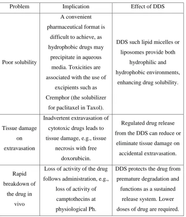
Many of the pharmacological properties of conventional drugs can be improved through the use of drug delivery systems, Table 1.1 [4] gives examples of problems exhibited by free drugs that can be ameliorated by the use of DDS.
example, many protein-based insulin and immunizations are often delivered by injection.
Table 1.1 Non-ideal properties and problems of drugs in common routes and their therapeutic effects of DDS [4].
Problem Implication Effect of DDS
Poor solubility
A convenient pharmaceutical format is
difficult to achieve, as hydrophobic drugs may
precipitate in aqueous media. Toxicities are associated with the use of
excipients such as Cremphor (the solubilizer
for paclitaxel in Taxol).
DDS such lipid micelles or liposomes provide both
hydrophilic and hydrophobic environments,
enhancing drug solubility.
Tissue damage on extravasation
Inadvertent extravasation of cytotoxic drugs leads to tissue damage, e.g., tissue
necrosis with free doxorubicin.
Regulated drug release from the DDS can reduce or
eliminate tissue damage on accidental extravasation. Rapid
breakdown of the drug in
vivo
Loss of activity of the drug follows administration, e.g.,
loss of activity of camptothecins at physiological Ph.
DDS protects the drug from premature degradation and
Unfavorable pharmacokinet
ics
Drug is cleared too rapidly, by the kidney, for example,
requiring high doses or continuous infusion.
DDS can substantially alter the PK of the drug and reduce clearance. Rapid renal clearance of small molecules is avoided.
Poor bio-distribution
Drugs that have widespread distribution in the body can
affect normal tissues, resulting in dose-limiting
side effects, such as the cardiac toxicity of
doxorubicin.
The particulate nature of DDS lowers the volume of
distribution and helps to reduce side effects in sensitive non-target tissue.
Lack of selectivity for
target tissues
Distribution of the drug to normal tissues leads to side
effects that restrict the amount of drug that can be
administered. Low concentrations of drugs in target tissues will result in suboptimal therapeutic
effects.
DDS can increase drug concentrations in diseased
tissues such as tumors by the EPR effect. Ligand-mediated targeting of the DDS can further improve
swelling-controlled systems, where the release rate of drug is based on the water swelling rate of the delivery system, swelling increases the flexibility of the polymer and makes larger pores, resulting in better drug mobility [17], such as PVA, PHEMA; (3) degradation-controlled systems, where the drug release rate depends on the level of chemical or physical degradation of the drug delivery matrix, it could be surface degradation or bulk degradation [18], such as PLGA, PCL; (4) external stimulus-controlled systems, where stimulus-controlled systems release therapeutic factors upon activation by a stimulus or multiple stimuli, leading to a physical or chemical change in the delivery system, these stimuli could be physical (e.g., pH, temperature) or chemical (e.g., the presence of glucose) [5], such as PNIPAM, PDMAEMA.
Among the four common mechanisms, initial drug burst was inevitable. The novel concept of using chemical affinity in drug delivery systems can reduce initial burst and control drug release availably. As defined, affinity is the tendency of a molecule to associate with another molecule. It may come from many chemical or physical interactions, such as covalent bond, charged interaction, hydrogen bonding or van der Waals forces. It has been reported that stable polyion complex can be formed by the electrostatical interaction between two polyelectrolytes with opposite charges. By the interaction or affinity between drug and polymeric matrices, drug loading and release could be controlled [11, 19].
Recently drug delivery system includes oral drug delivery system, trans-dermal drug delivery system, mucosal drug delivery system, targeted drug delivery system, cell encapsulated drug delivery system and micro-fabricated drug delivery system.
The main efforts in the field of drug delivery include the development of targeted delivery and sustained release formulations. To be specific, the drug would be only active in the target area of the body (for example, in cancerous tissues) in the targeted DDS and the drug would be released over a period of time in a controlled manner in the sustained DDS. In order to realize the efficiency of targeted delivery, the designed system must avoid the body's immune mechanisms and then manage to deliver the drugs to its intended site of action. Types of sustained release formulations include liposomes, drug loaded biodegradable microspheres and drug polymer conjugates.
materials. Nanotechnology has also contributed much to the development of DDS in the past decade. Since then when it was found that size and shape of nanoparticles (NPs) could help navigate biological carriers, the application of nanofabrication technologies has motivated to develop more effective particulate DDS, both top–down and bottom–up. As reported, the size of NPs regulates their bio-distribution. Particles less than 20 nm will be cleared from circulation via reticuloendothelial system (RES) within a few hours when injected intravenously, whereas larger ones will be trapped in the liver and the spleen within minutes [20, 21]. In a study of Kataoka and his colleagues, the results showed that polymeric micelles only less than 30 nm could effectively penetrate poorly permeable pancreatic tumor cells [22]. Fabricating techniques such as nano-precipitation, emulsion-based phase inversion, microfluidics-based self-assembly, layer-by-layer synthesis, and nano-imprinting have been used to generate particulate DDS to deliver a wide range of drugs. With the full understanding of the potential of particulate DDS in required size and shape, fabrication and nano-manufacturing will play a more and more prominent role in the future.
they could assemble to nanostructures in the form of micelles, electrostatic complexes, or polymersomes [25]; they could be polymer drug conjugates, conjugation to a polymeric carrier via a liable linker presents another attractive approach to alter and optimize the pharmacokinetics of therapeutic agents [6]; they could be natural polymers derived from biological systems including protein, DNA, and polysaccharides, they are biocompatible and biodegradable, moreover, they possess low toxicity and potentially favorable pharmacokinetics in the circulation [6]; they also could be recombinant protein-based drug carriers.
1.2
Gelatin
Nowadays, gelatin is utilized in practically all areas of modern life. However, interest in gelatin is not just restricted to classical applications; this natural product has numerous other application possibilities. New applications in health care and in specialized technical areas will result in gelatin and gelatin hydrolysates becoming a focal point of concern for a much wider pubic [26].
The first known gelatin by boiling animal tissues to use as glue should date back to about 8000 years ago. The revolution in the use of gelatin for biomedical applications occurred in 1833 when gelatin capsules were first fabricated to encapsulate drugs to prevent drugs from heat, humidity and to make drugs taste no longer bitter. In the 20th century, the application of gelatin in medical field exploded [26].
also bones, but it is more difficult; for economical and practical reasons, skin is usually preferred), either by partial acid or alkaline hydrolysis at a moderate temperature; the content of protein ranges from 85% to 92%, the remnant consists of salts and moisture remained after drying [26-28].
By controlling type and intensity of the hydrolysis, it is possible to control the molecular weight distribution. Gelatin hydrolyzed with alkaline treatment (type B) shows a narrower distribution, while type A gelatin has a broader distribution. Molecular weight will affect viscosity and gelling power. Both of them are increased by the increase in the molecular weight. The gel strength is the most important quality parameter for gelatin. The analytical measure of gelling power is the Bloom value, which is the weight required for a specified plunger to depress the surface of a standard, thermostatted gel to a defined depth under standard conditions.
and as carrier for drugs, gene therapeutic entities and cells [32, 34-38], although it is non-degradable and loses the ability for the sol-gel transition in vivo.
overcome immune problems and reducing the toxicity of high drug loading [42, 43].
1.3
Alginate
Commercial alginates are mainly extracted from three kinds of native brown seaweed: Laminaria hyperborean, Ascophyllum nodosum, and Macrocystis pyrifera [44]. Alginate, the primary polysaccharide in the these seaweed, is found in the intracellular matrix. The native alginate is mainly present as an insoluble Ca2+ cross-linked gel [45].
Figure 1.4: Structure of alginate [46]
shown in Figure 1.5. Among these cations, Ba2+ or Sr2+ ions produce stronger alginate gels than Ca2+ [49-51]. However, monovalent cations and Mg2+ ions do not induce gelation, other divalent cations also can cross-link alginate but their use is limited due to the toxicity.
Figure 1.5: Schematic representations of the poly-L-guluronate sequences of alginate cross-linked by calcium ions [49].
Alginate has been widely used in food and beverage industries as well as tablet manufacture for long time [52]. Due to the large availability in nature, the mildness of gelation conditions, limited toxicity, great biocompatibility, low immunogenicity, and low cost [47, 49, 52, 53], alginate has been widely proposed as a potential biopolymer for drug stabilization, tissue engineering, and controlled-release systems, as well as for cells encapsulation techniques [54-58].
gels in situ when placed on the surface of the eye [62]. Ito et al.[61] reported that cross-linking of alginate could occur spontaneously once injected into a living body because of divalent cations in the organism. In addition, although cross-linked alginate is non-biodegradable, degradation can occur by removal of the cross-linking ions[49], for instance, alginate gels will degrade in 0.1 M phosphate buffer solution[63]. Therefore, since the exit of HCO3- and HPO42- in the human body fluid[61], the degradation of alginate
gels can also take place in vivo. However, majority of them suffer the problem on fast release of encapsulated drugs which limits its application as the matrix of drug delivery system. To prolong the duration of drug release, it is a feasible way to immobilize drugs to charged polymer carriers by electrostatically interaction to form a polyion complex [64] or embed drug-loaded microbeads in polymeric matrix. Varied microbeads, micelle and beads were developed to trap drugs and delivery the drugs with assistance of polymeric matrix [65-69], and for the polyion complexation for sustained release, it is necessarily bio-safe and biodegradable.
1.4
Objectives
Gelatin Type A From Porcine Skin
Uncross-linked Gelatin Microbeads
With Sodium Alginate
With Agarose
Drug Delivery System
Dual Drug Delivery System
Multi-Drug Delivery System
Chapter 3 Chapter 4 Chapter 5 Chapter 2
Chapter 2
Preparation and characterization of
gelatin microbeads
2.1 Introduction
Microspheres with diameter in the range of 20-100 μm can be chosen for subcutaneous or intramuscular administration as sustained release depots. Smaller micro-particles have been considered for the treatment of infection and arthritis [72].
Embedding biodegradable particles into the formulation turns to be efficient to improve the therapeutic effect of various water soluble/insoluble medicinal drugs and bioactive molecules. In this way, absorption, bioavailability, intracellular penetration, solubility and retention time of drugs in the selected tissue are improved. The resulting formulation would reduce the risks of side effect and the cost of patients and protect the premature degradation and interaction with the biological environment [73-76].
Drugs or bioactive molecules related to some dreadful diseases like cancer, AIDS, diabetes, malaria and prion disease are successfully encapsulated into nano- or micro-particles to improve bioavailability, bioactivity and control delivery, some of them are already commercialized [77-81].
two ways to cross the most barriers are injection and carrying by a particular cell type.
In this chapter, gelatin microbeads would be prepared under all kinds of conditions by emulsification and the optimal parameter would be selected to obtain the suitable drug-loaded gelatin microbeads.
2.2 Materials
Gelatin (gelatin type A, gel strength ~300 g Bloom, from porcine skin), soybean oil, nonionic surfactant sorbitane monooleate (Span® 80) and n-Hexane were purchased from Sigma-Aldrich (Milan, Italy). All the materials were used as received without further purification.
2.3 Methods
2.3.1 Preparation of gelatin solution
2.3.2 Preparation of oily phase
Oily phase was a mixture of commercial soybean oil and Span® 80, prepared in a proportion of 20 mg of Span® 80 per 1 mL of soybean oil under mild stirring in a 40 °C water bath.
2.3.3 Preparation of gelatin microbeads
An emulsification-coacervation method was employed to prepare gelatin microbeads [84-87]. Briefly, a gelatin solution prepared as in section 2.3.1 was added dropwise into the oily phase prepared as in section 2.3.2 under continuous stirring in a 40 °C water bath to obtain a water-in-oil (W/O) emulsion. Different volume ratio between gelatin solution and oily phase was used. The effects of emulsifying time ranging from 2 to 15 min and stirring speed ranging from 200 to 1000 rpm on the W/O emulsion were also considered. After this stage, the emulsion was moved to an ice-bath and stirred for other 30 min at 200 rpm to complete the gelation of the gelatin microbeads inside the continuous oily phase. Oil was then removed with several washings in n-hexane and gelatin microbeads were placed in a vacuum drier overnight to remove hexane.
2.3.4 Characterization of gelatin microbeads
2.4 Results and Discussion
2.4.1 Characterization of gelatin microbeads
Emulsification method has been widely applied to the preparation of micrometric beads for its outstanding advantages such as easy-controlling and mild-condition operation, which was first proposed by Tanaka.[88] In this study a plant-derived soybean oil was chosen as the oily phase for safety considerations because it is often difficult to completely remove the oil after microbeads preparation.[89]
2.4.1.1 Effect of the concentration of gelatin solution on
the size of gelatin microbeads
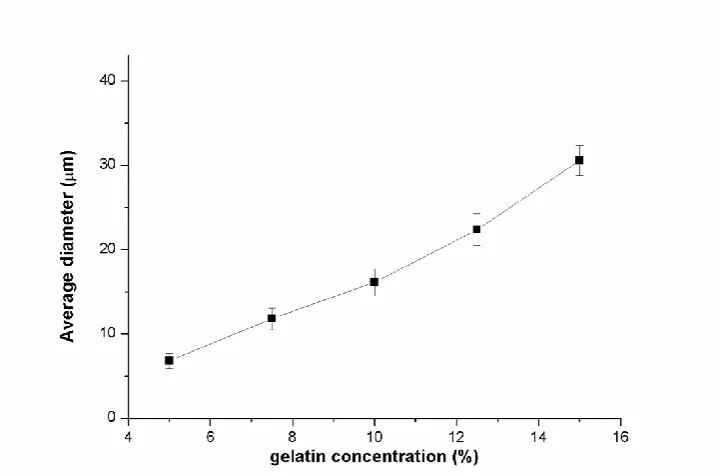
Figure 2.1 Average diameter of gelatin microbeads prepared from gelatin solution of different concentrations (ratio between water phase and oily phase: 1:5, stirring speed: 800 rpm and emulsifying time: 10 minutes)
proper to prepare gelatin microbeads by using low concentration gelatin solution.
In our case, five concentrations of 50 μg/mL, 75 μg/mL, 100 μg/mL, 125 μg/mL and 150 μg/mL (5%, 7.5%, 10%, 12.5%, 15% (w/v)) were chosen to obtain different gelatin microbeads for comparison by fixing the ratio of 1: 5 between gelatin solution and oily phase, stirring speed of 800 rpm and 10 min of emulsifying time.
As shown in Figure 2.1, the particle size of gelatin microbeads increased almost linearly from 6.8 ± 0.9 m to 30.6 ± 1.8 m with the concentration of gelatin solution ranging from 5% to 15%.
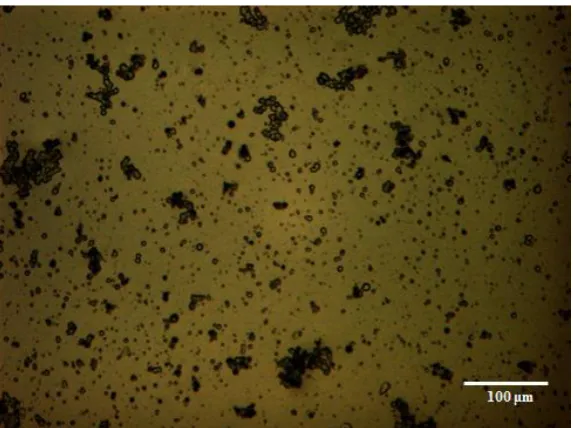
From Figure 2.2, it could be seen that when gelatin microbeads were prepared from low-concentration gelatin solution, the microbeads with smaller particle size had a strong tendency to agglomerate, which attributed to the high water content in the gelatin microbeads. During the stirring of preparation, it was easy to destroy the inner loose structure of gelatin microbeads, which would lead to the particles stick to each other.
Figure 2.2. Optical image of gelatin microbeads in oily phase prepared from 50 μg/mL gelatin solution (ratio between water phase and oily phase: 1:5, stirring speed: 800 rpm and emulsifying time: 10 minutes)
2.4.1.2 Effect of stirring speed on the characterization of
gelatin microbeads
Figure 2.4 Average diameter of gelatin microbeads prepared at different gelatin concentrations (ratio between water phase and oily phase: 1:5, stirring speed: 800 rpm and emulsifying time: 10 minutes)
Figure 2.5 Optical image of gelatin microbeads in oily phase prepared from 150 μg/mL gelatin solution (ratio between water phase and oily phase: 1:5, stirring speed: 400 rpm and emulsifying time: 10 minutes)
Figure 2.6 Optical image of gelatin microbeads in oily phase prepared from 100 μg/mL gelatin solution (ratio between water phase and oily phase: 1:5, stirring speed: 800 rpm and emulsifying time: 10 minutes)
Figure 2.7 Optical image of treated gelatin microbeads prepared from 100 μg/mL gelatin solution (ratio between water phase and oily phase: 1:5, stirring speed: 800 rpm and emulsifying time: 10 minutes)
After several washings with n-hexane and then putting in a vacuum drier for one hour, the optical image of treated medium-sized gelatin microbeads was taken and presented in Figure 2.7. These microbeads are small, round and opaque.
in the image was the residue of oil or n-hexane around the treated gelatin microbeads.
2.4.1.3 Effect of the ratio between gelatin solution and
oily phase on the characterization of gelatin microbeads
Figure 2.9 Average diameter of gelatin microbeads prepared at different ratio between gelatin solution and oily phase (concentration of gelatin solution: 100 μg/mL (10%), stirring speed: 800 rpm and emulsifying time: 10 minutes)
The same as above, fixing other parameters, such as 100 μg/mL (10%) of the concentration of gelatin solution, 10 min of emulsifying time, 800 rpm of stirring speed, three different volume ratio between water phase and oily phase of 1:8, 1:5 and 1:3 were chosen to obtain different gelatin microbeads for comparison.
As shown in Figure 2.9, with the increase of water phase from 1:8 to 1:3, the particle size of gelatin microbeads increased from 10.9 ± 1.1 m to 24.8 ± 1.4 m. Based on the results, it could be concluded that decreasing the ratio between water phase and oily phase, the ability of oily phase to disperse the gelatin solution would be increased, which would lead to the formation of small gelatin microbeads.
2.4.1.4 Effect of emulsifying time on the characterization
of gelatin microbeads
Figure 2.10 Optical image of gelatin microbeads in oily phase prepared under the following conditions: 100 μg/mL gelatin solution, volume ratio between water phase and oily phase: 1:5, stirring speed: 800 rpm and emulsifying time: 3 minutes.
2.5 Conclusions
A series of gelatin microbeads were prepared through emulsification-coacervation method in water-in-oil (w/o) emulsion. The influence of preparation parameters on particle size, surface morphology and dispersity of gelatin microbeads was examined. The studied parameters include concentration of gelatin solutions, water phase/ oil phase ratio, emulsifying time and stirring speed. The experimental results indicated that increasing the concentration of gelatin solution would increase the particle size of gelatin microbeads linearly. The water/oil ratio had the same influence on the particle size as that of gelatin solution concentration. In addition, with the increase of water/oil ratio, the surface of microspheres became smoother as well. The emulsifying time had little effect on the mean diameter of gelatin microbeads, but it affected the dispersity of particles apparently. When emulsifying time was shorter than 5 min or longer than 15 min, gelatin microbeads had bad dispersity. The stirring speed had the similar influence as that of emulsifying time. Slow stirring rate made large size distribution and bad sphericity; excessive stirring speed resulted in aggregation likewise. The smaller size distribution and better sphericity of gelatin microbeads were observed under the stirring speed between 500 rpm to 1200 rpm.
Chapter 3
Drug delivery system of gelatin
microbeads embedded in alginate
3.1 Introduction
The development of injectable drug-delivery systems has gained extensive attention over the last decade [91]. These systems possess unique advantages when compared to traditional administration routes, including localized and tissue-specific delivery, programmable release patterns, efficient loading and delivery, reduction of undesired side effects and minimally invasive administration [92, 93].
A wide range of synthetic and natural polymer-based materials have been proposed to form injectable hydrogels for drug delivery and cell encapsulation. Back to 1997, Jeong et al. synthesized a thermosensitive and biodegradable injectable hydrogel consisting of PEO and PLLA blocks as drug delivery systems [96]; since then, a variety of block copolymers of Poly(ethylene glycol) and degradable, biocompatible aliphatic polyesters with tailored degradation behavior and hydrophobicity were proposed as hydrogel-based drug carriers [97-99]. However, it is generally complicated to standardize the synthesis of these custom-made block copolymers; in addition, their synthesis methods often involve toxic solvents and reagents that are difficult and expensive to remove, instability of the functional groups and low coupling efficiency.
Naturally-derived polymers have been largely used as hydrogels for drug delivery due to inherent bio-compatibility, bio-resorbability, bio-adhesive properties, and the resemblance to natural extra cellular matrix. Hydrogel-forming biopolymers include collagen and cross-linked gelatin, chitosan, agarose, fibrin and alginate [34, 100-103].
in this chapter, the schematic representations of process was shown in Figure 3.1.
Figure 3.1 Schematic representations of the preparation of gelatin microbeads (Gm), encapsulation in cross-linkable alginate matrices and drug release analyses. A: preparation of liquid Gm loaded with FL by W/O emulsification in oil bath; B: cooling in ice-bath for complete gelation of Gm; C: extraction of solid Gm by hexane washing and release analysis; D: cross-linking of alginate gels encapsulated with solid Gm in CaCl2 solution;
3.2 Materials
Gelatin (gelatin type A, gel strength ~ 300 g Bloom, from porcine skin), Sodium Alginate (Alginic acid sodium salt from brown algae), Fluorescein (FL), Calcium Chloride, soybean oil, nonionic surfactant sorbitane monooleate (Span® 80) and n-Hexane were purchased from Sigma-Aldrich (Milan, Italy). All the materials were used as received without further purification.
3.3 Methods
3.3.1 Preparation of FL-loaded gelatin microbeads
FL-loaded gelatin microbeads were prepared with a similar process as described in section 2.3.3 under the optimal emulsification condition: 100 μg/mL (10%) gelatin solution, 1: 5 of water/oil volume ratio, 10 min of emulsifying time and 800 rpm of the stirring speed, where fluorescent molecules were mixed directly into the gelatin aqueous solution prior to the emulsification. Different gelatin solutions were prepared with FL concentrations equal to 5, 20 and 40 µg/mL.
3.3.2 Confocal images of FL-loaded gelatin
microbeads
3.3.3 Preparation of FL-loaded gelatin and in vitro
drug release from FL-loaded gelatin/gelatin
microbeads
A gelatin aqueous solution at a concentration of 10% (w/v) was prepared by dissolving gelatin powder in deionized (DI) water at 40°C for 30 min. Fluorescein (FL) was mixed into the solution to obtain a concentration of 20 µg/mL. 3 mL of the solution was filled into a 10-mm dialysis tubes (Regenerated cellulose dialysis membrane, MWCO 3500, Spectra/Por®, Spectrum Lab., CA, USA) and natural gelation was allowed at RT. Each dialysis tube was placed into a beaker with 60 mL of DI water under mild agitation to test the release of FL in time. The release experiment was performed both at room temperature (23 ± 1°C) and at body temperature (37 ± 1°C); three samples were tested for each experimental condition. For comparison, gelatin microbeads obtained from 10% (w/v) gelatin solution with 20 µg/mL of FL were also tested for the release in the same conditions. Every hour 1 mL of surnatant was extracted from each sample to determine the FL content, and replaced with fresh DI water. All the extracted samples were stored at 4°C in dark until all the time points were collected.
3.3.4 Preparation of FL-loaded cross-linked alginate
gels
microbeads were uniformly dispersed into the alginate solution by continuous stirring for 5 min as shown in Figure 3.1(d). The stirring was completed in the ice bath to avoid too much FL releasing from gelatin microbeads. Volumetric ratio of 3 to 1 for alginate solution to gelatin solution was used to prepare the samples. After stirring, the alginate solution with dispersed gelatin microbeads was immediately injected dropwise into a 3% (w/v) CaCl2 aqueous solution to induce alginate
cross-linking and to form macroscopic alginate beads carrying FL-loaded gelatin microbeads.
Table 3.1 Tested samples
Sample code Description
Gm Gelatin microbeads
Gb Gelatin directly loaded with fluorescein in bulk
Alg-1.25 Alginate macrobeads loaded with 1.25 g/mL fluorescein
Alg-5 Alginate macrobeads loaded with 5 g/mL fluorescein Alg-10 Alginate macrobeads loaded with 10 g/mL fluorescein Alg(Gm-5) Alginate macrobeads encapsuled with Gelatin
microbeads containing 5 g/mL fluorescein
Alg(Gm-20) Alginate macrobeads encapsuled with Gelatin microbeads containing 20 g/mL fluorescein
With this process, gelatin microbeads prepared from different FL concentrations (5, 20 and 40 µg/mL) were incorporated in alginate; the obtained composite gels are hereinafter named Alg(Gm-5), Alg(Gm-10), and Alg(Gm-20). For comparison, alginate macrobeads directly loaded with FL were also prepared. In this case, alginate was dissolved in aqueous solutions of FL at concentrations of 1.25, 5 and 10 µg/mL. FL-loaded alginate macrobeads with different content of FL were produced (hereinafter Alg-1.25, Alg-5 and Alg-10). These FL concentrations were specifically calculated to obtain the same absolute content of FL present in the previously described Alg(Gm-5), Alg(Gm-10), and Alg(Gm-20) samples. All the samples and compositions were summarized in Table 3.1.
3.3.5 In vitro drug release from FL-loaded
cross-linked alginate gels
3.3.6 Calibration curve of FL in DI water
The stock solution of FL was prepared by dissolving 50 mg of FL into 1000 mL of de-ionized water. Dilute the resulting solution into several different concentrations for the standard curve, then analyze with a Tecan Infinite 200 microplate reader (Tecan Group Ltd., Männedorf, Switzerland) (excitation wavelength: 494 nm, emission wavelength: 521 nm), using the de-ionized water as the blank sample. The calibration curve of FL is shown in Figure 3.2.
Figure 3.2 Standard curve of FL in DI water
3.3.7 FL release quantification and data analysis
Samples of 250 µL were placed in a 96-well microplate, and analyzed with a Tecan Infinite 200 microplate reader (Tecan Group Ltd., Männedorf, Switzerland) (excitation wavelength: 494 nm, emission wavelength: 521 nm) to quantify the concentration of released FL, according to the previously determined calibration curve. All measurements were performed in triplicates and release data were presented as mean ± standard deviation. Statistical analysis was performed using OriginPro software (OriginLab, Northampton, MA, USA) with significance level of p < 0.05.
3.4 Results and discussion
3.4.1 FL-loaded gelatin microbeads
Representative confocal image of FL-loaded gelatin microbeads prepared under those conditions was presented in Figure 3.3. FL-loaded microbeads did not reveal significant differences in particle size and shape (homogeneous, spherical and narrow distribution) when compared to FL-free microbeads.
3.4.2 In vitro FL release from gelatin gel and gelatin
microbeads
Figure 3.4 (a) FL-loaded gelatin gel, (b) after one-day release at RT, (c) after one-day release at 37°C
The images of FL-loaded gelatin gel before release test and after one-day release at RT and at 37°C were presented in Figure 3.4. After one-day release, the colour of FL-loaded gelatin gel changed from yellow to light yellow. Moreover, after 6-hour release, the colour of FL-loaded gelatin gels at both temperatures almost stopped changing and the extent of colour change of the gel at 37°C was deeper than the gel at RT, both of the phenomena were in accordance with the release profiles described as following.
release value at the plateau reached a similar value for both materials (about 48%) after 10 hours. We noted that at 37°C, gelatin microbeads underwent a gel-to-solution (gel-sol) transition in a matter of minutes; while gelatin in bulk was completely dissolved in few hours. Cumulative releases at RT were 40% and 37% after 10 hours for gelatin microbeads and gelatin gel in bulk, respectively. In all cases, gelatin constructs had been separated by the surrounding medium by means of a dialysis membrane with a cut-off of 3500 Da to prevent the dispersion of the solubilized gelatin in the release medium after gel-sol transition.
Even when the release profile reached the plateau, just about 50% of the loaded FL had been released in the surrounding medium; while more than 50% of the loaded FL remained bound to the gelatin proteins. The findings are in accordance with other works [35, 39]. Acidic FL and basic gelatin used in our study could form polyion complexation which limited the amount of the free drug and reduced the efficiency [32, 35]. In a previous study, Liao et al.[40] reported that a non-charged drug was completely released from alginate within 2 h, whereas charged drug compounds showed sustained release up to 3 weeks.
The possibility of an interaction between charged FL and gelatin is consistent with the high number of potential available basic sites in gelatin. Gelatin type A 300 Bloom has approximately an average molecular mass of 100 kDa, accounting for roughly 1100 total amino acids, and about 14% of the total amino acid residues are reported to have a basic character (positive charge) [26, 107]. On these bases, it is possible to estimate that the ratio between total number of basic gelatin sites and FL molecules can range from 1000 to 10000 depending on the FL concentration. However, as the number of really available basic sites depends on the acid-base equilibrium of the solution and can vary considerably with solution characteristics (pH and ionic strength), it is not possible to establish a univocal correlation between available sites and loaded FL molecules
3.4.3 FL-loaded cross-linked alginate hydrogels with
and without gelatin microbeads
(a) (b) (c)
(d) (e)
Figure 3.6 (a) blank alginate droplets after cross-linking, (b) 5, (c) Alg-10, (d) Alg(Gm-20), (e) Alg(Gm-40). Diameter of alginate droplets was about 3 mm.
Alginate was used as a carrier for gelatin microbeads. To model the release behavior of alginate in bulk, macroscopic spherical droplets of cross-linked alginate with a diameter of about 3 mm were prepared by adding dropwise alginate solution into a CaCl2 solution. In Figure 3.6, it is
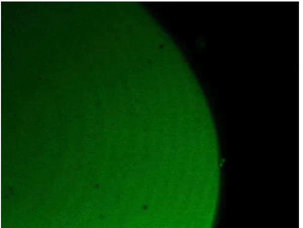
solution (Alg-5 and Alg-10) assumed a pale green color (Figure 3.6 (b) and (c)). Confocal image of the cross section of Alg-5 was presented in Figure 3.7; all the view was full of uniform green. Alginate gels encapsulated with FL-loaded gelatin microbeads (Alg(Gm-20) and Alg(Gm-40)) turned out to be opaque and whitish (Figure 3.6 (d) and (e)). As expected, by comparison between Alg-5 and Alg-10 or between Alg(Gm-20) and Alg(Gm-40), it was found that increasing FL content, the colour of alginate became more intensive.
Figure 3.7 Confocal image of the cross section of Alg-5
microbeads, which was confirmed by the confocal micrograph as shown in Figure 3.8 (a).
(a)
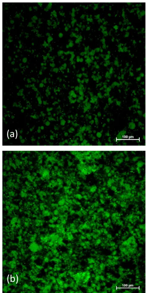
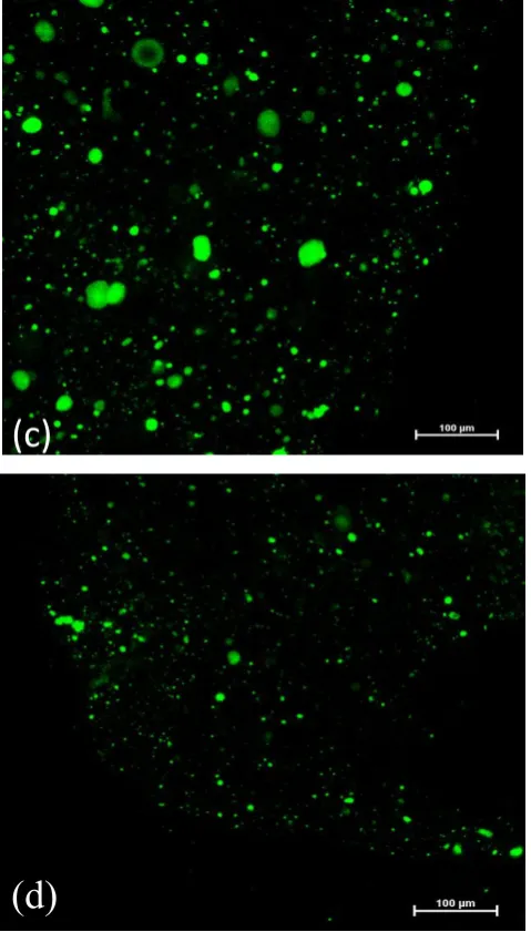
Figure 3.8 Confocal microscopy images of FL-loaded gelatin microbeads (Gm) encapsulated in alginate solution and cross-linked alginate gels: (a) Gm in liquid alginate solution before alginate cross-linking, (b) Gm in alginate hydrogel immediately after cross-linking, (c) Gm in alginate hydrogel after 24 hours of the release test at 37°C, (d) Gm in alginate hydrogel after 7 days of release at 37°C (scale bar =100 μm).
(d)
Drug loss from gelatin/alginate mixture to CaCl2 solution during alginate
cross-linking was equally inevitable, but accounted for less than 4% of the total loaded FL after 5-min of immersion. The washing stage in hexane did not involve any significant losses of loaded molecules, due to the limited solubility of FL in organic solvents.
3.4.4 In vitro drug release from FL-loaded
cross-linked alginate gel
The distribution of FL in the alginate/gelatin composite hydrogels after cross-linking was monitored by confocal microscopy. Representative confocal micrographs of sectioned alginate gels after different time intervals were shown in Figure 3.8. In the first stage after mixing and alginate cross-linking, a fraction of free FL was readily released from the gelatin microbeads and delivered to the surrounding alginate gel (Figure 3.8 (b)). However, as shown in Figure 3.8 (c), after just 24 hours of release, free FL was completely cleared out from the alginate gel, whereas gelatin microbeads retained a considerable amount of FL. Even after 7 days of release (Figure 3.8 (d)), the signal of FL could be detected very clearly in gelatin microbeads; while no FL was found in the alginate matrix. These qualitative results are confirmed also by the release profiles.
It is possible to see that composite gel allowed to significantly reducing the initial burst release. After 2 hours at 37 °C, Alg-5 presented an initial burst release of about 30% of the total loaded FL amount, while Alg(Gm-20) released less than 5% of the loeded FL in the same time span. A similar initial burst result was observed at RT.
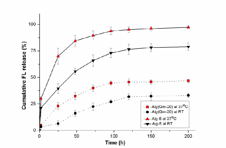
Figure 3.9 Release profiles of FL from FL-loaded gelatin microbeads encapsulated in cross-linked alginate (Alg(Gm-20)) and from cross-linked alginate directly loaded with FL in solution (Alg-5) both at 37°C and at RT. The net amount of FL initially loaded in both Alg(Gm-20) and Alg-5 was the same.
considerably lower in case of Alg(Gm-20), both at 37 °C and RT. At 37°C, the net release of FL from Alg-5 increased rapidly and about 70% of the loaded drug was released 24 h after the start of the release experiment, and more than 95% FL was released after 5 days. In case of Alg(Gm-20), the cumulative release was less than 25% after 24 hours release, and, even after one-week of release, less than 50% was delivered. The same tendencies were also observed at both temperature tested.
By comparing Figure 3.5 and Figure 3.9, it is possible to notice how the alginate matrix helped to extend the release time of FL from gelatin microbeads with a consequent marked reduction in the release kinetic. On the other hand, the total net cumulative release remained unchanged. At the same time, the addition of FL-delivering gelatin microbeads to the alginate drastically reduced the initial burst release found in case of alginate matrices loaded with free FL. According to our expectations, this reduction in burst release was specific to anionic drugs (or cationic drugs if gelatin type B is used); this does not apply to non-ionic drugs.
Additionally, since electrostatic attraction appears to be a dominant factor for lowering the initial burst, the ionic strength of the release medium could also affect the extent of drug/gelatin interaction. As a consequence, the high ionic strength of extracellular fluids in in vivo conditions could further reduce initial burst and release rate.
3.4.5 Effect of drug-loading concentration on the
release profiles
release profiles. As shown in Figure 3.10(a), alginate gels loaded with free FL (Alg-1.25, Alg-5, Alg-10) released about 80% of the total FL in 24 hours, and the remaining FL was almost completely delivered after 150 hours. The cumulative release after 150 hours was higher than 95% for all the initial FL-loading concentrations.
Figure 3.10 Effect of drug loading concentration on the release profiles of FL at 37 °C: (a) from cross-linked alginate directly loaded with FL in
solution (Alg-1.25, Alg-5, and Alg-10), (b) from cross-linked alginate gels encapsulated with FL-loaded gelatin microbeads (Alg(Gm-5), Alg(Gm-20), and Alg(Gm-40)).
The release efficiency of FL from cross-linked alginate encapsulated with gelatin microbeads (Alg(Gm-5), Alg(Gm-20), Alg(Gm-40)) was significantly lower (Figure 10(b)), and in all cases the total cumulative release of gelatin-loaded alginate gels after 7 days was reduced to about 50%. On the other hand, initial burst release dropped from about 30% to less than 5% in the paired case of Alg-5 vs Alg(Gm-20) and Alg-10 vs 40) and from 35% to about 15% in case of Alg-1.25 vs Alg(Gm-20). Sustained release was extended up to 200 hours. In general, comparing curves in Figure 10(b), it is possible to notice that higher loading concentrations resulted in lower drug release rate and efficiency; this fact is also in line with previous reports [39, 99].
Again, after the release process reached a steady state, just about 50% of the loaded FL had been released from the composite gels. This is consistent with the release profiles of FL from gelatin gels and microbeads presented in Figure 3.5. Confocal images in Figure 3.8(c) and (d) revealed that unreleased FL was still confined in the gelatin beads and probably bound to the gelatin proteins.
3.4.6 Versatility of composite gels and fate of the
system in vivo
Table 3.2, it was confirmed that more than 40% of the loaded FL was still trapped in the composite gels after the release. On the whole, more than 90% of the loaded FL was eventually detected. In addition, in this work a value of cumulative release at the plateau of about 50% of the loaded fluorescein was obtained. It is important to notice that this value is strictly dependent on the specific molecule that was used and the chemical affinity between gelatin and FL.
Table 3.2 Cumulative release after 7 days and delayed release after accelerated alginate dissolution in PBS from Alg(Gm-5), Alg(Gm-20), and Alg(Gm-40).
Sample
Cumulative release after 7
days (%)
FL detected after dissolution (%)
Total delivery (%) Alg(Gm-5) 51.3 ± 2.5 42.6 ± 2.1 93.9 ± 3.3 Alg(Gm-20) 47.3 ± 2.3 44.2 ± 1.8 91.5 ± 2.9 Alg(Gm-40) 43.1 ± 2.6 46.7 ± 1.9 89.8 ± 3.2
fluids. Uncross-linked gelatin then can be degraded by collagenase and other enzymes [108], and residual drugs bound to the gelatin can still be available for absorption.
In this study, we proposed to use a cross-linkable alginate solution as a carrier for drug-loaded gelatin microbeads. It has been previously reported that delayed cross-linking can be obtained with a number of different strategies [53, 60]. In some cases, alginate persists in a solution state long enough for the injection process to be completed; in other cases the forming alginate gel maintains a viscosity compatible with needle injection. In the present work, we used a fast gelation process to obtain consistently well-controlled alginate macroscopic spheres.
3.5 Conclusions
Chapter 4
Dual drug delivery system of gelatin
microbeads embedded in alginate
4.1 Introduction
For instance, after the orthopaedics and implant surgery, the therapies with multiple drugs are predominantly crucial to prevent bacterial infections, anti-inflammation, and improve osseo-integration or bone recovery [110-112]. Transporting multiple drugs in a system has been investigated increasingly and intensively. By employing various drugs at optimal dose and specific periods during the treatment, it would help DDS to reach the optimized effect and satisfy the needs in clinical therapies [113, 114]. Furthermore, different drugs for multiple-purpose therapy are also required in pharmaceutical and biomedical applications [66-69, 115]. In order to get optimal therapeutic effect of different drugs, the most important thing which should be considered is how to control release behaviour of each drug independently.
RhB-loaded gelatin microbeads were first prepared and then dispersed in the aqueous solution of sodium alginate and FL. The resulting suspension was cross-linked upon the addition of calcium chloride solution to obtain the cross-linked composite hydrogel for dual drug delivery. The effects of drug-loading method of gelatin microbeads and the ratio between gelatin microbeads and alginate solution on the release profiles were studied. This composite hydrogel may show a broad application in multiple-drug delivery systems.
4.2 Materials
Gelatin (type A, from porcine skin), Sodium Alginate (Alginic acid sodium salt from brown algae), Fluorescein (FL) with green fluorescence, Rhodamine-B (RhB) with red fluorescence, Calcium Chloride, Trizma base, hydrochloric acid, soybean oil, nonionic surfactant sorbitane monooleate (Span 80) and n-Hexane were all purchased from Sigma-Aldrich (Milan, Italy). All the materials were used as received without further purification.
4.3 Methods
4.3.1 Preparation of RhB-loaded gelatin microbeads
Gelatin microbeads loaded with RhB were prepared by an emulsification method as described previously [116]. Briefly, 1 mL of RhB-loaded aqueous gelatin solution at the concentration of 10% (w/v) was added to 5 mL of soybean oil containing 100 mg of Span 80 while stirring at 800 rpm for 10 min in a 10-mL beaker with an 8-mm magnetic stir bar in a 40°C water bath. The resulting water-in-oil emulsion was further stirred at 200 rpm for 30 min in an ice-bath for complete gelation of gelatin microbeads. The microbeads were washed five times with sufficient n-hexane and subsequently placed in a vacuum drier overnight to remove n-hexane. RhB-loaded microbeads produced with this process were identified as GM-1.
In addition, GM-1 microbeads were also frozen in liquid nitrogen and de-hydrated by freeze-drying to obtain RhB loaded gelatin microbeads in dry state (hereinafter identified as GM-2).
In alternative, also RhB-free gelatin microbeads were produced starting from 10% gelatin solution without any dyes. In this case, RhB was loaded into the microbeads by immersing blank freeze-dried gelatin microbeads into a 6 μg/mL aqueous solution of RhB overnight (hereinafter identified as GM-3). All the samples were summarized in Table 1.
4.3.2 Morphology of gelatin microbeads
microspheres were attached to the specimen stage with a double-side tape, and then spray-coated with gold at 0.6 kV before observation under the electron microscope.
4.3.3 Preparation of FL-loaded cross-linked alginate
gels
Table 4.1Samples prepared and their respective compositions Sample code Description
GM/GB gelatin microbeads/ gelatin gel in bulk GM-1 unfreeze-dried RhB-loaded GM GM-2 freeze-dried RhB-loaded GM
GM-3 RhB-loaded GM by immerse free-loaded freeze-dried GM into RhB solution
Alg FL-loaded alginate without GM
Alg(GM-1) FL-loaded alginate encapsulated with unfreeze-dried RhB-loaded GM
Alg(GM-2) FL-loaded alginate encapsulated with freeze-dried RhB-loaded GM
Alg(GM-3) Immerse free-loaded freeze-dried GM into RhB solution, then mix with FL-loaded alginate and crosslink
A 1.5% (w/v) alginate aqueous solution was prepared by dissolving sodium alginate powder in DI water at RT under mild agitation for 2 h and then moved to an ice bath.
FL was first mixed directly in the 1.5% (w/v) alginate solution to obtain a FL concentration of 5 µg/mL. Then, previously prepared RhB-loaded gelatin microbeads (GM-1, GM-2 and GM-3) were uniformly dispersed into the FL/alginate solution by continuous stirring for 5 min in the ice bath. After stirring, 3% (w/v) CaCl2 aqueous solution was immediately added on the top
of the suspension. This made alginate cross-link and generated FL-loaded macroscopic alginate bulk carrying RhB-loaded gelatin microbeads. Composite gels carrying different GMs in this process are hereinafter named Alg(GM-1), Alg(GM-2), and Alg(GM-3). For comparison, FL-loaded alginate gels without gelatin microbeads named Alg and double volume of FL-loaded alginate gels carrying the same amount of GM-1 as in Alg(GM-1) named 2-Alg(GM-1) were also prepared. All the samples and compositions were summarized in Table 4.1.
4.3.4 Preparation of Tris-HCl buffer (pH=7.4)
4.3.5 In vitro drug release from RhB-loaded gelatin
gel and gelatin microbeads
To study the different drug release behaviors of RhB-loaded gelatin gel and gelatin microbeads, both of them were prepared from 1 mL gelatin aqueous solution containing 6 µg/mL RhB and then were filled into a 3 mm dialysis tubes (Regenerated cellulose dialysis membrane, MWCO 3500, Spectra/Por®, Spectrum Lab., CA, USA). Each dialysis tube was immersed in 10 mL of Tris-HCl buffer solution (pH=7.4) under mild agitation. The release experiment was performed at 37 ± 1°C. At predetermined time intervals, 1 mL of release medium was extracted and replaced by 1 mL fresh medium. All the extracted samples were stored at 4°C in dark until all the time points were collected.
4.3.6 In vitro drug release from FL-loaded
cross-linked alginate gels
4.3.7 Calibration curve of FL and RhB in Tris-HCl
buffer (pH=7.4)
The equation of the curve is: I=81265C+1150 with the correlation coefficient of 0.9999, where I and C denote the intensity of fluorescence and the concentration of FL, respectively.
Figure 4.3 Standard curve of RhB in Tris-HCl buffer solution The equation of the curve is: I=222852C-255 with the correlation coefficient of 0.9999, where I and C denote the intensity of fluorescence and the concentration of RhB, respectively.
4.3.8 Drug release quantification
Switzerland; excitation wavelength: 494 nm, emission wavelength: 521 nm for FL; excitation wavelength: 560 nm, emission wavelength: 590 nm for RhB) to quantify the concentration of released FL and RhB, according to the previously determined calibration curves, using the Tris-HCl buffer solution as the blank sample. All measurements were performed in triplicates.
4.3.9 Data analysis
All release experiments were carried out with at least three samples and all data were presented as mean ± standard deviation. Statistical analysis was performed using OriginPro software (OriginLab, Northampton, MA, USA) with significance level of p < 0.05.
4.4 Results and Discussion
4.4.1 Characterization of gelatin microbeads
inter-one single gelatin particle. Due to freeze-drying treatment, the losing of water made gelatin particle become porous, which we assume would led to fast drug diffusion from gelatin matrix according to Patel’s study [119].
Figure 4.4 Scanning electron microscopic images of gelatin microbeads in low (a) and high magnification (b).
(a)
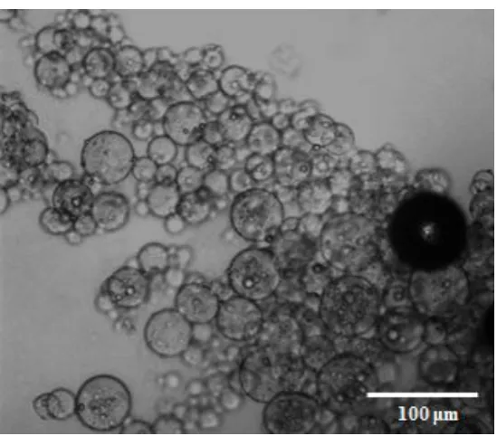
A representative confocal image of RhB-loaded gelatin microbeads in the oily phase was presented in Figure 4.5. From the figure we can see that the gelatin microbeads are spherical shaped in relatively uniform size distribution with an average diameter of 30 m and the intensity of fluorescence is very strong.
Figure 4.5 Confocal microscopy image of RhB-loaded gelatin microbeads (scale bar= 100 μm).
4.4.2 In vitro RhB release from gelatin gel and gelatin
microbeads
Figure 4.6 Release profiles of RhB directly from GM and GB at 37 °C.
4.4.3 In vitro dual drug release from alginate/gelatin
composite
strength of FL in alginate solution where more gelatin microbeads contained was much stronger than where fewer gelatin microbeads existed because free FL tend to absorb on the surface of gelatin microbeads. This happens due to the fact that acidic FL could form stronger bonding with basic gelatin compared with the interaction between acidic FL and acidic alginate [41]. However, Figure 4.7(A-2) shows that fluorescence of RhB could be observed only in the gelatin microbeads instead of
![Figure 1.1: Timeline showing FDA approved DDS in the market [6].](https://thumb-us.123doks.com/thumbv2/123dok_us/541785.2053598/20.595.116.479.410.639/figure-timeline-showing-fda-approved-dds-market.webp)