Doctoral School in Materials Science and Engineering
Bottom-up Tissue Engineering:
The Effect of 3D Tissue Fabrication Strategies on
Cellular Behavior.
Volha Liaudanskaya
XXV
II
cy
c
ii
iii
Bottom-up Tissue Engineering: the effect of 3D tissue
fabrication strategies on cellular behavior.
Volha Liaudanskaya
E-mail: [email protected]
Advisors Doctoral Committee
Prof. Claudio Migliaresi, Prof. Gian Domenico Soraru, Department of Industrial Engineering, Department of Industrial Engineering, Universita degli Studi di Trento, Italy. Universita degli Studi di Trento, Italy.
Prof. Antonella Motta, Prof. Ralf Riedel,
Department of Industrial Engineering, Institut für Materialwissenschaft, Universita degli Studi di Trento, Italy. Technische Universität Darmstadt,
Germany.
PhD internsip advisor: Prof. Sabine Fuchs, Prof. Utkan Demirci, Department of Traumatology, Stanford University School of Medicine, Experimental Trauma Surgery, Canary Center University Hospital Schleswig-Holstein, Department of Radiology, USA. Germany.
University of Trento,
iv
... To those who have dreams, but have doubts how to reach
them…. Never give up, never get discouraged, just keep work hard
and be dedicated to your dreams and life will reward you in the
v
Table of contents
List of abbreviations and acronyms xv
Abstract 2
1. General Introduction 4
1.1 Tissue engineering: general terms 4
1.1.1 Short history of tissue engineering 4 1.1.2 Progress, current state and problems of
tissue engineering 6
1.1.3 Tissue engineering approaches: advantages and open
questions 7
1.2 Organ printing: steps, current state and challenges 9
1.3 Cell encapsulation 13
1.3.1 General introduction 13
1.3.2 Methods for cell encapsulation 14
1.3.2.1Extrusion-based methods and state of the art 15 1.3.2.2Lithography-based methods 16
1.3.2.3Emulsion-based methods 17
1.3.2.4Microfluidics-based methods 17 1.3.2.5Bioprininting-based methods 18 1.3.2.6Superhydrophobic surfaces based methods 19 1.3.3 Materials selection for cell encapsulation 20
1.3.4 Cell source for encapsulation 23
1.4 Building blocks assembly techniques for 3D
tissue engineering 26
1.4.1 Magnetic and paramagnetic assembly 28
vi
1.4.3 Robotic 30
2. Research strategies and objectives 32
2.1. Thesis rationale 32
2.1.1 Building units fabrication 33
2.1.1.1 Materials 33
2.1.1.2 Cell encapsulation methods 37 2.1.1.3 Magnetic levitational assembly 40
2.1.2Cells 41
2.1.2.1 3T3 41
2.1.2.2 SHSY5Y 41
2.2 Outline and objectives 42
3. Assessing the impact of electro hydro-dynamic jetting on encapsulated cells viability, proliferation and ability to
self-assemble in 3D structures 44
3.1. Abstract 44
3.2. Introduction 46
3.3. Materials and methods 49
3.4. Results 55
3.5. Discussion 62
3.6. Conclusions 68
4. Homeostasis maintenance is critical parameter for application of
cell encapsulation in tissue fabrication 69
4.1. Abstract 69
4.2. Introduction 71
4.3. Materials and methods 75
4.4. Results 81
4.5. Discussion 91
vii
5. Fabrication of complex 3D structures with photolithographic
encapsulation and following magnetic levitational assembly 97
5.1. Abstract 97 5.2. Introduction 99
5.3. Materials and methods 102 5.4. Results and discussion 106
5.5. Conclusions 116
6. General conclusions 117
Bibliography 119
Scientific production 144
Participation to Congresses, Schools and Workshops 145
Other activities 147
viii
List of Tables
1.1 Cell encapsulation strategies [52-66]. 14
3.1 Primers sequence for RT-qPCR [177]. 53
4.1 Scheme of the conducted set of experiments for evaluation of cells viability, apoptosis/necrosis rate, activity and adhesion ability post-encapsulation.
ix
List of Figures
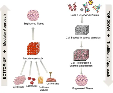
1.1 Tissue Engineering Approaches. Image adapted from R.
Tiruvannamalai-Annamalai et al. [12]. 8
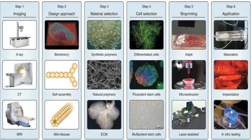
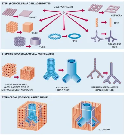
1.2 Organ printing process composed of three main steps. Image
adapted from Mironov et al [37]. 10
1.3 Bioprinting steps from the initial 3D computer modelling to the tissue printing and maturation. Images adapted from Atala et al
[27]. 19
1.4 Cell microencapsulation: nutrients, oxygen and stimuli diffuse across the membrane, whereas antibodies and immune cells are
excluded. Image adapted from Gorka O. et al [50]. 20 1.5 Stem cell source for tissue engineering applications. Image
adapted from Lutolf et al [130]. 25
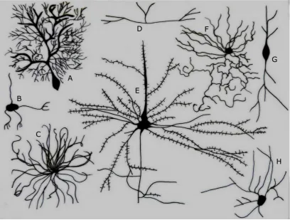
1.6 Different Types of Neurons. A. Purkinje cell B. Granule cell C. Motor neuron D. Tripolar neuron E. Pyramidal Cell F. Chandelier cell G. Spindle neuron H. Stellate cell. Images adapted from Ferris
Jabr; based on reconstructions and drawings by Cajal [131]. 26 1.7 Directed assembly methods in literature for microscale
hydrogels. A) Microfluidics assembly; B) Acoustic assembly; C)
Magnetic assembly; Images adapted from Gurkan et al [33]. 28 1.8 Building units assembly using acoustic waves. A) Diversity of
structures created by liquid-based templated assembly. B) Liquid-based templated assembly for tissue engineering. Images
adapted from Chen et al [26]. 29
1.9 Micro-robotic assembly of versatile in shape and size hydrogels: A) Two-dimensional micro-robotic coding of material
x
Images adapted from Tasoglu et al [36].
2.1 Sodium alginate structure. Image adapted from Lee at al [82]. 34 2.2 Sodium alginate sol-gel transition upon the contact with Ca2+
cations. 34
2.3 Synthesis of methacrylated gelatin. Gelatin macromers containing primary amine groups were reacted with methacrylic anhydride (MA) to add methacrylate pendant groups (A). To create a hydrogel network, the methacrylated gelatin was crosslinked using UV irradiation in the presence of a
photoinitiator (B). Images adapted from Nichol et al [103]. 36
2.4 Polyethylene glycol structure. 37
2.5 Electrohydrodynamic jetting encapsulation system. A. Encapsulation scheme; B. Beads view without cells; C. Beads with cells; D. Live/Dead analysis of cells encapsulated in the
beads. 38
2.6 Photolithographic encapsulation system. A. Encapsulation scheme; B. Hydrogel view with encapsulated cells; C. Live/Dead
analysis of cells encapsulated in the hydrogel [34]. 39 2.7 Paramagnetic levitational assembly of PEGDA hydrogels. A-H)
Concentric assembly of two hollow-disks and a solid-disk PEG
gel. Images adapted from Tasoglu et al [34]. 40 3.1 Beads with diameter consistently stable, about 200 µm were
made using 2% alginate ejected under 8kV from G33 stainless steel needle (I.D.: 0.108 mm) to CaCl2 solution for crosslinking.
(A) Optical microscope images represent cell behavior inside the beads at different time points: 3 hours, 7, 14, 21 and 28 days after encapsulation. Scale bar is 100µm. (B) Phenotypic transition in cells encapsulated in the beads and migrated
xi
3.2 (A) Live/Dead analysis with confocal microscopy of SHSY5Y cells encapsulated by EHDJ method in 200 µm 2% alginate beads: a) 3 hours; b) 1; c) 3; d)5; e) 7; f) 14; g) 21; h) 28 days; i) positive control (necrosis induced with 4% H2O2 overnight). Scale bar is 100µm. (B) Cell proliferation in the beads analyzed with DNA quantification assay within 4 weeks of encapsulation. Circles represent the beads border. Dead cells are evidenced by arrows except than in i). Error bars represent Mean±SD (n=3).
**p<0.01, ****p<0.0001. 57
3.3 RT-qPCR analysis of stress/apoptotic/necrotic markers in SHSY5Y cells encapsulated in 2% alginate beads by EHDJ method and evaluated at 8 time points. Expression of all targeted genes in studying samples were compared to the negative control samples (cells grown in tissue culture plate) and to the positive control samples (with induced hypoxia, apoptosis, necrosis and heat shock) [171, 172, 173, 174]. A) Expression level of HSP70B’- stress marker; B)CASP3, HMGB1 – apoptotic/necrotic markers;
C)HYOU1,GAPDH - hypoxia markers; D)CDH2 – adherence
marker; E)COL1A1 – extracellular matrix marker. Error bars represent Mean±SD (n=3). *p<0.05, **p<0.01, ***p<0.001,
****p<0.0001. 59
3.4 Theoretical model of cell response to stress exposure. In red
boxes, factors that were evaluated in this article. 63
4.1 Apoptotic/necrotic cell death evaluation. Cells were stained with DAPI for detection of the total amount of cells. An example of
Promikine kit stainings with experimental sample of Day 7. 82 4.2 Cell death type and rate evaluation with PromoKine in 8
studying groups and the reference control group. Experimental
xii
detection at 4 time points: day 1, 3, 5 and 7. The results were evaluated with confocal microscope, and following calculations with ImageJ cell counter plugin. A. Total amount of cell death; B. Apoptosis rate; C. Necrosis rate. Statistical analysis was performed comparing results of experimental groups to the control one within in vitro evaluation days: 1, 3, 5 and 7. Error bars represent meanSD (n=3). *p<0.05, **p<0.01, ***p<0.001, and ****p<0.0001.
4.3 Cell proliferation rate measured with DNA quantification assay. All experimental groups and control were divided in 2 subgroups: cells adhered to the surface of tissue culture plate, and cell in suspension that were not able to adhere. Experimental and reference groups were subjected to DNA quantification test at 4 time points: day 1, 3, 5 and 7. A. Cells adhered to the surface of tissue culture plates; B. Cells in suspension. Statistical analysis was performed comparing results of experimental groups to the control one within in vitro evaluation days: 1, 3, 5 and 7. Error bars represent meanSD
(n=3). *p<0.05, **p<0.01, ***p<0.001, and ****p<0.0001. 85 4.4 Cell activity rate measured with Alamar Blue assay.
Experimental and reference groups were subjected to Alamar Blue assay at 4 time points: day 1, 3, 5 and 7. Statistical analysis was performed comparing results of experimental groups to the control one within in vitro evaluation days: 1, 3, 5 and 7. Error bars represent meanSD (n=3). *p<0.05, **p<0.01, ***p<0.001,
and ****p<0.0001. 87
4.5 The ability to communicate and self-assemble in groups was evaluated with N-cadherin/DAPI expression at Day 1 on cells
xiii
and reseeded at 8 experimental time points in control conditions, and compared to the group of cells that were not encapsulated.
4.6 The ability to communicate and self-assemble in groups was evaluated with N-cadherin/DAPI expression at Day 7 on cells encapsulated in 2% alginate beads by EHDJ method, released and reseeded at 8 experimental time points in control conditions, and compared to the group of cells that were not
encapsulated. 89
4.7 Phenotypic changes and growth patterns were evaluated with brightfield imaging of cells encapsulated in 2% alginate beads by EHDJ method, released and reseeded at 8 time points in control conditions and compared to the group of cells that was not
encapsulated. 90
5.1 Example of GelMA hydrogels for GD salt toxicity evaluation. 106 5.2 Cell viability after GD exposure: A) at different concentration; B)
at different time. Long-term viability results of 50mM GD treated/not treated hydrogels for 10 minutes. Lines connecting individual groups indicate statistically significant difference.
One-way ANOVA with Tukey’s post-hoc tests, *p<0.05, **p<0.01. 108 5.3 Long-term effect evaluation of GD exposure on cells proliferation
capacity and ability to produce ECM with immunocytochemistry
to ki67 and collagen type 1. 109
5.4 Brightfield images of not treated/treated with GD cell-laden 5%
GelMA hydrogels at days 7 and 14. 110
5.5 Levitational self-assembly of soft micro-components. Concentric
assembly of two hollow-disk and a solid-disk PEG gels. 111 5.6 A) Live/Dead test before and immediately after the levitational
xiv
type 1 markers.
5.7 A) Cell-seeded beads before the assembly; B) Beads assembly
process by the levitational set-up. 113
5.8 A) Viability with Live/Dead kit; B) Viability assay with Alamar Blue assay; C) Immunocytochemistry at day 1; and D) day 7 to
xv
List of abbreviations and acronyms
TE Tissue Engineering
PGA Poly (glycolic acid)
UV Ultraviolet light
3D bioprinting Three-dimensional bioprinting
ECM Extracellular matrix
TCP Tissue culture plate
Gd Gadolinium salt
EHDJ Electro-hydro dynamic jetting
PI Photoinitiator
hESCs Human embryonic stem cells
PHEMA Poly (hydroxyethyl metacrylate)
PVA Poly (vinyl alcohol)
HA Hyaluronic acid
GelMA Gelatin metacrylate
2
Abstract
Organ failure is one the biggest problems, doctors face every day. Many patients are not able to get a transplant, but even those who recieved it, may undergo painful process of organ rejection and be on the transplant waiting list again. Organ transplants shortage is severe problem in current medicine that has many ethical and medical issues. To solve this problem, the new direction in regenerative medicine was formed, organ prinitng. The main goal of organ printing is fabrication of organ replacements that would mimic the original ones in terms of complexity and functionality. By direct fabrication and maturation of organs in vitro, the problem of organ shortage can be solved, moreover, based on the advances in cell therapy, these organs can be printed with patients own cells, which will eliminte the problem of transplant rejection.
Organ printing is multistep and complex process, composed of three main steps: tissue design, or theoretical modelling of replacement composition; tissue fabrication, or direct cell encapsulation and controlled assembly of building units; at last, tissue maturation to reach desirable functionality of the replacement. In the past decade, there was developed a variety of methods for the second step of organ printing, cell encapsulation, which is practicaly the main procedure for tissue fabrication. However, all these methods of cell encapsulation are complex and they might affect cells viability and functionality, which will result in changed tissue function.
3
The encapsulation step was performed with two different methods based on chemical or photo crosslinking of the material. Cell parameteres were evaluated on the molecular level for variety of parameters, including viability, activity, proliferation, stress markers expression, at last ability to adapt artificial environment to the cell functional niche with extracellular matrix markers expression, and proteoglycans.
The innovation of the presented study consists in the developing a unique protocol for detailed cell functionality evaluation during the organ printing procedures. In fact, based on the conducted study, it was proved the safety of the encapsulation methods. Moreover, based on the cell parameters post-encapsulation, there was suggested the optimal time for tissue maturation for application of the fabricated structures in organ printing, but also in other fileds, like developmental and pathological biology, or drug screening. Eventully, a novel way of simple blocks assembly into 3D complex structures was developed and proved to be safe for cell parameters.
4
Chapter 1.
General Introduction
1.1
Tissue engineering: general terms
1.1.1
Short history and definition of tissue
engineering
Tissue engineering (TE) as independent field of science was formed around 40 years ago, however the first historical note about tissue engineering can be found in Genesis I: 1: “The Lord, breathed a deep sleep on the man and while he was asleep he took out one of his ribs and closed up this place with flesh. The Lord God then built up into a woman the rib that he had taken from the man”[1]. Moving through the ages many references of TE applications were found. Recently, a women mummy was found aged around 3000 years which had a wooden toe prosthesis that replaced the amputated one [2]. In India in 600 B.C. there was a first written reference about the rotational flaps application for reconstruction of amputated noses [3]. In Italy in 15-16th
centuries many talented surgeons tried to improve the indian first version of rhinoplasty. Since that time, rhinoplasty procedure was drastically improved, however, the main principle remains the same as it was invented thousands of years ago.
Around 30 years ago, the term tissue engineering was mainly referred to the use of prosthetic devices and the surgical manipulation of tissues. The first experiments in TE though were conducted in 1970th in Boston, city that may be
5
generate a viable tissue. Based on this conclusion, couple of years later in Massachusetts Institute of Technology a group of scientists developed a novel skin draft by using collagen matrix and dermal fibroblasts. However, the real inception of current tissue engineering field started in mid-1980s by two talented scientists Dr. Joseph Vacanti and Dr. Robert Langer, when they came up with the idea of designing scaffolds for in vivo cell delivery. The results were unexpected and hardly explainable but they lay to the groundwork of the extensive research in the field of tissue engineering. Even though, in 1988 during the meeting organized by National Science Foundation Tissue Engineering field was first introduced and described within the article of keynote presentation, till today the article that was published a little while after in 1993 in Science considered as the most-cited paper that describes tissue engineering discipline. In this paper Dr. Vacanti and Dr. Langer define Tissue Engineering as an “interdisciplinary field that applies the principles of engineering and life sciences toward the development of biological substitutes that restore, maintain, or improve tissue function or a whole organ” [4].
Since the time those first small, but so valuable experiments were conducted, many scientific groups focused their attention on tissue engineering field and drastic progress was achieved.
6
1.1.2
Progress, current state and problems of
tissue engineering
During the first meeting in 1988 tissue engineering field was discussed as an alternative method for some injuries treatment by manipulating with prosthetic materials combined with existing tissues. The actual organ or tissue production using the basic concept of combining materials with cells wasn’t even considered at this memorable meeting, but since that time a lot has changed and significant progress was made in the field. One of the greatest scientists who made a priceless contribution in the field of tissue engineering is Dr. Anthony Atala and his group. At present his group is focused on growing tissues and organs of more than 30 parts of the body. Back in 2006 in the Lancet journal Atala published his first results of human bladder transplantation in 7 patients and their post-operative control [5]. The results were very promising, all patients recovered from the surgery and were able to improve the quality of their lives significantly. Since that publication many more successful bladder transplantations were made. Great success was also made in some other areas like skin regeneration [6, 7, 8], cartilage and bone tissue engineering [9, 10], corneal regeneration [11] and in many other fields.
7
Each direction of tissue engineering like neural, cartilage or heart faces specific set of challenges that need to be solved, like cell source, scaffold modelling and proper biocompatible material selection, cell seeding and growth factors effect, following cell matrix analysis and mechanical testing, and finally animal testing before implementation on patients [2].
1.1.3
Tissue
engineering
approaches:
advantages and open questions.
8
Figure 1.1: Tissue Engineering Approaches. Image adapted from R. Tiruvannamalai-Annamalai et al. [12].
9
1.2
Organ printing: challenges, steps and current
state.
Organ printing technology can be defined as biomedical application of rapid prototyping or layered additive biofabrication by using capable to self-assemble cell-laden building blocks or cell aggregates [27, 28, 29, 30]. The fundamental principal of organ printing technology implicates tissue capability for fusion to closely placed cell aggregates or building blocks. This principal was taken from natural embryonic development, where fusion is very common event during the layers formation and body parts development [31].
The ultimate goal of organ printing is large scale fabrication of 3D vascularized functional living organs for clinical transplantation and in vitro implementation for drug screening as well as platform for disease diagnostics [22, 24, 26, 29, 32]. Organ printing technology still has a lot of challenges, but with research and advanced improvements this developed technology will allow production of 3D functional living organs suitable for clinical implementation. Rapid prototyping is already well established and includes many methods such as stereolithography, selective laser sintering (SLS), fused deposition modeling (FDP), ballistic particles manufacturing (BPM) and others [29, 33, 34, 35, 36].
10
Organ printing composed of three main steps (Figure 1.2) [37]. Proper parameters and material selection for each of this step are essential for success in the 3D structure fabrication. Dr. Mironov defined 10 top challenges in organ printing technology [29, 30]. Below all these challenges are described in terms of organ printing steps: pre-processing, printing and post-processing [30].
11
1. Pre-processing is challenged by design, theoretical modeling and
in silico tissue self-assembly of the desired organ. This is the first, and the most
important set of challenges, without a precise design and theoretical modelling of cell behavior after printing, all the following steps will be useless. Initially obtained with computer tomography scanning picture of the organ can’t be used directly for organ printing. In silico tissue self-assembly will calculate and predict cells behavior, and which changes might happen during the organ maturation, such as cell fusion, shrinkage, matrix degradation by cells activity and many other parameters that have to be taken under control.
2. Printing step or direct cell encapsulation and controlled placement in defined order is challenged by the proper selection of biopaper, bioink and bioprinter, which in biological terms refer to the proper selection of biomaterials, cell source and encapsulation or printing conditions. Bioink refers to the cell-laden building blocks [41, 42, 43]. As far as the fundamental principal of organ printing based on tissue fusion process, the building blocks has to be located and prepared in the way that would stimulate and guide tissue fusion [40]. Thus, cells functionality, so as materials properties (biopaper) will play here a critical role. Biopaper can be defined as biodegradable, biomimetic hydrogels specifically designed for the bioprinting process with tissue fussion-permessive properties [31, 44, 45]. At last, bioprinter will have a crucial role on future 3D bioprinted organ. Bioprinter may affect significantly cell viability and vital characteristics during the following maturation step. Cell entrapment in 3D condition is stressful by itself, plus an effect of some encapsulation steps might affect cells viability, ability to proliferate and differentiate and many other characteristics might be affected, thus bioprinter selection so as direct encapsulation process has to be deeply evaluated before organ production.
12
desirable behavior of printed organ it has to be placed in conditions that would mimic in vivo, like pressure, growth factors supply, stress resistance and others. At last, during the maturation process, so as before implantation or printed organ implementation its main parameters have to be evaluated in non-invasive monitoring. Viability, vascularization, main organ function have to be perfect, otherwise implantation of not properly prepared organ may cause severe complications [48].
13
1.3
Cell encapsulation
1.3.1
General introduction.
In 1933 Bisceglie made a simple experiment, he enclosed tumor cells in polymer membrane and implanted them into pig’s abdominal cavity. Results showed, that cells survived long enough, to make a conclusion that immune system didn’t damage or destroy them [49, 50, 57]. Three decades later Chang introduced cell encapsulation as promising method for protecting cells from immune aggressiveness by encapsulating them in ultrathin polymeric membranes, and subsequently evoked term “artificial cells” [51]. This idea was widely applied in 70-80th for diabetes control in small animals by immobilizing
xenograft islet cells. Since then, a dramatic progress was made in understanding materials science, immunology, genetics and pharmaceutical technology that are so crucial for successful cell encapsulation [49]. Microencapsulation found many applications in different fields, such as therapeutic treatment for diabetes, cancer, hemophilia and renal failure [50, 52, 53], stem cell research [22] and others [54, 55, 56]. Around a decade ago, cell encapsulation received enormous scientific attention as the main method for the second step of organ printing technology, as it described above.
Cell encapsulation methods consist in the entrapment of cells in microcapsules or microbeads starting from a suspension of cells in polymeric solution that can be solidified by chemical or physical methods [50-57]. Generally cell encapsulation involves fabrication of three-dimensional (3D) scaffolds, with either seeded cells on top or with cells directly encapsulated with aim to use this fabricated 3D structure for healthy replacement of damaged tissues, or for guidance and support for tissue recovery.
14
restoration but also for transplantation and substitution of damaged or diseased tissues and organs.
1.3.2
Methods for cell encapsulation
With microencapsulation methods cells can be entrapped in various controllable forms such as beads, sheets, or fibers [52, 57]. In the recently published review Gasperini [52] described main cell encapsulation strategies that are widely used. Table 1 summarizes and compares several cell encapsulation techniques according to the final scaffold size and shape, but also the scalability of these methods and type of gelation mechanism applied for encapsulation.
Name Control of
size
Minimum Size (d in
µm)
Size dispersion
Best gelling mechanism
Extrusion Medium 80 Medium/
Low
Fast/ Ionotropic
Lithography Very good 50 Low All
Emulsion Low 10 High/
Medium Thermal
Microfluidic Very good 50 Low All
Bioprinting Very good 100 Low
All (more complex with thermal) Superhydrophobic
surfaces Very good 1000 Low All
15
1.3.2.1
Extrusion methods and state of the art.
The simplest and the most applicable method for cell encapsulation is extrusion [52, 56, 57, 58, 59]. This method is based on gravitational dripping properties of liquids. The suspension of pre-polymer solution mixed with cells extruded through the syringe or a needle. On the top of the needle drops form and when they reach a critical mass they fell freely in the collecting bath with the crosslinking solution. The final size of beads fabricated with extrusion methods depend on many factors, such as ionic concentration of the crosslinking bath, the surface tension of the drop, and the diameter of the pendant droplet neck [22, 52, 60]. Generally, the beads size is around 1mm, and the shape of the beads is not perfectly spherical, but rather droplet-like shape.
Many extrusion methods were advanced for obtaining the control over the final scaffold or bead size and shape, but also for improving cell viability and functionality [61, 62, 63]. For example, wet spinning, was modified extrusion method for final fabrication of fibers instead of beads. This method allows production of fibers, by extruding pre-polymer solution directly into the collecting bath with crosslinking solution without an air gap between the needle and a bath.
One else example of extrusion method modification is for generation of smaller size beads with controllable shape [52, 62]. The idea for production of smaller beads is to break down the extrusion jet before the drop will reach the critical mass and fells by itself. One of the applying methods is coaxial air flow [54, 62]. This method applies a compressed gas that directed through the extruding droplet and it causes the drop detachment faster than the droplet critical mass is reached. In the same mechanism beads can be produced but implying another liquid instead of gas.
16
size droplets by applying vibration to the nozzle. By manipulating with flow and vibration parameters it is possible to obtain beads size around 100 µm.
At last, by applying an electrical charge to the extruding needle it is also possible to obtain smaller beads with controlled size, by so-called bio-electrospraying method [22, 52, 60, 62, 66]. The extruding droplet reacts to the presence of electrical charge by accumulating ions of opposite sign on its surface. As the result, ions accumulation creates stress that force droplet to detach from the needle and drop into the crosslinking bath [66].
1.3.2.2
Lithography-based methods
Lithography methods include two subtypes: stereolithography and photolithography [41-43, 50, 52, 55, 56, 62, 67, 68, 69]. Stereolithography is based on the hydrogels production using replica moulding. At first, replicas produced by spin-coating or pouring and following UV crosslinking. Next, replicas filled with cells suspended in prepolymer solution and exposed to UV
light hydrogel crosslinking [67].
17
tissue engineering application, it has to have low cytotoxicity. A separate attention must be paid for combination of factors like UV time exposure during photopolymerization, and PI concentration, both these factors can affect cell viability. At last, a photomask is important as well, it has to be made in the way to protect unwanted areas from UV light, and those that exposed, has to be permeable for UV rays and initiate the crosslinking.
Polymer networks can be synthesized using various chemical methods, such as photo-, chemical, mechanical- or thermal-initiated polymerization. Advances in materials science made it possible to design and synthesize polymer networks with molecular-scale control over the structure such as crosslinking density and with tailored properties, such as biodegradation, mechanical strength, and chemical and biological response to stimuli.
1.3.2.3
Emulsion-based methods
Fabrication of spherical capsules by emulsion-based method found a wide application in pharmaceutical industry [53, 70, 71]. This strategy is based on the mixing of two liquids that are immiscible (for tissue engineering application one of the liquids is hydrogel precursor), the surfactants applied to the mixture will stabilize the emulsion and allows preparation of smaller droplets. After the suspension reaches the equilibrium precursor droplets undergoes sol-gel transition and become solid [26].
1.3.2.4
Microfluidics-based methods
18
applying flow of liquids to control the shape and size of produced microdroplets. Droplets that fabricated by microfluidics can be considered as bottom-up manufacturing, whereas the classical emulsion approach is top-down. The generation of microfluidics droplets happens through injecting the mixture of hydrogel precursor and cells into a microchannel and the droplets form when it reaches the continuous phase of non-miscible solution coming from the other inlets.
1.3.2.5
Bioprinting-based methods
3D bioprinting idea was taken from the paper printing technology. Like industrial printing technology had a revolutionary effect on people education, politics, religion and language across the globe, 3D printing affects dramatically science and research. Using 3D printing technology archeologists reproduce replicas of ancient and rare artifacts or some fossils for art, education and exhibitions. This technology allows design and work with three-dimensional spatial models [27]. But one of the most important applications 3D bioprinting found in organ or tissue fabrication.
19
Figure 1.3: Bioprinting steps from the initial 3D computer modelling to the tissue printing and maturation. Images adapted from Atala et al [27].
There were developed many biofabrication technologies such as organ printing or directed tissue self-assembly, solid scaffold-based biofabrication, embedding and molding technology, cell sheet technology, digital and inkjet bioprinting and many others [27-28, 32, 39, 40, 79, 80].
1.3.2.6
Superhydrophobic
surfaces
based
methods
20
1.3.3
Material selection for cell encapsulation
Cell encapsulation process starts from the suspension of buffer-based pre-polymer solution with cells, so called the sol flowing phase. After applying chemical, physical or biochemical stimuli liquid pre-polymer solution undergoes a transition to gel form, so called non-flowing phase. All the stages of sol-gel transition have to be well adjusted for cell viability, means they have to be as close as possible to the cell physiological conditions [52, 82, 83, 86, 87]. Over the years of materials studying and application of cell encapsulation for organ printing technology, list of desirable materials properties become well-formed and clear. Materials for cell encapsulation must have suitable crosslinking mechanism for successful encapsulation process, biocompatible but most importantly it has to support cells viability, activity, proliferation and main functions [22, 24, 43].
Figure 1.4: Cell microencapsulation: nutrients, oxygen and stimuli diffuse across the membrane, whereas antibodies and immune cells are excluded. Image adapted from Gorka O. et al [50].
21
characteristics, depends on the future application [84]. It has to be supportive for inflowing nutrients, oxygen and growth factors, and outflowing toxic waste products and cells activity products, for example insulin for diabetes treatment encapsulated cell delivery [82]. But, crucially it has to protect cells against antibodies and immune cells penetration, for cell protection inside the beads [84]. The ideal bead/ hydrogel schematically presented in Figure 1.4.
Materials currently used in the field of tissue engineering for regeneration and repair are mainly naturally derived polymers, such as alginate [14, 82, 83, 102], gelatin and its modifications [86, 90], collagen [15, 87], silk fibroin [18, 19, 20, 88], hyaluronic acid [16, 17, 89]; but there are also synthetic polymers that are widely used for some applications, such as PGA, polyethylene glycol (PEG) [21, 91, 92]. Both of the material types have its advantages and disadvantages. Natural polymers have similar to human extra cellular matrix (ECM) composition and structure [93, 94, 95, 96]. They are also very biocompatible and supportive for cells growth and activity. Whereas synthetic polymers have good repeatability characteristics and good control over gelation, degradation processes. Another advantage of synthetic polymers, that they can be easily modified or tailored to reach desirable mechanical or physical properties, which is complicated with natural polymers [27]. However, synthetic polymers generally have poor biocompatibility characteristics, toxic metabolites and loss of mechanical properties during degradation.
22
they are prevent protein adsorption. Because of easy manipulation and inertness properties, PEG polymers were modified in different ways for cell application to improve adhesion and proliferation, for example peptide sequence was incorporated into PEG structure to induce degradation, and cell adhesion [43].
PHEMA is another hydrogel that has been extensively studied and used in biomedical applications such as contact lenses and drug delivery. PHEMA hydrogels have good mechanical properties, optical transparency, and stability in water. PHEMA hydrogels are easily modified as well, to modulate the properties for different applications [97, 98, 99]. At last, PVA is another synthetic polymer. PVA hydrogels are stable, with crosslinks through the repeated thawing-freezing procedure or chemically crosslinked [99, 101].
Another group of polymers are naturally derived, such as collagen, silk, hyaluronic acid (HA), agarose, alginate, or gelatin and their modifications [13-20, 43]. Depending on their origin and composition, these materials can be applied for different applications. An advantage of natural polymers over synthetic is their natural low toxicity and biocompatibility.
Collagen and other mammalian cell derived protein-based polymers are effective matrices for cellular growth because they contain many cell-signalling domains present in vivo ECM. Collagen hydrogels can be easily obtained through the thermal crosslinking at 37°C degree, however they have relatively weak mechanical properties. Thus, many modifications were done with collagen that improved their mechanical characteristics, and crosslinking process.
HA is glycosaminoglycan (GAG) that is composed of repeated disaccharide units, and most abundant in wound healing and in joints, however it is still can be found in all cells types, but in less amount.
23
described, cell entrapment in GelMA hydrogels showed good results in cells long-term viability, and high proliferative potential [103, 104]. Thus, his group made an advanced study over 3D hydrogel binding surface, cells elongation and migration within the GelMa hydrogels. And they demonstrated that GelMA allows rapid cell adhesion, cell phenotype elongation, high proliferation potential, and migration in 2D and 3D. GelMA is promising material for variety of applications in tissue engineering [90, 105].
Another advanced research over GelMA application was made within Ali Khademhosseini group, by mixing GelMA with the FDA approved and described above PEG polymer [105]. By mixing these two polymers they wanted to obtain highly biocompatible, non-toxic, mechanically stable and easily manipulated hydrogel for cell encapsulation and micro patterned 3D hydrogels fabrication. Thus, they synthesized a mixed of PEG/GelMA hydrogel that is inexpensive, easily produced, and most importantly biologically and mechanically tunable.
1.3.4
Cell source for encapsulation
24
protocols for induction of pluripotent stem cells [118, 119, 120, 121, 122, 123]. These cells can solve both complications: differentiation potential and immune inertness. In many cases less potent autologous stem cells can be a suitable source for tissue engineering, exception will be perfect when patience cells are damaged or patient doesn’t have enough cells for therapy. Mesenchymal [124, 125], adipose [126, 127], blood, bone marrow derived stem cells [128, 129] are the most commonly used source for adult autologous transplantation and tissue engineering. The variety of stem cells that can be used for tissue engineering purposes is presented in Figure 1.5.
Any cell type chosen for cell encapsulation as building blocks for organ printing, has to have a high proliferation potential, to reach sufficient amount of cells for transplantation. Precise control over differentiation and proliferation in vitro and more importantly in vivo, is crucial for cell encapsulation. As it was mentioned above ECs and iPSCs are capable of teratoma formation which has to be eliminated and controlled. Too slow proliferation rate may result in the loss of viability and as a result cell death, whereas too high rate may cause hyperplasia and apoptosis. Timing for cell proliferation important as well, initial high cell proliferation rate is important to reach a proper cell density. But after some time the rate has to be equal to in vivo conditions to achieve cellular homeostasis, albeit without hyperplasia. This can be controlled with growth factors delivery, in some cases viral transfection is used for control over cell proliferation and senescence.
25
26
1.4
Building blocks assembly techniques for 3D
tissue engineering.
Human tissues and organs are complex, some of them like skin, or bladder are relatively simply organized, with small variety of cell types composed in the formation of these organs, whereas heart, kidney, and least understood brain are very complex, many different cells compose these organs and participate in the proper functioning. For example brain is composed of over 1 billion of different types of neurons, and 10 to 50 times more of glial cells, that act as servants for proper neural work (Figure 1.6) [131, 132].
27
neuron H. Stellate cell. Images adapted from Ferris Jabr; based on reconstructions and drawings by Cajal [131].
The complex tissue organization makes tissue engineering field is very challenging. As it was described above in section 1.4 there are many ways to fabricate cell-laden hydrogels in different shapes and sizes for variety of applications. However, many of them are capable of producing small size hydrogels, and more importantly they are limited to encapsulation of the poor variety of cell types into one hydrogel, which in many cases will be a serious limitation for tissue/organ fabrication [33, 34, 35, 36, 133, 134, 135, 136, 137].
Thus, properly prepared and laden with different cell types hydrogels have to be assembled in more complex structure that would mimic in vivo tissue in size and complexity.
28
Figure 1.7: Directed assembly methods in literature for microscale hydrogels. A) Microfluidics assembly; B) Acoustic assembly; C) Magnetic assembly; Images adapted from Gurkan et al [33].
1.4.1
Magnetic and paramagnetic assembly
29
inexpensive. There are some ways of producing hydrogels or building blocks with magnetic properties. The most common and originally proposed was loading hydrogels with magnetic nanoparticles of different kind, such as gold, iron oxide or bacteriophage. However, magnetic nanoparticles are potentially dangerous because of tissue cytotoxicity. Thus, new approaches that do not require nanoparticle presence are in great demand. Tasoglu and his group developed a new method for magnetic assembly by loading hydrogels with free radicals and vitamin E. These easy modifications allowed to perform guided self-assembly of materials with heterogeneous properties such as porosity, mass density and stiffness into the 3D structures [24, 34, 133, 134].
1.4.2
Acoustic assembly
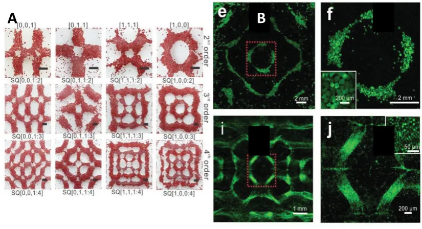
Figure 1.8: Building units assembly using acoustic waves. A) Diversity of structures created by liquid-based templated assembly. B) Liquid-based templated assembly for tissue engineering. Images adapted from Chen et al [26]. Acoustic fields have been used for in vivo imaging in medicine for quite some time, lately it has been applied in many other fields like particle
30
microcentrifugation, aggregation and trapping, cells manipulations like sorting and separation, and droplet concentration and mixing [26, 33, 135, 136].
Recently, acoustic field was applied for hydrogels assembly in bottom-up tissue engineering technology. This approach is based on the acoustic waves properties to organize cells or hydrogels of different size and weight on different layers of places. This approach shows high cell biocompatibility and versatile final structure manipulation as it shown in Figure 1.8 [26].
1.4.3
Robotic assembly
In the present world more and more fields are becoming mechanized, means industry is trying to use machines instead of humans work. This allows fabrication of many products in large-scale and in short time. Tissue engineering technology as well tries to mechanize the process of tissue/organ fabrication in many different ways. One of them is hydrogels self-assembly using micro-robots [36, 137].
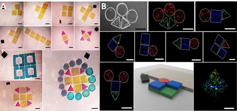
Figure 1.9: Micro-robotic assembly of versatile in shape and size hydrogels: A) Two-dimensional micro-robotic coding of material composition.
31
B) Spatially coded constructs for tissue culture. Images adapted from Tasoglu et al [36].
32
Chapter 2.
Research strategies and objectives
2.1
Thesis rationale
In the previous chapter there was introduced the concept of tissue engineering, its main approaches, advantages and open questions. After, there was presented a concept of organ printing with particular attention to its second step, cell encapsulation. There were described many methods for scaffolds or beads fabrication with focus on their complexity, proper cell type and material selection. At last, there was described in details a review on the cutting edge technology of building blocks assembly methods for complex 3D structures fabrication.
The final objective of the presented research work was detailed analysis of the organ printing strategies impact on cells viability, functionality and ability to maintain homeostasis in artificial conditions during the encapsulation process with following self-assembly into 3D complex structures (as the second step of organ printing), and during the tissue maturation period (as the third step of organ printing).
33
Furthermore, different tissue engineering applications have set of crucial and unique requirements for fabricated structures that have to be met. Based on this knowledge, we evaluated the optimal maturation time referring to cells activity and functionality for variety of applications, such as organ printing, drug screening and 3D disease models. At last, we developed a simple contactless method for self-assembly of fabricated building blocks into 3D structures to mimic the tissue complexity, and evaluated its effect on cell behavior parameters.
In the presented research work, different techniques and materials were used to evaluate the most favorable and safe conditions for functional 3D living microtissues fabrication.
In the next section materials, structures and setups introduced and decribed.
2.1.1
Building units fabrication
2.1.1.1
Materials
To reach the main goal of the presented work there were used two natural polymers: alginate, gelatin methacrylate, and one synthetic polymer polyethylene glycol (PEG). For the levitational assembly part there were also used manufactured beads modified for cell application.
Alginate
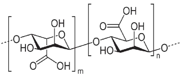
34
Figure 2.1 Sodium alginate structure. Image adapted from Lee at al [82]. Alginate composed 2 copolymeric blocks of (1,4)-linked β-D-mannuroic (M block) and α-L-guluronic (G block) acids as it showed in Figure 2.1 [52, 82]. The crosslinking process of sodium alginate happens upon the contact with multi-valent cations (Ca2+), they bind adjacent sodium alginate chains forming
ionic interchain bridges (Figure 1.4.3)
Figure 2.2: Sodium alginate sol-gel transition upon the contact with Ca2+
cations.
Alginate is lack of cell adhesion motifs, though a lot of research was done in tailoring alginate with RGD peptides sequence, which is crucial for cell adhesion and differentiation [102].
35
well as for hydrogels rigidity and mechanical properties. But also, there was found a strong correlation between the alginate permeability and crosslinking ions; higher the concentration of cations, creates tighter alginate structure, especially on the outer part of hydrogel which is in direct contact with crosslinking solution.
The other very important characteristic of alginate, it’s the ability to dissolve in vivo condition, as a result of bivalent ions exchange with monovalent ions from the body liquids. It will gradually dissolve calcium alginate, and transform it back to sodium alginate which is not degradable in vivo.
Alginate has become one of the most studied materials for cell encapsulation, and widely used. It was adjusted and modified for many different applications, such as immunoprotection of pancreatic islets, treatment of brain tumours, treatment of anemia and cryopreservation [52, 82, 102].
Gelatin methacrylate (GelMA)
The profound attention in photolithographical cell encapsulation received naturally-derived polymer gelatin, modified with methacrylate sites [103]. As it presented in Figure 2.3, gelatin was chemically modified with methacrylic anhydride, to obtain functional sites for photo crosslinking. Gelatin methacrylate (GelMA) is a photopolymerizable hydrogel comprised of modified natural ECM components, what makes it biocompatible and promising material for tissue engineering application.
36
Figure 2.3: Synthesis of methacrylated gelatin. Gelatin macromers containing primary amine groups were reacted with methacrylic anhydride (MA) to add methacrylate pendant groups (A). To create a hydrogel network, the methacrylated gelatin was crosslinked using UV irradiation in the presence of a photoinitiator (B). Images adapted from Nichol et al [103].
Polyethylene glycol (PEG)
Polyethylene glycol s a polyether compound with many applications from industrial manufacturing to medicine. The structure of PEG is (note the repeated element in parentheses): H-(O-CH2-CH2)n-OH.
37
inertness properties, PEG polymers were modified in different ways for cell application to improve adhesion and proliferation, for example peptide sequence was incorporated into PEG structure to induce degradation, and cell adhesion [34, 43, 91, 92].
Figure 2.4: Polyethylene glycol structure.
2.1.1.2
Cell encapsulation methods
Electrohydrodynamic jetting
A custom-designed electrically insulated holder was placed under a biological hood. Sterile 3 mL syringe was filled with cells suspended in the alginate solution, connected to a polytetrafluoroethylene tube and placed on the pump set to a fixed flux of 0.05 mL/min. The other side of the tube was connected to a stainless steel needle and placed in the holder under the hood. The positive cathode of a generator (ES30, Gamma High Voltage Research Inc, USA) was connected to the needle while the anode was connected to a metallic plate placed under a Petri dish containing the sterile calcium chloride solution and placed below the needle at a distance of 5 cm. Once the flux of solution was established and the generator was switched on (V=8kV), droplets of alginate solution containing cells were ejected from the needle directly to CaCl2 solution.
38
Figure 2.5: Electrohydrodynamic jetting encapsulation system. A. Encapsulation scheme; B. Beads view without cells; C. Beads with cells; D. Live/Dead analysis of cells encapsulated in the beads.
Photolithographic crosslinking system
Photolithography is another widely applied method for cell encapsulation and based on placing a photo-mask on top of the photocrosslinking material mixed with cells [34, 41, 42, 43, 68, 90, 104, 105, 106]. Photo mask can be designed to produce a hydrogels in variety of sizes and shapes. But also, the hydrogels can be produced with different cell types by mimicking tissue/organ complexity. This strategy is widely used for 3D model
A
Live/Dead
B
C
D
Scale bar: 100µM
39
fabrication for diseases investigation or general science.
Figure 2.6: Photolithographic encapsulation system [34]. Photolithographic encapsulation system. A. Encapsulation scheme; B. Hydrogel view with encapsulated cells; C. Live/Dead analysis of cells encapsulated in the hydrogel.
There are several important requirements for fabrication of patterned biocompatible cell-laden hydrogels by photolithography [42]. First requirement is proper photocurable material amenable to polymerization caused by photosensitive free radicals. Another important requirement is photoinitiator (PI), that must not only initiate the reaction, but also eliminate the reaction as soon as UV light exposure is stopped, but most importantly for tissue engineering application, it has to have low cytotoxicity. A separate attention must be paid for combination of factors like UV time exposure during photopolymerization, and PI concentration, both these factors can affect cell viability. At last, a photomask is important as well, it has to be made in the way to protect unwanted areas from UV light, and those that exposed, has to be permeable for UV rays and initiate the crosslinking.
Hydrogel Fabrication Process
Scale bar 200 µm
40
2.1.1.3
Magnetic levitational assembly
Magnetic levitation is contactless and nanoparticle-free approach for tissue self-assembly [24, 34, 133, 134]. This method is based on the idea of levitating subjects in paramagnetic suspension. Salt ions of gadolinium (Gd3+)
and magnesium (Mn2+) or radicals have paramagnetic properties. Cells were
encapsulated in photo-crosslinkable polymers of gelatin (GelMA) and polyethylene glycol diethacrylate (PEGDA) and levitated in the media suspension with ions of Gd3+ salt. This approach shows high scaling potential,
good cell viability and functionality, but also easy to manipulate and guide the hydrogels assembly makes it very promising for bottom-up tissue engineering.
41
2.1.2
Cells
In this work two main cell lines were used: m3T3 and hSHSY5Y.
2.1.2.1
NIH3T3
NIH 3T3 mouse embryonic fibroblast cells come from a cell line isolated and initiated in 1962 at the New York University School of Medicine Department of Pathology. 3T3 refers to the cell transfer and inoculation protocol for the line, and means “3-day transfer, inoculum 3 x 10^5 cells.” Using this protocol, the immortal cell line begins to thrive and stabilize in cell culture after about 20-30 generations of in vitro growth. The cell line has since become a standard fibroblast cell line and one the most commonly used cell models for tissue engineering methods testing.
2.1.2.2
SHSY5Y
42
2.2
Outline and objectives
Organ printing as promising direction of tissue engineering tends to fabricate precisely designed with controlled shape and composition 3D tissue, which after a short maturation process would be capable of performing its main function and be ready for implantation. To reach this goal the process of tissue fabrication has to be evaluated in detailes from different aspects, fabrication methods, materials selection, at last, cells viability and functionality.
In general all tissue/organ fabrication methods are multistep and complex, thus they might affect cells viability and functionality during the fabrication process. There were developed and studied variety of methods for tissue fabrication, with detailed and precise research in materials or methods, but not that much attention was paid to cell molecular mechanisms stability after encapsulation process, and culturing in artificial 3D environment.
Thus, the overall objective of the presented work was evaluation of the effect of organ fabrication methods on cell parameters, such as viability, activity, proliferation and ability to maintain homeostasis in artificial 3D environment.
The main objective was reached with following aims.
Chapter 3 – AIM1
To evaluate the effect of EHDJ encapsulation system on cells behavior.
Cells were encapsulated in alginate beads by EHDJ method and cultured up to 4 weeks to evaluate the short- and long-terms effect on cell parameters. At defined time points cells encapsulated in the beads were analyzed on their main characteristics. It was shown, that cells are mildly stressed first week after encapsulation, however towards the 4th week of culturing in the beads, cells
43
Chapter 4 – AIM 2
To evaluate the most favorable encapsulation time for variety of applications.
Cells were encapsulated in alginate microbeads by EHDJ method and cultured up to 4 weeks, at the established time points, cells were released from the beads and reseeded in tissue culture plates for following experiments. This work is targeted for the last step of organ fabrication, maturation. At this stage, cells have to replace the material used for fabrication and be able to produce ECM and maintain 3D structure without an artificial matrix. Results showed that most suitable for tissue maturation is to keep cells in the beads for 21-28 days to achieve the highest rate of their functionality in organ printing application. For drug screening application though, it was suggested to use cells that were encapsulated for 7-21 days.
Chapter 5 - AIM 3
Fabrication of building units with photolithographic encapsulation system, evaluation of cells parameters, and following assembly of fabricated building blocks into 3D complex structures.
44
Chapter 3
Assessing the impact of electro hydro-dynamic
jetting
on
encapsulated
cells
viability,
proliferation and ability to self-assemble in 3D
structures.
The main part of the presented in this chapter work was published in Tissue Engineering: Part C journal.
Liaudanskaya V., Gasperini L., Maniglio D., Motta A., Migliaresi C. Assessing the impact of electro hydro-dynamic jetting on encapsulated cells viability, proliferation and ability to self-assemble in 3D structures. Tissue Engineering: Part C (2014).
3.1
Abstract
In this chapter, systemic approach is propose to investigate the impact of electrohydrodynamic jetting (EHDJ) encapsulation on viability, proliferation, and functionality of the encapsulated cells. EHDJ consists in applying a high-voltage electrical field between a target substrate and a jetting needle, which is fed with a suspension of cells in a polymeric solution undergoing a sol-gel transition upon contact with the target. The viability, proliferation, and self-assembling ability of SHSY5Y human neuroblastoma cell line encapsulated in 2% alginate microbeads were analyzed by confocal microscopy and DNA quantification assays. In addition, the expression of stress (HSP70B’), apoptotic
(CASP3), necrotic (HMGB1), hypoxic (HYOU1, GAPDH), adhesion (NCDH) and
45
initial upregulation of the HSP70B’ expression within 24 h, its expression decreased to the negative control level together with a decrease in the expression of CASP3. Any increase in necrotic or hypoxic marker expression was not detected, while a slight upregulation of NCDH was observed in the first days after encapsulation, followed by its downregulation and stabilization to the control level. Furthermore, cell-laden beads started to self-assemble in three-dimensional (3D) constructs from the 3rd week after encapsulation. The results
46
3.2
Introduction
Cell encapsulation methods consist in the entrapment of cells in microcapsules or microbeads starting from a suspension of cells in polymeric solution that can be solidified by chemical or physical methods [64]. The technology has already found several applications, such as for the long-term encapsulation of insulin-producing cells for the diabetes treatment [82], chondrocytes phenotype retention [138], stem cells research [32]. Recently, cell encapsulation has been investigated for neural regeneration and for the treatment of the central nervous system malignancies [139, 140].
In regenerative medicine, cell encapsulation has received attention for the reconstruction of tissues or organs, following an early work of Mironov [29] who introduced the concept of organ printing. The procedure consists in the layer-by-layer deposition of encapsulated cells that are assembled in 3D structures [37, 38] to reproduce the organ/tissue architecture. 3D cell-laden structures may also be a suitable model for the fundamental science, drugs screening, as well as platform for diseases diagnostics [29, 37, 38, 39, 40].
Many methods have been proposed for cell encapsulation, such as microfluidics, emulsion, stereo/photolitography and extrusion [64]. These methods require a fine tuning of several parameters and an accurate selection of the encapsulating materials to keep cells alive, active, and capable of carrying out their main functions, such as the ability to self-assemble [44] or to produce biomolecules [141]. In the previous years, cell encapsulation has been proposed for applications in different fields, like bioprinting or skeletal tissue engineering. Agarose, gelatin, chitosan, collagen, polyethylene glycol, hyaluronic acid, alginate and other materials have been considered for the encapsulation of cells [31, 83-84].
47
expression in bovine aortic endothelial cells (BAEC) printed by laser pulses [64]; the analysis of gene expression on some house-keeping, non- and blood-specific genes in bio-electrospraying assay of whole human blood [143]; the determination of the immune response on the presence of allogeneic/xenogeneic hepatoma cells encapsulated in the beads and transplanted in rats [85].
One still open issue is whether encapsulated cells keep their original functionality, gene expression profile and ability to self-organize in tissues/organs.
In this study cells were encapsulated in alginate by electrohydrodynamic jetting (EHDJ) technology [64, 141, 144].
48
extacellular matrix was analysed with COL1A1 (COLlagen 1A1) expression [168, 169, 170]. The expression of all above genes in the samples were compared to the negative control (cells grown in tissue culture plate (TCP) till 85-90% confluence) and to the positive control (with induced hypoxia, apoptosis, necrosis and heat shock) [171, 172, 173, 174]. Gene expression level was normalized to the house-keeping control gene RPS18 (40S ribosomal protein S18) expression [175].
49
3.3
Materials and methods
Materials
Alginic acid sodium salt from brown algae (alginate) and calcium chloride dehydrate were purchased from Sigma-Aldrich (USA). Calcein-AM, Propidium Iodide (PI), Phosphate buffer saline without calcium and magnesium (PBS), Dulbecco’s modified eagle medium (DMEM) were purchased from Invitrogen (USA). The encapsulation system consists of a generator (ES30, Gamma High Voltage Research Inc., USA), a pump (NE-300, New Era Pump Systems, USA), a polyetrafluorethylene tube and a gauge 33 stainless steal needle (outer diameter: 0.210 mm, inner diameter: 0.108 mm Hamilton, Bonaduz/Switzerland). SHSY5Y human neuroblastoma cell line (ATCC®
CRL-2266™, UK), Sonicator (UP400S Heilscher, Germany), Quant-iT PicoGreen dsDNA
Assay Kit (Invitrogen, catalog number: P11496), 0.05% Triton-X 100 in PBS, RNeasy Plus Mini Kit (QIAGEN, catalog number: 74134) have been used.
Encapsulation process
Hydrogel preparation and sterilization.
50
Cell culture.
SHSY5Y human neuroblastoma cell line was expanded in 25-175mm tissue-treated culture flasks as monolayer (passages
12-of CO2 in high glucose Dulbecco’s Modified Eagle Medium (DMEM) with 10%
fetal bovine serum (GIBCO), 2mM Glutamine and 1% penicillin-streptomycin mixture (Sigma, USA). DMEM medium was changed every third day. The cells were cultured to 90-95% confluence before encapsulation [176].
Preparation of the alginate suspension with cells.
At 90-95% confluence cells were detached and moved to a 15 ml vial. Cells inside the vial were stirred at 1000 rounds/min for 10 minutes and the surnatant was removed. Cells on the bottom of the vial were resuspended in PBS and stirred again to remove any residues of medium containing cations that could crosslink the alginate solution. Cells were dispersed by vibration inside the buffer and an aliquot of the solution was taken to count the number of cells using a Cellometer Auto T4 (Nexcelom, USA) and Trypan Blue 0,4% (Life technologies, USA) as contrast agent. Cells were stirred again, and after removing the surnatant the alginate solution was added to obtain an alginate suspension containing 5 millions cells for milliliter. Medium, PBS, Trypsin/EDTA and the alginate suspension used were warmed to 37°C.
Cell encapsulation by electrohydrodynamic jet method.
51
and the anode to the metallic plate under the Petri dish. The Petri dish containing sterile calcium chloride solution was placed below the needle at a distance of 5 cm. Once the flux of solution was established and the generator was switched on (V=8kV), droplets of alginate solution containing cells were ejected from the needle directly to C
![Table 1.1: Cell encapsulation strategies [52-66].](https://thumb-us.123doks.com/thumbv2/123dok_us/537674.2053397/28.595.97.515.343.645/table-cell-encapsulation-strategies.webp)