R E S E A R C H
Open Access
Epigenetic marking of sperm by post-translational
modification of histones and protamines
Andrea M Brunner
1,3*, Paolo Nanni
2and Isabelle M Mansuy
1*Abstract
Background:The concept that individual traits can be acquired and transmitted by the germline through epigenetic mechanisms has gained recognition in the past years. However, epigenetic marks in sperm have not been are not well identified.
Results:Using a novel proteomic approach that combines peptide-based bottom-up and intact protein top-down tandem mass spectrometry, we report the identification of epigenetic marks on histones and protamines in adult mouse sperm. We identified a total of 26 post-translational modifications (PTMs) on specific residues of the core histones H2B, H3 and H4, and the linker histone H1, four of which had not been described previously in any tissue or cell line. We also detected 11 novel PTMs on the protamines PRM1 and PRM2 and observed that they are present in specific combinations on individual protamines.
Conclusions:Both histones and protamines carry multiple PTMs in the adult mouse sperm. On protamines, specific PTM combinations might form a‘protamine code’similar to the‘histone code’. These findings suggest a potential role for PTMs on sperm histones and protamines in epigenetic signatures underlying transgenerational inheritance.
Keywords:Epigenetics, Mouse sperm, Histones, Protamines, Post-translational modifications, Mass spectrometry, Electron transfer dissociation, Intact protein analysis, Top-down, Proteoforms
Background
The epigenetic status of the genome in eukaryotes strongly influences chromatin structure and remodeling, and determines the level of gene regulation. Typically, the epigenetic profile of a cell is conferred by DNA methylation and post-translational modifications (PTMs) of histones H1, H2A, H2B, H3 and H4, which together form a code that controls gene expression. These epi-genetic marks are specific to each gene, and are dynam-ically regulated during development and adulthood. They are also influenced by various factors throughout life, in particular by environmental conditions. In sperm cells, these marks are extremely important because they provide an identity to each cell and, as they can carry in-formation from parent to offspring, may be involved in
the maintenance and the inheritance of innate or ac-quired epigenetic signatures [1].
Sperm cells are highly specialized cells, produced by spermatogenesis, a process that involves extensive cellular, epigenetic and chromatin changes. Spermatogenesis starts with the replication and differentiation of spermatogonial stem cells into primary spermatocytes, which through genetic recombination during meiosis develop into hap-loid secondary spermatocytes [2]. In the haphap-loid phase of spermatogenesis, round spermatids mature into spermato-zoa. During this process, nucleosomes are disassembled upon histone H4 hyperacetylation and incorporation of non-canonical histone variants. Histones are widely re-placed by highly basic proteins, first by transition proteins and subsequently by the two protamines PRM1 and PRM2 [3,4]. In contrast to PRM1, PRM2 associates with the DNA in a precursor form that is processed proteo-lytically to give rise to the mature PRM2 protamine with approximately 40% of the N-terminus cleaved off [5]. In human sperm, protamines have been shown to be phosphorylated at PRM1S9, PRM1S11, and PRM2S59. However, the function of this phosphorylation remains * Correspondence:[email protected];[email protected]
1Department of Health Science and Technology of ETH Zürich, Neuroscience
Centre Zürich, Brain Research Institute, Medical Faculty of the University of Zürich, Winterthurerstrasse 190, Zürich CH-8057, Switzerland
3Biomolecular Mass Spectrometry and Proteomics, Utrecht University,
Padualaan 8, 3584 CH, Utrecht, The Netherlands
Full list of author information is available at the end of the article
© 2014 Brunner et al.; licensee BioMed Central Ltd. This is an open access article distributed under the terms of the Creative Commons Attribution License (http://creativecommons.org/licenses/by/2.0), which permits unrestricted use, distribution, and reproduction in any medium, provided the original work is properly cited.
unknown. Further post-translational processing of PRM1 and PRM2 occurs during transit of the spermatozoa through the epididymis. Protamines form disulfide bonds and zinc bridges, both within individual proteins and among different proteins [6,7].
The association between DNA and protamines leads to substantial molecular remodeling and ultimately to 10-fold compaction of the male genome in toroidal nucleoprotamine structures. The association with prot-amines might facilitate the hydrodynamic shape of the sperm head and protect the paternal genome from phys-ical and chemphys-ical damage, while the protamines them-selves could play a role in epigenetic processes [8]. In mature spermatozoa, about 99% of histones are replaced by the protamines PRM1 and PRM2 in mice, and about 85% in humans.
Intriguingly, the genome-wide distribution of protamine-and nucleosome-associated DNA regions is not rprotamine-andom, but nucleosomes retained in sperm are significantly enriched at loci important for embryogenesis, at specific promoters including those of miRNAs, and at imprinted genes. Furthermore, histone PTMs localize to specific de-velopmental loci: H3K4me2/3 is enriched at certain devel-opmental promoters; H3K4me3 localizes to HOX gene clusters, noncoding RNAs, and paternally expressed imprinted loci; and H3K27me3 is enriched at develop-mental promoters, which are repressed in early embryos [9,10]. Most recently, it was shown that throughout the genome, the retained nucleosomes are enriched at CpG-rich sequences that lack DNA methylation. The non-canonical histone H3.3 variant was shown to be abundant and trimethylated at K4 in these nucleosomes, while the canonical histones H3.1 and H3.2 were trimethylated at K27 [11]. Other non-canonical histone variants had been reported previously to be present in the retained nucleo-somes of mature sperm: TH2B was observed in human, H2A-Bbd, H2AL1/L2, and H2BL1 in mammalian sperm [8]. Further, overall acetylation of histones H3 and H4, ubiquitination of H2A and H2B, and H3K9me3 had been described [12].
After fertilization, histones seem to remain associated with the paternal genome and to contribute to zygotic chromatin despite the extensive reorganization of the sperm chromatin [13]. In postfertilization, the sperm DNA is decondensed from its highly compacted and transcrip-tionally quiescent state, and expands to the inducible state found in the paternal pronucleus [14]. The paternal his-tones could therefore serve as a template for the incorpor-ation of newly synthesized histones during replicincorpor-ation in the zygote. Accordingly, the programmatic chromatin packaging in sperm could potentially deliver epigenetic information to the oocyte and the zygote postfertilization.
While histone PTMs have been described in develop-ing male germ cells previously [15,16], little is known
about the histone PTM status in mature sperm. Further, it is also not known whether protamines are subjected to PTMs in mouse sperm. Here, using a novel proteomic approach combining peptide-based bottom-up and in-tact protein top-down mass spectrometry (MS), we qualitatively characterize mouse sperm cells. These ana-lyses reveal novel PTMs on histones and protamines, and are the first to show distinct combinations of pro-tamine PTMs, reminiscent of the histone code.
Results
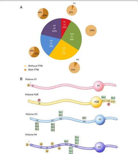
To identify chromatin PTMs in mouse sperm, we used a peptide-based bottom-up MS/MS strategy. We devel-oped a protocol for isolating sperm histone and protam-ine peptides. It consists of nuclear isolation from whole sperm cells, acid and high-salt extraction of basic nuclear proteins, digestion with various enzymes to optimize MS sequence coverage, and strong cation exchange chroma-tography (SCX) to enrich for acetylated and phosphory-lated peptides. Peptides and PTMs were then identified using a high mass accuracy LTQ-Orbitrap XL mass spec-trometer with a combination of electron transfer dissoci-ation (ETD) and collision-induced dissocidissoci-ation (CID) for peptide fragmentation. The CID and ETD fragmentation spectra of all identified histone peptides with PTM are shown in Additional file 1: Figure S2. With this workflow, we identified all five histone types, and detected 176 different peptides from these histones, including 40 to 45 peptides from H2A, H2B and H4, 33 from H3 and 14 from H1, with a false discovery rate (FDR) of less than 5% (Figure 1A and [see Additional file 2: Table S1]). On these peptides, the extent of PTM varied greatly depending on the histone type, and ranged from over 60% for H3 and H4 peptides to 7% for H1 peptides and 0% for H2A pep-tides (Figure 1A). Overall, a total of 26 PTMs was detected on H1, H2B, H3 and H4, including lysine and arginine methylation, N-terminal and lysine acetylation and threo-nine phosphorylation (Figure 1B).
*
*
Figure 1Histone peptides and post-translational modifications (PTMs) detected in mouse sperm. A)Proportion of detected histone peptides. The number of different peptides identified for each histone type, and sequence coverage (%) observed. The percentage of peptides with and without PTM is indicated.B)27 PTMs identified on histones H1, H2B, H3 and H4. PTMs are indicated by A for acetylation, Me1, Me2 and Me3 for mono-, di- and trimethylation, and P for phosphorylation. Residues are numbered starting with the first residue after the cleaved methionine according to histone field standard. * indicates that the acetylation site could not be assigned to a single residue, (that is, N-terminus or serine acetylation).
Brunneret al. Epigenetics & Chromatin2014,7:2 Page 3 of 12
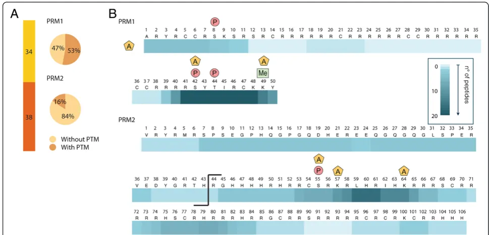
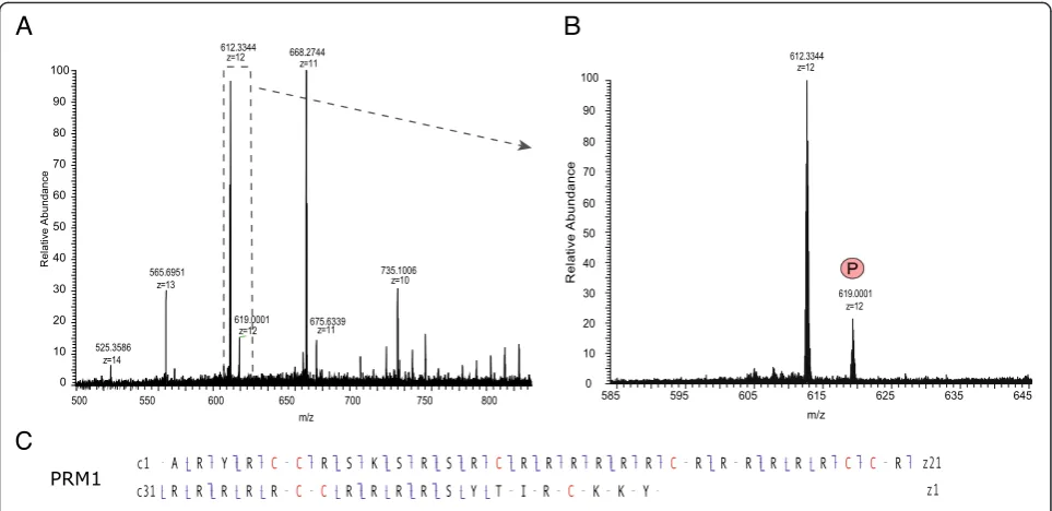
In these analyses, the ensemble of PRM2 peptides spanned nearly the entire sequence, resulting in good PRM2 sequence coverage. The sequence coverage for PRM1, however, was limited, due to long stretches of ar-ginine in the core region of this protamine (residues 13 to 41) that were difficult to sequence (Figure 2). To improve sequence coverage and identify PTMs in the unsequenced regions, we developed a top-down ap-proach based on the analysis of intact nuclear sperm proteins by liquid chromatography coupled to an LTQ-Orbitrap Velos ETD. Using this approach, we could identify full-length PRM1 and PRM2 proteins. PRM1 was detected both as unmodified and phosphorylated (Figure 4 and [see Additional file 5: Figure S3]) while PRM2 was mainly present as a processed form with the first 44 amino acids cleaved off. Various modified forms of processed PRM2 were also detected including mono-methylated, acetylated and phosphorylated forms, and various combinations of these PTMs such as one ation combined with one methylation site, two acetyl-ation sites together, and two acetylacetyl-ation and one methylation sites (Figure 5B and C, and [see Additional file 3: Figure S3]). The PTMs could be assigned to spe-cific PRM2 residues on the basis of spespe-cific fragment ions. Thus, PRM2 di-acetylation was on S55 and K57, S55 and K64, or S55 and S90, while di-acetylation and mono-methylation occurred on S55ac, S90ac and R88/ 89me1, or K57ac, S90ac and R88/89me1 (Figure 5C).
These results reveal that multiple PTMs co-occur on in-dividual protamines.
Discussion
Sperm cell specificity of novel histone post-translational modifications?
Figure 3(See legend on next page.)
Brunneret al. Epigenetics & Chromatin2014,7:2 Page 5 of 12
PTMs were not reported previously could be that three of the four PTMs (H2BK117me3, H2BK121me3, and H3R83me1) occur in the core regions of histones H2B and H3, which are less well studied than their terminal tails.
Functional roles of newly identified sperm histone post-translational modifications
In the case of sperm histones, only overall acetylation of histones H3 and H4, and ubiquitination of H2A and H2B, and H3K4me2/3, H3K9me3 and H3K27me3 have been reported previously [12]. Our data confirm the methylation of H3K9 and H3K27, but newly show that these residues can be mono-, di- or tri-methylated. Our data also reveal additional methylation sites on H3 (H3K36me1/2, H3R83me1). Moreover, H3K27 and H3K36 methylation frequently co-occur on the same peptide, consistent with previous reports [18]. Interestingly in our data, H3K36 methylation is only observed when H3K27 is also methylated, indicating potential crosstalk in H3 methylation (that is, that H3K27me contributes to the control of H3K36me). Regarding the functional role of these methylation sites, H3K9me2, H3K9me3 and
H3K27me3 are known to be involved in transcriptional re-pression. In contrast, H3K36me3 is a transcriptional acti-vator [19]. In sperm in particular, H3K27me3 was shown to mark developmental regulators [9], which are repressed in early embryos [10]. The specific roles of the mono- and di-methylated forms in sperm, and whether the PTMs are enriched at distinct genetic loci, remain to be investigated. It is interesting to note that while the core PTM H3R83me1 was only detected on the H3 variant H3.3, all N-terminal tail PTMs (H3K9me1/2/3, H3K27me1/2/3, and H3K36me1/2) and combinations thereof were identi-fied on both canonical H3 (H3.1/H3.2) and the H3.3 vari-ant [see Additional file 2: Table S1].
In addition to H3, we identified methylation sites on H4 (R19me2, K20me1/2/3, R23me1/2/3) and H2B (K117me3, K121me3). H4K20me is known to be impli-cated in chromatin compaction and transcriptional re-pression [19]. Recently, H4R19me and H4R23me, and in particular their combination, have been suggested to regu-late the interaction of H4K20me with its binding proteins [20]. Correspondingly, we detected H4K20me1R23me1, HK29me1R23me2 and H4K20me2R23me1 peptides [see Additional file 2: Table S1]. The functional role of the (See figure on previous page.)
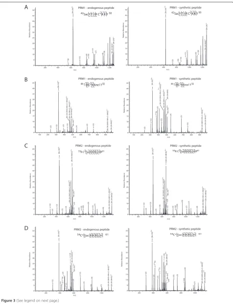
Figure 3Protamine post-translational modification (PTM) site validation by spectral comparison with synthetic peptides.Mass spectra of identified endogenous protamine peptides with novel PTM sites and their synthetic counterparts. Major peaks are labeled in the spectra and the fragment ions indicated in the peptide sequence.A)A novel site of serine acetylation on residue S42 of PRM1.B)A novel site of lysine methylation on residue K49 of PRM2.C)A novel site of serine phosphorylation on residue S55 of PRM2.D)A novel site of serine acetylation on residue S55 of PRM2.
Figure 5(See legend on next page.)
Brunneret al. Epigenetics & Chromatin2014,7:2 Page 7 of 12
identified H2B methylation sites remains to be elucidated, as methylation in general can be associated with both transcriptional repression and activation.
Our data also newly reveal multiple sites of modification including N-terminal or S1, K5, K8, K12 and K16 acetyl-ation on H4, N-terminal or S1 acetylacetyl-ation on H1, and T9 and T120 phosphorylation on H2B. Both acetylation and phosphorylation are prominent activation mark on his-tones. Acetylation is known to neutralize the positive charge of the lysine residue, weakening the DNA-histone interaction. It is also involved in DNA repair, replication and condensation [21]. Similarly, phosphorylation creates a repellent force to the DNA, opening the chromatin and activating gene transcription [22]. The identification of PTMs associated with transcriptional activation is unex-pected, as sperm cells are thought to be transcriptionally inert. However, these marks in paternal chromatin could contribute to the rapid activation of specific genes after fertilization. Alternatively, the presence of H4K8ac and H4K12ac could indicate centromeric heterochromatin [23]. N-terminal acetylation of H4 has been shown to regulate arginine methylation and chromatin silencing [24], while more generally, N-terminal protein acetylation is known to regulate protein-protein interactions, protein stability and protein localization [25].
The absence of PTMs on H2A corroborates a previous study reporting a low level of H2A modification site oc-cupancy in somatic cells [26]. This suggests the possi-bility that H2A, if not regulated by PTM, may be controlled by exchange of variants such as H2AX, H2AL1, H2AL2 and H2BL1 previously reported [27,28]. Indeed, we detected multiple peptides unique for the testis-specific expressed gene 1 protein (H2A-Bbd), an atypical histone H2A variant associated with active tran-scription and mRNA processing [see Additional file 2: Table S1]. This variant was recently shown to be highly expressed in adult testis, mainly in spermatocytes [29].
Overall, this MS-based study provides a novel map of histone PTMs in adult mouse sperm, generating a global picture of possible modified histone forms. Further stud-ies on the presence of sperm-specific histone PTMs and their combinatorial patterns in specific gene and promoter regions are needed to better understand the contribution of these histone forms to epigenetic inheritance. The importance of this endeavor is supported by two recent studies providing evidence that histone PTMs are in-volved in the transgenerational transmission of acquired traits [30,31].
Evidence for a‘protamine code’?
Our findings are the first to report the identification of multiple PTMs on protamines in mature mouse sperm (Figure 2B). They reveal that PRM1 is phosphorylated on S8 and PRM2 on S55, confirming human data (based on sequence homology) [32-34]. The data also identify unpredicted PTMs such as S42 and T44 phosphorylation on PRM1, N-terminal, S42 and K49 acetylation on PRM1, S55, K57, and K64 acetylation on PRM2, and K49 methy-lation on PRM1. The function of these PTMs is not known, but as histone PTMs, they may modulate protamine-DNA interactions [5]. On histones, PTMs are known to change protein structure and DNA binding by altering the electrostatic properties of histones. Acetyl-ation, for instance, counteracts the positive charge of the epsilon-amino group of the lysine. Similarly, phosphoryl-ation adds a negative charge to the histone. Both PTMs reduce the overall charge of histones and decrease their affinity for the negatively charged DNA, leading to chro-matin opening and facilitation of recruitment of the tran-scriptional machinery [22]. The fact that protamines are modified by PTMs classically associated with transcrip-tional activation is, however, intriguing because these pro-teins are thought to ensure tight packaging of the DNA in sperm cells, and contribute to transcriptional silencing. This apparent contradiction can be reconciled by the pos-sibility that PTMs on protamines may be involved in other regulatory functions. For instance, since PTMs in sperm are thought to provide paternal contribution to the epi-genetic reprogramming of the zygote [13], protamine PTMs could control the incorporation of maternal his-tones after fertilization, with specific combinations of PTMs favoring the recruitment of selected histones. Inter-estingly in these combinations, while acetylation and methylation can co-occur on a given protamine, acetyl-ation and phosphorylacetyl-ation appear to be exclusive. We did not detect proteins that were both acetylated and phos-phorylated. Our data also suggest that the acetylation of residues PRM1 S42 and PRM2 S55, which can be modi-fied by both PTMs, likely prevents their phosphoryl-ation, consistent with a previous report [35]. Finally, the possibility that a protamine code plays a role in sperm cells and in the zygote shortly after fertilization is ap-pealing, but needs to be investigated. It would be inter-esting for instance, to examine whether as is the case for histones, protamines with PTMs are enriched at spe-cific promoters or loci important for early embryonic development [9].
(See figure on previous page.)
Analysis of protamine forms by high mass accuracy mass spectrometry and electron transfer dissociation
fragmentation
An important parameter for the efficient detection and identification of protamines and PTMs in this study was the use of ETD fragmentation. ETD is a powerful method for the analysis of peptides with multiple basic residues such as protamines (rich in arginines), and in contrast to CID is not restricted to low charged peptides [36,37]. However, despite ETD, some arginine-rich re-gions, such as the PRM1 core (residues 13 to 41) that contains roughly 75% arginines, could not be sequenced using the bottom-up approach. We thus developed a top-down method for intact protein analysis to obtain sequence coverage of these regions, as well as to investi-gate the presence of specific modified protamine forms. This method allowed us to demonstrate the presence of both the full-length and processed PRM2 protein in ma-ture mouse sperm. As expected, PRM2 was mainly present in its mature processed form with the first 44 amino acids cleaved off. This form originates by associ-ation of the PRM2 full-length precursor with the DNA and successive proteolytic cleavage. The cleaved mature PRM2 protein comprises the amino acid residues 45 to 107 and has been shown to be the predominant form in the mature sperm head [5]. Furthermore, the four PTMs identified with the peptide-based approach (that is, S55, K57 and K64 acetylation, and S55 phosphorylation) could also be confirmed.
Conclusions
The presented study introduces a novel proteomic work-flow for an aspect of epigenetic marking that had remained unexplored to date: the characterization of chromatin PTMs in adult mouse sperm. The workflow allowed the identification of novel histone PTMs in the male germline, and, for the first time, of PTMs on prot-amines, including serine, lysine and N-terminal protein acetylation, serine and threonine phosphorylation, and lysine methylation. Only three phosphorylation sites had been described in human sperm before, but no PTM on mouse protamines was known. Furthermore, combina-tions of PTMs on individual protamines were identified, providing an unexpected picture of the epigenetic land-scape in sperm chromatin, These results suggest the ex-istence of a‘protamine code’ in addition to the‘histone code’ in sperm, the role of which in transgenerational epigenetic inheritance remains to be investigated.
Methods C57BL/6 mice
Mature sperm was collected from the caudal epididymis of adult 3- to 12-month-old C57BL/6 males as previ-ously described [38]. Mice were housed under a reversed
light cycle (dark phase, 7:00 to 19:00) in standard condi-tions. All experiments were ethically approved by the Swiss Cantonal Veterinary Office under license No. 55– 2012, Title ‘Study of the impact of early trauma on be-havior across generations in the mouse’.
Isolation of nuclear proteins from mouse sperm
Histones and protamines were purified from adult sperm as previously described for brain histones [26]. In brief, sperm cells from the sperm of 5 to 15 mice per sample were homogenized in lysis buffer (Sigma Nuclei Pure isolation kit, Sigma-Aldrich, Buchs, Switzerland) with 1 × protease inhibitor cocktail and phosphatase inhibitor cocktail I and II (Sigma-Aldrich). Nuclei were isolated by sucrose gradient centrifugation. Two volumes of 1.8 M sucrose were added to the homogenates and the mixture was layered on top of one volume of 1.8 M sucrose. Nu-clei were then pelleted by centrifugation at 30,000 g for 45 min, and snap frozen at−80˚C until analyzed. To sep-arate histones from other chromatin components, acid and high-salt extraction was performed as described pviously [39]. For acid extraction, isolated nuclei were
re-suspended in 0.2 M sulphuric acid (H2SO4) and
incubated for >2 h at 4˚C with end-over-end rotation. For high salt extraction, isolated nuclei were lysed by re-suspension in 3 mM EDTA, 0.2 mM EGTA and incu-bated at 4°C for 30 min with end-over-end rotation. After centrifugation at 6,500 g for 5 min, the nucleoplasm-containing supernatant was removed and snap frozen at−80˚C until analyzed. The chromatin pellet containing DNA, histones and protamines was re-suspended in solubilization buffer containing 50 mM Tris-Cl pH 8.0, 2.5 M NaCl, 0.05% NP40, and incubated for 30 min at 4°C with end-over-end rotation. After centrifugation at 16,000 g for 10 min, both acid- and salt-extracted histones and protamines were precipitated with trichloroacetic acid (TCA) followed by a 30-min incubation on ice. After cen-trifugation at 16,000 g for 10 min, the pellet containing histones and protamines was washed twice with ice-cold acetone and centrifuged a second time.
Reduction of disulphide bridges and alkylation of cysteines Histones and protamines were resuspended in an appro-priate buffer for subsequent enzymatic digestion (see below) or in 100 μl 25 mM NH4HCO3, pH 8 for intact
protein analysis. Disulphide bridges were reduced by incu-bation with 10 mM dithiothreitol (DTT) for 45 minutes at 50°C, and cysteines alkylated by incubation with 50 mM iodoacetamide (IAA) for 1 hr at room temperature in the dark. The reaction was blocked with 50 mM DTT. Sam-ples for intact protein analyses were desalted with ZipTip C18 columns (Millipore, Billerica, MA, USA) and lyophi-lized dry prior to MS analysis. Samples for peptide-based analyses were enzymatically digested (see below).
Brunneret al. Epigenetics & Chromatin2014,7:2 Page 9 of 12
In-solution digestion of histone proteins
Proteins were digested into peptides with trypsin (Pro-mega, Madison, WI, USA) in 50 mM ammonium bicar-bonate, pH 8.0 at 37˚C for 2 h (1:200 enzyme:substrate), with Glu-C (Roche Applied Science, Penzberg, Germany) in 25 mM ammonium carbonate, pH 7.8, at 24˚C for 18 h (1:20 enzyme:substrate), and with AspN (Roche Applied Science) and chymotrypsin (Roche Applied Science) in 100 mM Tris–HCl, 10 mM CaCl2, pH 7.8, at 25˚C for
25 h (1:200 enzyme:substrate), as described previously [26]. Enzymatic digests were stopped by adding 10% tri-fluoroacetic acid (TFA) to a final pH <3. Peptides were then desalted with ZipTip C18 columns (Millipore) and lyophilized dry prior to analysis by ETD/CID-MS/MS.
Acetylated and phospho-peptide enrichment
To enrich for acetylated and phospho-peptides, digested peptides were fractionated by SCX [40]. Peptides were loaded onto a 12μm, 300 Å pore size ZipTip SCX column (Millipore) and eluted in four fractions with increasing KCl concentration (50 mM, 150 mM and 300 mM KCl in 0.1% TFA, and 5%NH4OH).
Synthetic peptides
Lyophilized synthetic peptides were ordered from 21st Century Biochemicals, Boston, MA, USA, resolubilized in 50% acetonitrile (ACN), 0.1% formic acid (FA) and analyzed by direct infusion.
Peptide-based bottom-up collision induced dissociation/ electron transfer dissociation mass spectrometry/mass spectrometry analysis
Peptide samples were analyzed on an LTQ-Orbitrap XL-ETD mass spectrometer (Thermo Scientific, Germany) coupled to an Eksigent Nano-HPLC system (Eksigent, AB Science, Redwood City, CA, USA). Solvent compos-ition at the two channel was 0.2% formic acid, 1% ACN for channel A and 0.2% formic acid, 80% ACN for chan-nel B. Peptides were resolubilized in 3% ACN and 0.2% formic acid and loaded on a 10 cm fused silica column packed with 3 μm 200 Å pore size C18 resin. Peptides were eluted with a flow rate of 200 nl/min by an ACN gradient of 5-30% ACN over 35 min and 30 to 80% ACN over the subsequent 13 min. Full-scan mass spec-tra (m/z 300 to 2000) were acquired in the Orbispec-trap with a resolution of 60,000 at 400 m/z, after accumulation to a target value of 2e5. Six sequential CID and ETD MS/ MS scans were acquired in the ion trap on the three most intense signals above a threshold of 500. The AGC target value for ion trap MSn scans was set to 1e4. CID was performed using a normalized collision energy of 35 and activation time of 30 ms. The ETD reaction time was 120 ms and isolation width was 2 m/z. The ETD anion target value was set at 1e6 and the activation time
at 100 ms. Supplementary activation was employed and charge state dependent ETD time enabled. For all exper-iments, the precursor masses already selected for MS/ MS were excluded for further selection for 30 s. The ex-clusion window was set to 20 ppm and the size of the exclusion list was set to 500. Samples were acquired using internal lock mass calibration set on m/z 429.0877 and 445.1200 m/z. The synthetic peptides were analyzed by direct infusion on an LTQ-Orbitrap XL-ETD. For every peptide, fragmentation was performed by CID and ETD, and spectra acquired in both the linear ion trap and the FT-Orbitrap. Twenty scans were collected for every acquisition. For CID/ETD fragmentation and for the acquisition of ion trap MS/MS spectra the same pa-rameters used for LC-MS analysis were applied. For FT MS/MS spectra an AGC target value of 5E5 and an in-jection time of 200 ms were set.
Peptide and post-translational modification identification Mascot generic format (mgfs) files were generated from MS and MS/MS raw data. Mgfs were searched against a mouse protein database from the European Bioinformatics Institute (EBI, 48,564 sequences) using Mascot version 2.3 (Matrix Science, London, UK) with a peptide mass tolerance of 6 ppm and a frag-ment mass tolerance of 0.6 Da. The following PTMs were included in the searches: carbamidomethylation (C, fixed, 57.021464 Da), phosphorylation (S, T, and Y, variable, 79.966331 Da), acetylation (protein N-term, S, T, Y and K, variable, 42.010565 Da), mono-, di- and tri-methylation (R and K, variable, 14.015650 Da, 28.031300 Da and 42.046950 Da), and oxidation (M, variable, 15.994915 Da). Only peptides with the Mascot parameter rank 1 were ac-cepted. In the case of histone peptides, a peptide expect cut-off of 0.05 and an FDR <5% was applied, and all PTMs identified by only one spectrum were discarded. For spectra of all peptides with PTM(s), confident PTM site placement was based on the Mascot site analysis probability [41] and manual validation. When alternative site localizations were possible for a given PTM, the ambiguous sites were indi-cated by parentheses. All identified peptides with PTMs are listed in Additional file 2: Tables S1 and Additional file 3: Table S2. Residues are numbered starting with the first residue after the cleaved methionine according to histone field standard.
Intact protein top-down electron transfer tandem mass spectrometry analysis
an ACN gradient of 1 to 55% ACN over 36 min in a buffer containing 0.2% FA at flow rate of 250 nl/min. Both full scan MS and MS/MS spectra were acquired in the Orbi-trap analyzer, at a resolution of 60,000 for MS1 and 30,000 for MS2 scans. The AGC target values were set to 1E6 for MS1 and 1E5 for MS2. The maximum injection time was 250 ms for MS1 and 200 ms for MS2 scans. Fragmenta-tion of the three most intense ions above a threshold of 5,000 was performed by ETD with an activation time of 15 ms. In order to improve the efficiency of ETD fragmen-tation, the precursor ions were isolated in a 10 Da m/z window. For each fragmentation event, supplementary ac-tivation was employed and 5μscans were summed.
Protein and proteoform identification
Spectra were deconvoluted with Mascot Distiller v 2.3.2. Protein and PTM identification was performed with Mascot version 2.3 and ProSight PTM 2.0 [42]. Mascot searches were performed against a protein database con-taining histones, protamines and their processed forms, and E. coli protein sequences. Peptide and fragment mass tolerance were set to 2 Da to account for incorrect isotope picking. The same PTMs as in the peptide-based approach were included in the searches: carbamidometh-ylation (C, fixed), phosphorcarbamidometh-ylation (S, T, and Y, variable), acetylation (protein N-term, S, T, Y and K, variable), mono-, di- and tri-methylation (R and K, variable), and oxidation (M, variable). In ProSight PTM 2.0, the intact and fragment ion masses of selected scans were searched using the single protein search. PTM sites were manually placed and scored using sequence gazer.
Additional files
Additional file 1: Figure S2.MS2 spectra of all modified histone peptides obtained from histone digests across all experiments. The Mascot peptide view, fragment ion table and the annotated peptide sequence with post-translational modification (PTM) are shown.
Additional file 2: Table S1.List of all histone peptides derived from mouse sperm found in the peptide-based bottom-up experiments. In the peptide sequence the site/s of N-terminal acetylation are designated by
‘ac-’, before the modified residue, acetylation by‘ac’, phosphorylation by
‘p’, and mono-/di-/tri-methylation by‘me1, me2 or me3’respectively. Percentages from the Mascot site analysis indicate the Mascot Delta score as a post-translational modification (PTM) site probability when alternative site localizations are possible for given PTM(s). For four peptides, the PTM is in parenthesis because the PTM site could not be assigned to a single residue.
Additional file 3: Table S2.List of all protamine peptides derived from mouse sperm found in the peptide-based bottom-up experiments. In the peptide sequence the site/s of N-terminal acetylation are designated by
‘ac-’, before the modified residue, acetylation by‘ac’, mono-methylation by‘me1’, and phosphorylation by‘p’.
Additional file 4: Figure S1.Protamine post-translational modification (PTM) site validation by spectral comparison with synthetic peptides. Mass spectra of identified endogenous protamine peptides with novel PTM sites and their synthetic counterparts. Major peaks are labeled in the spectra and the fragment ions indicated in the peptide sequence. A)
A novel site of acetylation at the N-terminus of PRM1. B) A novel site of serine phosphorylation on residue S8 of PRM1. C) A novel site of serine phosphorylation on residue S42 of PRM1. D) A novel site of threonine phosphorylation on residue T44 of PRM1. E) A novel site of lysine acetylation on residue K49 of PRM1. F) A novel site of lysine acetylation on residue K57 of PRM2. G) A novel site of lysine acetylation on residue K64 of PRM2.
Additional file 5: Figure S3.MS2 spectra of intact PRM1 and PRM2 forms.
Abbreviations
Ac:acetylation; ACN: acetonitrile; CID: collision induced dissociation; Da: Dalton; ETD: electron transfer dissociation; FA: formic acid; FDR: false discovery rate; H2SO4: sulphuric acid; IAA: iodoacetamide; me1/me2/ me3: mono-/di-/tri-methylation; mgf: Mascot generic format; MS: mass spectrometry; p: phosphorylation; PTM: post-translational modification; SCX: strong cation exchange; TFA: trifluoroacetic acid.
Competing interests
The authors declare that they have no competing interests.
Authors’contributions
AMB conceived the project, planned and performed experiments, did data analysis and wrote the manuscript. AMB and PN planned and performed MS analyses. PN and IMM critically read the manuscript. IMM directed and supervised experiments, and contributed reagents, materials and analysis tools. All authors read and approve the final manuscript.
Acknowledgments
We thank Dr. Jonas Grossmann and Dr. Christian Panse, Functional Genomics Centre Zurich for IT support. Katharina Gapp for helpful discussions and Dr. Ry Y. Tweedie-Cullen for critical reading of the manuscript. The lab of IM Mansuy is funded by the University of Zürich, the Swiss Federal Institute of Technology, The Swiss National Foundation the National Centre of Competence in Research‘Neural Plasticity and Repair’, and Roche. AM Brunner is funded by an iPhD grant from SystemsX.ch.
Author details 1
Department of Health Science and Technology of ETH Zürich, Neuroscience Centre Zürich, Brain Research Institute, Medical Faculty of the University of Zürich, Winterthurerstrasse 190, Zürich CH-8057, Switzerland.2Functional Genomics Centre Zürich, University of Zürich and ETH Zürich,
Winterthurerstrasse 190, Zürich CH-8057, Switzerland.3Biomolecular Mass Spectrometry and Proteomics, Utrecht University, Padualaan 8, 3584 CH, Utrecht, The Netherlands.
Received: 28 August 2013 Accepted: 20 December 2013 Published: 20 January 2014
References
1. Rando OJ:Daddy issues: paternal effects on phenotype.Cell2012, 151:702–708.
2. Oliva R, Castillo J:Proteomics and the genetics of sperm chromatin condensation.Asian J Androl2011,13:24–30.
3. Govin J, Caron C, Lestrat C, Rousseaux S, Khochbin S:The role of histones in chromatin remodelling during mammalian spermiogenesis.Eur J Biochem2004,271:3459–3469.
4. Steger K:Transcriptional and translational regulation of gene expression in haploid spermatids.Anat Embryol (Berl)1999,199:471–487.
5. Balhorn R:The protamine family of sperm nuclear proteins.Genome Biol
2007,8:227.
6. Aitken RJ, Nixon B, Lin M, Koppers AJ, Lee YH, Baker MA:Proteomic changes in mammalian spermatozoa during epididymal maturation.
Asian J Androl2007,9:554–564.
7. Bjorndahl L, Kvist U:Human sperm chromatin stabilization: a proposed model including zinc bridges.Mol Hum Reprod2010,16:23–29. 8. Rathke C, Baarends WM, Awe S, Renkawitz-Pohl R:Chromatin dynamics
during spermiogenesis.Biochim Biophys Acta2013. doi: 10.1016/j.bbagrm. 9. Brykczynska U, Hisano M, Erkek S, Ramos L, Oakeley EJ, Roloff TC, Beisel C,
Schubeler D, Stadler MB, Peters AH:Repressive and active histone
Brunneret al. Epigenetics & Chromatin2014,7:2 Page 11 of 12
methylation mark distinct promoters in human and mouse spermatozoa.
Nat Struct Mol Biol2010,17:679–687.
10. Hammoud SS, Nix DA, Zhang H, Purwar J, Carrell DT, Cairns BR:Distinctive chromatin in human sperm packages genes for embryo development.
Nature2009,460:473–478.
11. Erkek S, Hisano M, Liang CY, Gill M, Murr R, Dieker J, Schubeler D, van der Vlag J, Stadler MB, Peters AH:Molecular determinants of nucleosome retention at CpG-rich sequences in mouse spermatozoa.Nat Struct Mol Biol2013,20:868–875.
12. Carrell DT:Epigenetics of the male gamete.Fertil Steril2012,97:267–274. 13. van der Heijden GW, Ramos L, Baart EB, van den Berg IM, Derijck AA, van der Vlag J, Martini E, de Boer P:Sperm-derived histones contribute to zygotic chromatin in humans.BMC Dev Biol2008,8:34.
14. Jenkins TG, Carrell DT:Dynamic alterations in the paternal epigenetic landscape following fertilization.Front Genet2012,3:143.
15. Tan M, Luo H, Lee S, Jin F, Yang JS, Montellier E, Buchou T, Cheng Z, Rousseaux S, Rajagopal N, Lu Z, Ye Z, Zhu Q, Wysocka J, Ye Y, Khochbin S, Ren B, Zhao Y: Identification of 67 histone marks and histone lysine crotonylation as a new type of histone modification.Cell2011,146:1016–1028.
16. Lu S, Xie YM, Li X, Luo J, Shi XQ, Hong X, Pan YH, Ma X:Mass spectrometry analysis of dynamic post-translational modifications of TH2B during spermatogenesis.Mol Hum Reprod2009,15:373–378.
17. Montellier E, Boussouar F, Rousseaux S, Zhang K, Buchou T, Fenaille F, Shiota H, Debernardi A, Hery P, Curtet S, Jamshidikia M, Barral S, Holota H, Bergon A, Lopez F, Guardiola P, Pernet K, Imbert J, Petosa C, Tan M, Zhao Y, Gérard M, Khochbin S:Chromatin-to-nucleoprotamine transition is controlled by the histone H2B variant TH2B.Genes Dev2013, 27:1680–1692.
18. Jung HR, Sidoli S, Haldbo S, Sprenger RR, Schwammle V, Pasini D, Helin K, Jensen ON:Precision mapping of coexisting modifications in histone H3 tails from embryonic stem cells by ETD-MS/MS.Anal Chem2013, 85:8232–8239.
19. Kouzarides T:Chromatin modifications and their function.Cell2007, 128:693–705.
20. Hirano Y, Hizume K, Kimura H, Takeyasu K, Haraguchi T, Hiraoka Y:Lamin B receptor recognizes specific modifications of histone H4 in
heterochromatin formation.J Biol Chem2012,287:42654–42663. 21. Campos EI, Reinberg D:Histones: annotating chromatin.Annu Rev Genet
2009,43:559–599.
22. Bannister AJ, Kouzarides T:Regulation of chromatin by histone modifications.Cell Res2011,21:381–395.
23. Puri D, Dhawan J, Mishra RK:The paternal hidden agenda: epigenetic inheritance through sperm chromatin.Epigenetics2010,5:386–391. 24. Schiza V, Molina-Serrano D, Kyriakou D, Hadjiantoniou A, Kirmizis A:
N-alpha-terminal acetylation of histone H4 regulates arginine methylation and ribosomal DNA silencing.PLoS Genet2013,9:e1003805.
25. Hollebeke J, Van Damme P, Gevaert K:N-terminal acetylation and other functions of Nalpha-acetyltransferases.Biol Chem2012,393:291–298. 26. Tweedie-Cullen RY, Brunner AM, Grossmann J, Mohanna S, Sichau D, Nanni
P, Panse C, Mansuy IM:Identification of combinatorial patterns of post-translational modifications on individual histones in the mouse brain.
PLoS One2012,7:e36980.
27. Li Z, Yang J, Huang H:Oxidative stress induces H2AX phosphorylation in human spermatozoa.FEBS Lett2006,580:6161–6168.
28. Govin J, Escoffier E, Rousseaux S, Kuhn L, Ferro M, Thevenon J, Catena R, Davidson I, Garin J, Khochbin S, Caron C:Pericentric heterochromatin reprogramming by new histone variants during mouse spermiogenesis.
J Cell Biol2007,176:283–294.
29. Gu C, Tong Q, Zheng L, Liang Z, Pu J, Mei H, Hu T, Du Z, Tian F, Zeng F: TSEG-1, a novel member of histone H2A variants, participates in spermatogenesis via promoting apoptosis of spermatogenic cells.
Genomics2010,95:278–289.
30. Zeybel M, Hardy T, Wong YK, Mathers JC, Fox CR, Gackowska A, Oakley F, Burt AD, Wilson CL, Anstee QM, Barter MJ, Masson S, Elsharkawy AM, Mann DA, Mann J:Multigenerational epigenetic adaptation of the hepatic wound-healing response.Nat Med2012,18:1369–1377.
31. Vassoler FM, White SL, Schmidt HD, Sadri-Vakili G, Pierce RC:Epigenetic inheritance of a cocaine-resistance phenotype.Nat Neurosci2013,16:42–47. 32. Papoutsopoulou S, Nikolakaki E, Chalepakis G, Kruft V, Chevaillier P,
Giannakouros T:SR protein-specific kinase 1 is highly expressed in testis and phosphorylates protamine 1.Nucleic Acids Res1999,27:2972–2980.
33. Chirat F, Arkhis A, Martinage A, Jaquinod M, Chevaillier P, Sautiere P: Phosphorylation of human sperm protamines HP1 and HP2: identification of phosphorylation sites.Biochim Biophys Acta1993, 1203:109–114.
34. Hornbeck PV, Kornhauser JM, Tkachev S, Zhang B, Skrzypek E, Murray B, Latham V, Sullivan M:PhosphoSitePlus: a comprehensive resource for investigating the structure and function of experimentally determined post-translational modifications in man and mouse.Nucleic Acids Res
2012,40:D261–D270.
35. Mukherjee S, Hao YH, Orth K:A newly discovered post-translational modification–the acetylation of serine and threonine residues.
Trends Biochem Sci2007,32:210–216.
36. Syka JE, Coon JJ, Schroeder MJ, Shabanowitz J, Hunt DF:Peptide and protein sequence analysis by electron transfer dissociation mass spectrometry.Proc Natl Acad Sci U S A2004,101:9528–9533.
37. Mikesh LM, Ueberheide B, Chi A, Coon JJ, Syka JE, Shabanowitz J, Hunt DF: The utility of ETD mass spectrometry in proteomic analysis.Biochim Biophys Acta2006,1764:1811–1822.
38. Franklin TB, Russig H, Weiss IC, Graff J, Linder N, Michalon A, Vizi S, Mansuy IM: Epigenetic transmission of the impact of early stress across generations.
Biol Psychiatry2010,68:408–415.
39. Shechter D, Dormann HL, Allis CD, Hake SB:Extraction, purification and analysis of histones.Nat Protoc2007,2:1445–1457.
40. Beausoleil SA, Jedrychowski M, Schwartz D, Elias JE, Villen J, Li J, Cohn MA, Cantley LC, Gygi SP:Large-scale characterization of HeLa cell nuclear phosphoproteins.Proc Natl Acad Sci USA2004,101:12130–12135. 41. Savitski MM, Lemeer S, Boesche M, Lang M, Mathieson T, Bantscheff M,
Kuster B:Confident phosphorylation site localization using the Mascot Delta Score.Mol Cell Proteomics2011,10:M110 003830.
42. Zamdborg L, LeDuc RD, Glowacz KJ, Kim YB, Viswanathan V, Spaulding IT, Early BP, Bluhm EJ, Babai S, Kelleher NL:ProSight PTM 2.0: improved protein identification and characterization for top down mass spectrometry.Nucleic Acids Res2007,35:W701–W706.
doi:10.1186/1756-8935-7-2
Cite this article as:Brunneret al.:Epigenetic marking of sperm by post-translational modification of histones and protamines.
Epigenetics & Chromatin20147:2.
Submit your next manuscript to BioMed Central and take full advantage of:
• Convenient online submission
• Thorough peer review
• No space constraints or color figure charges
• Immediate publication on acceptance
• Inclusion in PubMed, CAS, Scopus and Google Scholar
• Research which is freely available for redistribution