O R I G I N A L A R T I C L E
Open Access
Hepatitis B virus reactivation and hepatitis in
diffuse large B-cell lymphoma patients with
resolved hepatitis B receiving rituximab-containing
chemotherapy: risk factors and survival
Kai-Lin Chen
1,2†, Jie Chen
3†, Hui-Lan Rao
1,4, Ying Guo
1,5, Hui-Qiang Huang
1,2, Liang Zhang
6, Jian-Yong Shao
1,7,
Tong-Yu Lin
1,2, Wen-Qi Jiang
1,2, De-Hui Zou
8,9, Li-Yang Hu
1,2, Michael Lucas Wirian
1,2and Qing-Qing Cai
1,2*Abstract
Introduction:Hepatitis B virus (HBV) reactivation has been reported in B-cell lymphoma patients with resolved hepatitis B (hepatitis B surface antigen [HBsAg]-negative and hepatitis B core antibody [HBcAb]-positive). This study aimed to assess HBV reactivation and hepatitis occurrence in diffuse large B-cell lymphoma (DLBCL) patients with resolved hepatitis B receiving rituximab-containing chemotherapy compared with HBsAg-negative/HBcAb-negative patients to identify risk factors for HBV reactivation and hepatitis occurrence and to analyze whether HBV reactivation and hepatitis affect the survival of DLBCL patients with resolved hepatitis B.
Methods:We reviewed the clinical data of 278 patients with DLBCL treated with rituximab-containing therapy between January 2004 and May 2008 at Sun Yat-sen University Cancer Center, China. Predictive factors for HBV reactivation, hepatitis development, and survival were examined by univariate analysis using the chi-square or Fisher’s exact test and by multivariate analysis using the Cox regression model.
Results:Among the 278 patients, 165 were HBsAg-negative. Among these 165 patients, 6 (10.9%) of 55 HBcAb-positive (resolved HBV infection) patients experienced HBV reactivation compared with none (0%) of 110 HBcAb-negative patients (P= 0.001). Patients with resolved hepatitis B had a higher hepatitis occurrence rate than HBsAg-negative/HBcAb-negative patients (21.8% vs. 8.2%,P= 0.013). HBcAb positivity and elevated baseline alanine aminotransferase (ALT) levels were independent risk factors for hepatitis. Among the 55 patients with resolved hepatitis B, patients with elevated baseline serum ALT or aspartate aminotransferase (AST) levels were more likely to develop hepatitis than those with normal serum ALT or AST levels (P= 0.037,P= 0.005, respectively). An elevated baseline AST level was an independent risk factor for hepatitis in these patients. Six patients with HBV reactivation recovered after immediate antiviral therapy, and chemotherapy was continued. HBcAb positivity, HBV reactivation, or hepatitis did not negatively affect the survival of DLBCL patients.
Conclusions:DLBCL patients with resolved hepatitis B may have a higher risk of developing HBV reactivation and hepatitis than HBsAg-negative/HBcAb-negative patients. Close monitoring and prompt antiviral therapy are required in these patients.
Keywords:Diffuse large B-cell lymphoma, Hepatitis, Hepatitis B virus reactivation, Resolved hepatitis B
* Correspondence:caiqq@sysucc.org.cn †Equal contributors
1Sun Yat-sen University Cancer Center; State Key Laboratory of Oncology in
South China; Collaborative Innovation Center of Cancer Medicine, Guangzhou 510060, Guangdong, P. R. China
2
Department of Medical Oncology, Sun Yat-sen University Cancer Center, Guangzhou 510060, Guangdong, P. R. China
Full list of author information is available at the end of the article
Background
Hepatitis B virus (HBV) infection is a serious global public health problem. It is common in China and other parts of Southeast Asia as well as in the Western Pacific regions [1]. HBV reactivation is a well-recognized complication in cancer patients with chronic HBV infection undergoing immunosuppressive or cytotoxic chemotherapy.
Rituximab is a chimeric mouse/human anti-CD20 monoclonal antibody. Rituximab in combination with cyclophosphamide, hydroxydaunomycin (doxorubicin), vincristine, and prednisone (R-CHOP) is the current standard chemotherapy regimen for diffuse large B-cell lymphoma (DLBCL) [2,3]. Recent evidence has shown that HBV reactivation is associated with the use of rituximab [4]. Without prophylaxis, hepatitis B surface antigen (HBsAg)-positive patients receiving rituximab-containing therapy show a high incidence of HBV re-activation and HBV-related liver failure and death [5-7]. Antiviral prophylaxis is therefore currently rec-ommended for these patients [8]. HBV reactivation can also be observed in lymphoma patients with resolved HBV infection (HBsAg-negative and hepatitis B core antibody [HBcAb]- and/or hepatitis B surface antigen antibody [HBsAb]-positive) during the course of rituximab-containing therapy and may prove to be fatal [4,7,9-13]. The data on the incidence of HBV reacti-vation and its risk factors as well as the effects of HBV reactivation and hepatitis on the survival of HBsAg-negative DLBCL patients after rituximab-containing therapy are limited in China.
This retrospective study therefore aimed to deter-mine the occurrence rates of HBV reactivation and hepatitis in DLBCL patients with resolved hepatitis B compared with HBsAg-negative/HBcAb-negative pa-tients, to identify risk factors for HBV reactivation and hepatitis in HBsAg-negative patients and patients with resolved hepatitis B, and to analyze whether HBV reactivation or hepatitis could affect the survival of patients with resolved hepatitis B after rituximab-containing therapy.
Methods Patient selection
Between January 2004 and May 2008, patients diagnosed
with CD20+ DLBCL who were treated with
rituximab-containing chemotherapy at Sun Yat-sen University Can-cer Center, China were screened for this study. Their HBsAg status was determined before they were adminis-tered anticancer therapy.
Patients who were negative for HBsAg underwent HBV serology tests, including those for HBsAb, hepatitis B e antigen (HBeAg), hepatitis B e antibody (HBeAb), and HBcAb. In addition, HBV serology, HBV DNA, and
liver function (alanine aminotransferase [ALT], aspar-tate aminotransferase [AST], and total bilirubin [TB] levels) were tested before each chemotherapy cycle and at least every 3 months during the follow-up period. HBsAg or HBV DNA tests were performed if abnormal liver function was observed or if hepatitis was sus-pected. Patients enrolled in this study had no evidence of hepatitis A virus (HAV), hepatitis C virus (HCV), hepatitis D virus, hepatitis E virus, or human immuno-deficiency virus infection and had adequate available clinical information and follow-up data. The exclusion criteria were the coexistence of another type of lymph-oma, associated chronic inflammation, and a previous malignancy or second primary tumor. Hepatitis ser-ology was tested for all patients before they started chemotherapy. HBV DNA was tested using a polymer-ase chain reaction assay (ABI 7900; Applied Biosystems, Foster City, CA, USA). The lower detection limit for HBV DNA was 100 IU/mL.
This study was approved by the Institutional Review Board of Sun Yat-sen University Cancer Center. In-formed consent for the collection of medical information was obtained from all patients at their first visit. All pathologic specimens were reviewed and reclassified according to the World Health Organization (WHO) criteria for pathological diagnosis [14].
Definitions
Hepatitis and HBV reactivation have been defined previ-ously [15,16]. Hepatitis was defined as a 3-fold or greater increase in serum ALT levels that exceeds the upper limit of normal (ULN) or as an absolute increase in ALT levels to >100 U/L. The ULN of ALT in our hospital is 40 U/L. Hepatitis was attributed to HBV reactivation when there was evidence of HBsAg seroreversion (reappearance of HBsAg), with an increase in HBV DNA levels compared with baseline, in the absence of clinical or laboratory fea-tures of acute infection with HAV, HCV, or other systemic infections.
The international prognosis index (IPI) included five fac-tors: age (≤60 years vs. >60 years), lactate dehydrogenase (LDH) value (≤245 U/mL vs. >245 U/mL), Eastern Co-operative Oncology Group (ECOG) performance status (PS) (0–1 vs. >1), Ann Arbor stage (I/II vs. III/IV), and the number of extranodal involvements (0–1 vs. >1). IPI scores were separated based on the number of factors present [17].
Statistical analysis
Multivariate logistic regression analysis was performed to identify predictors of hepatitis or HBV reactivation.
Survival curves were drawn by the Kaplan–Meier
method and compared using the log-rank test. The prognostic importance of factors was analyzed using the Cox regression model [18]. Multivariate analysis was carried out using a forward stepwise procedure. Factors with a P value < 0.2 in the univariate analysis were incorporated into the multivariate analysis. Statistical significance was defined asP< 0.05 (two-tailed). Statis-tical analyses were performed with PASW version 18.0 software (IBM, Armonk, NY, USA).
Results
Patient characteristics
Between January 2004 and May 2008, 278 DLBCL patients were treated with rituximab-containing che-motherapy (Figure 1). Among them, 165 were negative for HBsAg. Of these 165 patients, 55 (33.3%) were positive for HBcAb, 80 (48.5%) were positive for HBsAb, and 150 (90.9%) were negative for HBeAb. All patients were negative for HBV DNA before rituximab-containing therapy. The HBsAg-negative pa-tients were predominantly males, with a median age of 54 years (range, 8–83 years). Most of these patients had a favorable PS, no bulky mass, no evidence of B symptoms, no liver, spleen, or bone marrow involve-ment, and normal liver function. There were no signifi-cant differences in the baseline characteristics between
HBcAb-positive and -negative patients, except for the positive rate of HBsAb (94.5% vs. 25.5%, P< 0.001). The median age was 56 years (range, 8 to 83 years) for HBcAb-negative patients and 58 years (range, 18 to 79 years) for HBcAb-positive patients (P =0.798). The median number of rituximab-containing chemotherapy cycles was 4 (range, 1 to 8) for HBcAb-negative pa-tients and 4 (range, 1 to 7) for HBcAb-positive papa-tients (P =0.343). The detailed clinical characteristics are listed in Table 1.
HBV reactivation rate and its risk factors in DLBCL patients with resolved hepatitis B after rituximab-containing chemotherapy
Overall, 6 patients developed HBV reactivation after rituximab-containing chemotherapy, and they all had re-solved HBV infection. Among the 110 HBsAg-negative/ HBcAb-negative patients, 9 developed hepatitis, but none were associated with HBV reactivation (Figure 1). Univariate analysis showed that HBsAg-negative/HBcAb-positive patients had a greater likelihood of developing HBV reactivation than HBsAg-negative/HBcAb-negative patients (10.9% vs. 0%,P= 0.001). There were no signifi-cant difference between patients with and without HBV reactivation in terms of age, sex, ECOG PS, Ann Arbor disease stage, bulky mass presence, B symptoms, IPI score, liver, spleen, extranodal site, or bone marrow involvement, and ALT, AST, TB, or LDH levels (allP> 0.05) (Table 2). Among the 55 patients with resolved hepatitis B, there
Figure 1Hepatitis B virus (HBV) status and hepatitis outcome in 278 hepatitis B surface antigen (HBsAg)-negative patients with CD20+diffuse
were no significant differences between patients with and without HBV reactivation in terms of the above men-tioned variables (allP> 0.05).
Hepatitis rate and its risk factors in DLBCL patients with resolved hepatitis B after rituximab-containing
chemotherapy
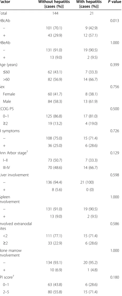
Hepatitis was observed in 21 (12.7%) of the 165
HBsAg-negative CD20+ DLBCL patients, 6 of whom had
HBV-related hepatitis. Other possible causes of hepatitis were chemotherapy (n= 7), tumor progression (n= 4), heart failure (n= 1), infection (n= 2), and septic shock (n= 1). For the 3 HBsAg-, HBcAb-, and HBsAb-negative patients, the possible causes of hepatitis were chemotherapy (n= 2) and tumor progression (n= 1). Univariate analysis identi-fied a higher incidence of hepatitis in the HBcAb-positive group compared with the HBcAb-negative group (21.8% vs. 8.2%, P= 0.013). Moreover, patients with elevated serum ALT levels at baseline were more likely to develop hepatitis than those with normal serum ALT levels (30.4% vs. 9.9%,P= 0.013). The risk of hepatitis was also higher in patients with elevated serum AST levels at baseline than in those with normal serum AST levels (31.6% vs. 10.3%,P= 0.019). The univariate analysis results of poten-tial prognostic factors associated with hepatitis are shown in Table 3. Multivariate logistic regression analysis re-vealed that HBcAb positivity and elevated serum ALT levels at baseline were independently associated with the development of hepatitis in HBsAg-negative DLBCL pa-tients (bothP< 0.05) (Table 4).
For the 55 patients with resolved hepatitis B, 22 (40%) had elevated serum ALT levels during the chemotherapy and follow-up period, and 12 (21.8%) experienced hepa-titis. The possible causes of hepatitis were HBV-related disease (n= 6), chemotherapy (n = 1), tumor progression (n= 2), heart failure (n= 1), and infection (n= 2). Patients with elevated serum ALT or AST levels at baseline were more likely to develop hepatitis than those with normal
serum ALT or AST levels (P= 0.037 and P= 0.005,
Table 1 Baseline clinical characteristics according to HBcAb status in HBsAg-negative DLBCL patients Characteristic HBcAb-negative
[cases (%)]
HBcAb-positive [cases (%)] P
value
Total 110 55
Sex 0.434
Male 67 (60.9) 30 (54.5)
Female 43 (39.1) 25 (45.5)
ECOG PS 0.525
0–1 96 (87.3) 46 (83.6)
≥2 14 (12.7) 9 (16.4)
Bulky mass 0.619
Yes 28 (25.5) 16 (29.1)
No 82 (74.5) 39 (70.9)
B symptoms 0.448
− 80 (72.7) 43 (78.2)
+ 30 (27.3) 12 (21.8)
Ann Arbor stage† 0.698
I–II 52 (47.3) 28 (50.9)
III-IV 57 (51.8) 27 (49.1)
Liver involvement 0.720
− 104 (94.5) 53 (96.4)
+ 6 (5.5) 2 (3.6)
IPI score† 0.851
0–1 48 (43.6) 21 (38.2)
2 28 (25.4) 17 (30.9)
3 23 (20.9) 11 (20.0)
4-5 10 (9.1) 6 (10.9)
ALT 0.874
≤40 U/L 95 (86.4) 47 (85.5)
>40 U/L 15 (13.6) 8 (14.5)
AST 0.863
≤45 U/L 97 (88.2) 49 (89.1)
>45 U/L 13 (11.8) 6 (10.9)
TB 0.852
≤20.5μmol/L 99 (90.0) 50 (90.9)
>20.5μmol/L 11 (10.0) 5 (9.1)
LDH 0.910
≤245 U/mL 67 (60.9) 33 (60.0)
>245 U/mL 43 (39.1) 22 (40.0)
HBsAb <0.001
– 82 (74.5) 3 (5.5)
+ 28 (25.5) 52 (94.5)
Table 1 Baseline clinical characteristics according to HBcAb status in HBsAg-negative DLBCL patients(Continued)
Characteristic HBcAb-negative [cases (%)]
HBcAb-positive [cases (%)] P
value
HBeAb 0.251
– 102 (92.7) 48 (87.3)
+ 8 (7.3) 7 (12.7)
respectively). The univariate analysis results of potential prognostic factors associated with hepatitis in patients with resolved hepatitis B are shown in Table 5. Multivari-ate logistic regression analysis revealed that elevMultivari-ated serum AST levels at baseline were independently associ-ated with the development of hepatitis in DLBCL patients with resolved hepatitis B (odds ratio [OR] = 10.25, 95% confidence interval [CI] = 1.60–21.73,P= 0.014).
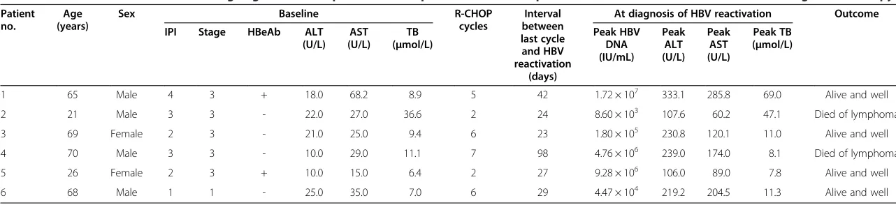
Details of 6 patients who developed HBV reactivation Details of the patients who developed HBV reactivation are shown in Table 6. Most of these patients were males, were older (>60 years), and had bulky masses, advanced Ann Arbor stage, and HBsAb positivity at baseline. No patient showed detectable serum HBV DNA levels at baseline. Most of the patients had normal liver function at baseline, except for Patients 1 and 2. None of the pa-tients received antiviral prophylaxis.
HBV reactivation was more likely to occur after an average of 5 R-CHOP cycles (range, 2–7 cycles) at a me-dian interval of 27 days (range, 24–98 days). Antiviral drug administration was immediately started after HBV DNA detection. Three patients developed severe hepa-titis. Hepatoprotective drugs were used when necessary, and HBV reactivation was managed with lamivudine (100 mg/day). Rituximab-containing chemotherapy was continued after serum HBV DNA became undetectable and liver function had improved.
Survival of DLBCL patients with resolved hepatitis B after rituximab-containing chemotherapy
Among patients with resolved hepatitis B, the median follow-up duration was 61 (1–93) months, the median 1-year OS rate was 85.2% (95% CI, 75.8%–100.0%), the Table 2 Univariate analysis of potential risk factors for
HBV reactivation in HBsAg-negative DLBCL patients
Factor Without HBV
reactivation [cases (%)]
With HBV reactivation
[cases (%)]
Pvalue
Total 159 6
HBcAb 0.001
– 110 (69.2) 0 (0)
+ 49 (30.8) 6 (100.0)
Age (years) 0.237
≤60 65 (40.9) 4 (66.7)
>60 94 (59.1) 2 (33.3)
Sex 1.000
Female 66 (41.5) 2 (33.3)
Male 93 (58.5) 4 (66.7)
ECOG PS 1.000
0–1 137 (86.2) 5 (83.3)
≥2 22 (13.8) 1 (16.7)
Bulky mass 0.343
Yes 115 (72.3) 6 (100.0)
No 44 (27.7) 0 (0)
B symptoms 1.000
– 118 (74.2) 5 (83.3)
+ 41 (25.8) 1 (16.7)
Ann Arbor stage† 0.211
I–II 79 (49.7) 1 (16.7)
III-IV 79 (49.7) 5 (83.3)
Liver involvement 1.000
– 151 (95.0) 6 (100.0)
+ 8 (5.0) 0 (0)
Spleen involvement 0.441
– 145 (91.2) 5 (83.3)
+ 14 (8.8) 1 (16.7)
Involved extranodal sites 0.627
<2 122 (76.7) 4 (66.7)
≥2 37 (23.3) 2 (33.3)
Bone marrow involvement 1.000
– 148 (93.1) 6 (100.0)
+ 11 (6.9) 0 (0)
IPI score† 0.402
0–1 68 (42.8) 1 (16.7)
2–5 90 (56.6) 5 (83.3)
HBeAb 0.094
– 146 (91.8) 4 (66.7)
+ 13 (8.2) 2 (33.3)
Table 2 Univariate analysis of potential risk factors for HBV reactivation in HBsAg-negative DLBCL patients(Continued)
Factor Without HBV
reactivation [cases (%)]
With HBV reactivation
[cases (%)]
Pvalue
ALT 0.597
≤40 U/L 136 (85.5) 6 (100.0)
>40 U/L 23 (14.5) 0 (0)
AST 0.526
≤45 U/L 141 (88.7) 5 (83.3)
>45 U/L 18 (11.3) 1 (16.7)
TB 0.463
≤20.5μmol/L 144 (90.6) 5 (83.3)
>20.5μmol/L 15 (9.4) 1 (16.7)
LDH 0.681
≤245 U/mL 97 (61.0) 3 (50.0)
>245 U/mL 62 (39.0) 3 (50.0)
3-year OS rate was 79.5% (95% CI, 68.7%–90.2%), and the 5-year OS rate was 75.7% (95% CI, 64.1%–87.2%). Among these patients, no significant difference was ob-served in OS between patients with and without HBV
reactivation (66.7% [95% CI, 29.0%–100%] vs. 73.2%
[95% CI, 59.9%–86.5%], P= 0.682) (Figure 2A) or be-tween patients with and without hepatitis (75.0% [95% CI, 50.5%–99.5%] vs. 71.4% [95% CI, 56.5%–86.3%], P= 0.927) (Figure 2B). OS was similar in patients with re-solved hepatitis B and HBsAg-negative/HBcAb-negative patients (75.7% [95% CI, 64.1%–87.2%] vs. 68.5% [95% CI,
57.8%–76.2%], P= 0.274) (Figure 2C). Among
HBsAg-negative patients, no significant difference was observed in OS between patients with and without HBV reactivation (66.7% vs. 70.2%,P= 0.908) or between patients with and without hepatitis (75.2% vs. 71.3%,P= 0.512).
Discussion
This study demonstrated that CD20+ DLBCL patients
with resolved HBV infection had a significantly higher risk of HBV reactivation and hepatitis compared with HBsAg-negative/HBcAb-negative patients after rituximab-containing chemotherapy. Baseline HBcAb positivity and ele-vated baseline serum ALT levels were independent risk factors for hepatitis in HBsAg-negative patients. An elevated baseline AST level was an independent risk factor for hepatitis in pa-tients with resolved hepatitis B. HBV reactivation could be managed with prompt antiviral therapy followed by the con-tinuation of chemotherapy. HBcAb positivity, HBV reactiva-tion, or hepatitis did not negatively affect patient survival.
Several studies have consistently demonstrated that the incidence of HBV infection is higher in patients with B-cell lymphoma than in the general population [19-22]. Chronic HBV infection was considered to be associated with lymphomagenesis [23], especially for Table 3 Univariate analysis of potential prognostic
factors associated with hepatitis in HBsAg-negative DLBCL patients
Factor Without hepatitis [cases (%)]
With hepatitis [cases (%)] P
value
Total 144 21
HBcAb 0.013
– 101 (70.1) 9 (42.9)
+ 43 (29.9) 12 (57.1)
HBeAb 1.000
– 131 (91.0) 19 (90.5)
+ 13 (9.0) 2 (9.5)
Age (years) 0.399
≤60 62 (43.1) 7 (33.3)
>60 82 (56.9) 14 (66.7)
Sex 0.756
Female 60 (41.7) 8 (38.1)
Male 84 (58.3) 13 (61.9)
ECOG PS 0.500
0–1 125 (86.8) 17 (81.0)
≥2 19 (13.2) 4 (19.0)
B symptoms 0.726
− 108 (75.0) 15 (71.4)
+ 36 (25.0) 6 (28.6)
Ann Arbor stage† 0.129
I–II 73 (50.7) 7 (33.3)
III-IV 70 (48.6) 14 (66.7)
Liver involvement 0.598
− 136 (94.4) 21 (100)
+ 8 (5.6) 0 (0)
Spleen involvement
1.000
− 131 (91.0) 19 (90.5)
+ 13 (9.0) 2 (9.5)
Involved extranodal sites
0.586
<2 111 (77.1) 15 (71.4)
≥2 33 (22.9) 6 (28.6)
Bone marrow involvement
1.000
− 134 (93.1) 20 (95.2)
+ 10 (6.9) 1 (4.8)
IPI score† 0.180
0–1 63 (43.8) 6 (28.6)
2–5 80 (55.8) 15 (71.4)
Table 3 Univariate analysis of potential prognostic factors associated with hepatitis in HBsAg-negative DLBCL patients(Continued)
Factor Without hepatitis [cases (%)]
With hepatitis [cases (%)]
Pvalue
ALT 0.013
≤40 U/L 128 (88.9) 14 (66.7)
>40 U/L 16 (11.1) 7 (33.3)
AST 0.019
≤45 U/L 131 (91.0) 15 (71.4)
>45 U/L 13 (9.0) 6 (28.6)
TB 0.696
≤20.5μmol/L 129 (89.6) 20 (95.2)
>20.5μmol/L 15 (10.4) 1 (4.8)
LDH 0.728
≤245 U/mL 88 (61.1) 12 (57.1)
>245 U/mL 56 (38.9) 9 (42.9)
B-cell lymphoma [24]. In this study, 33.3% (55/165) of pa-tients had resolved HBV infection, which was consistent with the 20%–40% incidence reported in endemic areas [25]. The incidence of HBV reactivation was 3.6% (6/ 165) in HBsAg-negative patients in this study, but was reported as high as 80% (12/15) in HBsAg-positive
lymphoma patients receiving rituximab-containing
chemotherapy without prophylaxis [5].
Compared with HBsAg-negative/HBcAb-negative pa-tients, HBsAg-negative/HBcAb-positive patients had a greater likelihood of developing HBV reactivation (10.9% vs. 0%,P= 0.001). However, Huiet al. [9] found no sig-nificant difference between the two groups in a study of
non-Hodgkin’s lymphoma and Hodgkin lymphoma
pa-tients, of whom only 20.1% (49/244) received R-CHOP therapy. The incidence of HBV reactivation varied from 2.3% to 23.8% of patients with resolved HBV infection [4,10-13]. The reasons for this discrepancy remain to be elucidated. However, treatment intensity, patient charac-teristics, and geographic HBV differences may be re-sponsible. Among the patients with resolved hepatitis B, no significant risk factors for HBV reactivation were found. Prospective studies with larger sample sizes are required to investigate the risk factors for resolved hepatitis B.
Hepatitis can be caused by a range of factors, includ-ing tumor progression, drug-related factors, heart failure, and severe sepsis. In this study, hepatitis was caused by HBV reactivation in 28.5% (6/21) of patients, and HBV reactivation was accompanied by hepatitis in all of the 6 patients, given that HBV reactivation often causes a dis-ease flare that leads to liver dysfunction. However, HBV reactivation can also be transient and clinically silent if the viral load is low [26]; thus, HBV reactivation can also occur in the absence of hepatitis [27,28].
Resolved hepatitis B involves interaction between the virus and the immune system. A variety of mechanisms may be involved. Low-level HBV virus replication is the main cause of resolved HBV infection. Other causes in-clude HBV gene mutation, HBV integration into the host chromosome, HBV infection of peripheral mononuclear cells, host immune response abnormalities, and interfer-ence by other viral infection. Persistent HBV infection
can promote liver disease, thereby leading to hepatitis and cirrhosis [29].
ALT is primarily localized in the liver, with lower en-zymatic activities found in skeletal muscle and heart tis-sues. AST is localized in heart, brain, skeletal muscle, and liver tissues. Damaged hepatocytes release their con-tents, including ALT and AST, into the extracellular space [30]. Serum ALT or AST levels are generally con-sidered sensitive indicators of liver cell injury. However, in addition to HBV infection, elevated baseline serum ALT levels can be caused by various other factors, in-cluding tumor progression, fatty liver, and diabetes [31]. In contrast to our results, Yeo et al. [4] found that all the patients who developed HBV reactivation had nor-mal baseline serum ALT levels and that the incidence of hepatitis was similar in HBsAg-negative/HBcAb-negative patients and in patients with resolved HBV infection. However, 53.7% (43/80) of patients in that study were treated without rituximab. Further studies are therefore needed to clarify the relationship between elevated base-line serum ALT levels and the hepatitis risk, as well as the possible mechanisms involved.
Among the 55 HBcAb-positive patients in our study, 8 (14.5%) had elevated baseline serum ALT levels. During the chemotherapy and follow-up period, 22 (40%) of the 55 patients had elevated serum ALT levels. Data on the proportion of HBcAb-positive patients with elevated serum ALT levels were limited. In a study by Yeoet al. [4], the incidence of elevated serum ALT levels in HBcAb-positive patients was 19.6% during chemother-apy. The reasons for these differences remain unclear. However, differences in patient characteristics and treat-ment intensities may be responsible. Therefore, there is no obvious association between HBcAb-positive patients and ALT levels, and further studies are warranted.
Several studies demonstrated that rituximab obviously improved outcomes in patients with B-cell lymphoma [2,3]. However, rituximab has been reported to increase the HBV reactivation rate in patients with chronic HBV infection or resolved HBV infection. Although HBV in-fection control is mediated mainly by HBV-specific cyto-toxic T lymphocytes, B lymphocytes are still required for antigen presentation. Rituximab is a chimeric murine/ human anti-CD20 monoclonal antibody that alters the activity of T lymphocytes and destroys B lymphocytes, resulting in the failure of antigen presentation and the sub-sequent expansion of HBV infection in hepatocytes [32,33]. HBV-related hepatitis during rituximab-containing chemo-therapy can be severe and fatal [34-36].
According to previous data and our experience, HBV-related hepatitis often develops after 5 or 6 cycles of chemotherapy at an interval of 1 to 13 months after chemotherapy. Antiviral drugs successfully controlled HBV reactivation in most patients, although 2 elderly Table 4 Multivariate analysis of potential risk factors
associated with hepatitis in HBsAg-negative DLBCL patients
Variate OR 95% CI Pvalue
HBcAb (+) 3.27 1.25-8.59 0.016
ALT (>40 U/L) 4.22 1.43-12.49 0.009
AST(>45 U/L) NA NA 0.129
patients (ages 77 and 84 years) developed fatal HBV-related disease [4,13,37]. In this study, it was reasonable for such patients to undergo close monitoring of HBV serology, HBV DNA, and liver function before each chemotherapy cycle and at least every 3 months during the follow-up period; this is consistent with the monitor-ing frequency of 1–3 months recommended by the latest European Association for the Study of the Liver (EASL) Clinical Practice Guidelines [8] and the consensus on the management of lymphoma with HBV infection in China [38]. In addition, in this study, HBsAg and HBV DNA testing was performed in the event of abnormal liver function or suspected hepatitis. Lamivudine successfully controlled HBV reactivation in all 6 affected patients. Prophylactic agents thus may not be recommended for HBsAg-negative/HBcAb-positive patients if close monitor-ing of HBV DNA is guaranteed.
However, regular monitoring may not be suitable for all patients. Some experts recommend prophylaxis with antiviral drugs in all HBsAg-negative/HBcAb-positive patients who receive rituximab-containing regimens for hematologic malignancies with a high risk of HBV re-activation and/or if close monitoring of HBV DNA is not guaranteed [8]. Lamivudine is widely used for prophylaxis; however, its efficacy is hampered by the de-velopment of viral mutations that result in drug resist-ance [39]. The prophylactic entecavir was found to be effective and associated with minimal resistance in lymphoma patients with resolved hepatitis B [40], but its long-term use is expensive. More large-scale studies or meta-analyses are required to identify host and viral factors that can help to predict the occurrence of HBV-related hepatitis and thus allow the design of individualized strat-egies for preventing HBV-related hepatitis. Different approaches based on cost-effectiveness, particularly in HBV-endemic areas, could be used for patients with differ-ent risk levels, and antiviral prophylaxis should be contin-ued indefinitely.
In HBsAg-positive patients with non-Hodgkin’s lymph-oma, the HBV reactivation-associated mortality was 30%– 50% without antiviral prophylaxis [5]. The HBV-related Table 5 Univariate analysis of potential prognostic
factors associated with hepatitis in DLBCL patients with resolved hepatitis B
Factor Without hepatitis [cases (%)]
With hepatitis [cases (%)] P
value
Total 43 12
HBeAb 0.639
– 38 (88.4) 10 (83.3)
+ 5 (11.6) 2 (16.7)
Age (years) 0.831
≤60 23 (53.5) 6 (50.0)
>60 20 (46.5) 6 (50.0)
Sex 0.340
Female 21 (48.8) 4 (33.3)
Male 22 (51.2) 8(66.7)
ECOG PS 0.974
0–1 36 (83.7) 10 (83.3)
≥2 7 (16.3) 2 (16.7)
B symptoms 0.763
− 34 (79.1) 9 (75.0)
+ 9 (20.9) 3 (25.0)
Ann Arbor stage 0.168
I–II 24 (55.8) 4 (33.3)
III-IV 19 (44.2) 8 (66.7)
Liver involvement 0.447
− 41 (95.3) 12 (100)
+ 2 (4.7) 0 (0)
Spleen involvement 0.873
− 40 (93.0) 11 (91.7)
+ 3 (7.0) 1 (8.3)
Involved extranodal sites
0.275
<2 35 (81.4) 8 (66.7)
≥2 8 (18.6) 4 (33.3)
Bone marrow involvement
0.594
− 42 (97.7) 12 (100.0)
+ 1 (2.3) 0 (0.0)
IPI score 0.288
0–1 18 (41.9) 3 (25.0)
2–5 25 (58.1) 9 (75.0)
ALT 0.037
≤40 U/L 39 (90.7) 8 (66.7)
>40 U/L 4 (9.3) 4 (33.3)
AST 0.005
≤45 U/L 41 (95.3) 8 (66.7)
>45 U/L 2 (4.7) 4 (33.3)
Table 5 Univariate analysis of potential prognostic factors associated with hepatitis in DLBCL patients with resolved hepatitis B(Continued)
Factor Without hepatitis [cases (%)]
With hepatitis [cases (%)]
Pvalue
TB 0.918
≤20.5μmol/L 39 (90.7) 11 (91.7)
>20.5μmol/L 4 (9.3) 1 (8.3)
LDH 0.894
≤245 U/mL 26 (60.5) 7 (58.3)
>245 U/mL 17 (39.5) 5 (41.7)
Table 6 Details and outcomes of 6 HBsAg-negative/HBcAb-positive DLBCL patients who developed HBV reactivation after rituximab-containing chemotherapy
Patient no.
Age (years)
Sex Baseline R-CHOP
cycles
Interval between last cycle and HBV reactivation
(days)
At diagnosis of HBV reactivation Outcome
IPI Stage HBeAb ALT
(U/L) AST (U/L)
TB (μmol/L)
Peak HBV DNA (IU/mL)
Peak ALT (U/L)
Peak AST (U/L)
Peak TB (μmol/L)
1 65 Male 4 3 + 18.0 68.2 8.9 5 42 1.72 × 10⁷ 333.1 285.8 69.0 Alive and well
2 21 Male 3 3 - 22.0 27.0 36.6 2 24 8.60 × 10³ 107.6 60.2 47.1 Died of lymphoma
3 69 Female 2 3 - 21.0 25.0 9.4 6 23 1.80 × 10⁵ 230.8 120.1 11.0 Alive and well
4 70 Male 3 3 - 10.0 29.0 11.1 7 98 4.76 × 10⁶ 239.0 174.0 8.1 Died of lymphoma
5 26 Female 2 3 + 10.0 15.0 6.4 2 27 9.28 × 10⁶ 106.0 89.0 7.8 Alive and well
6 68 Male 1 1 - 25.0 35.0 7.0 6 29 4.47 × 10⁴ 219.2 204.5 11.3 Alive and well
R-CHOP, cyclophosphamide, hydroxydaunomycin (doxorubicin), vincristine, and prednisone regimen. Other abbreviations as in Tables1and2.
Journal
of
Cancer
(2015) 34:18
Page
9
of
morbidity and overall mortality remained high in lymph-oma patients treated with antiviral prophylaxis [41]. In this study, OS was similar in HBsAg-negative/HBcAb-positive patients and HBsAg-negative/HBcAb-negative patients after rituximab-containing chemotherapy. Moreover, no significant difference was observed in OS between patients with and without HBV reactivation or between patients with and without hepatitis. Fukushima et al. [11] found similar survival rates in HBcAb-positive or HBcAb-negative patients, although their study included other types of lymphoma in addition to DLBCL, and 26.4% (19/72) of pa-tients had not received rituximab-containing therapy. In addition, DLBCL is a heterogeneous disorder with varied clinical outcomes. Hsuet al. [28] also demonstrated that re-solved HBV infection with HBV reactivation was associated with low OS and progression-free survival rates, although the differences were not significant. Further studies are needed to determine the impact of HBV reactivation on the clinical outcomes of lymphoma patients.
Our study had several key points. First, all patients had
newly diagnosed DLBCL and received
rituximab-containing therapy. Second, we compared the incidence of HBV reactivation or hepatitis between HBcAb-positive and HBcAb-negative patients and identified risk factors for the occurrence of HBV reactivation and hepatitis in HBsAg-negative patients and patients with resolved hepa-titis B. Third, we analyzed the effects of HBcAb positivity, HBV reactivation, and hepatitis on survival, which are cur-rently unknown. Furthermore, South China is a highly en-demic HBV area, and this is the first to confirm these findings in the southern Chinese population in the largest cancer center in South China.
However, this study was limited by its retrospective nature. First, the assignment of rituximab-containing chemotherapy was not randomized in HBcAb-positive or HBcAb-negative patients but was instead based on
the consideration of individual patients, leading to inev-itable bias in terms of which patients received rituxi-mab. Second, this study was limited to the analysis of patients from a single institute. All of the patients were Chinese, and our findings therefore must be confirmed in patients from other parts of Asia and in other ethnic groups.
Conclusions
In conclusion, this study clearly indicates that patients with resolved hepatitis B are at a higher risk of developing HBV reactivation and hepatitis after rituximab-containing chemotherapy compared with HBsAg-negative/HBcAb-negative patients. Close monitoring of HBV DNA levels and liver function and prompt antiviral therapy are re-quired in these patients. Prospective studies including more patients are required to confirm our findings and to determine the most effective monitoring and therapeutic strategies.
Competing interests
The authors declare that they have no competing interests.
Authors’contributions
QC designed research; QC, KC, JC, and DZ performed research and analyzed data; QC and KC wrote the article; YG helped to analyze data; HR, HH, LZ, JS, TL, and WJ conceived study and provided clinical data; LH and MW helped to collect data. All authors read and approved the final manuscript.
Acknowledgments
This work was supported by the National Natural Science Foundation of China (No. 81372883, No. 81001052), the Science and Technology Planning Project of Guangdong Province, China (No. 2011B031800222), the Young Talents Project of Sun Yat-sen University Cancer Center (to QC), the Young Talents Project of Sun Yat-sen University (to QC), the Natural Science Foundation of Guangdong Province, China (No. 8151008901000043), and the Sister Institution Network Fund of MD Anderson Cancer Center (to HR). This research was also partly supported by the NIH through MD Anderson’s Cancer Center Support Grant (No. CA016672).
Author details 1
Sun Yat-sen University Cancer Center; State Key Laboratory of Oncology in South China; Collaborative Innovation Center of Cancer Medicine, Guangzhou 510060, Guangdong, P. R. China.2Department of Medical Oncology, Sun Yat-sen University Cancer Center, Guangzhou 510060, Guangdong, P. R. China.3Guangdong Province Key Laboratory of Arrhythmia and
Electrophysiology, Radiotherapy Department, Sun Yat-sen Memorial Hospital of Sun Yat-sen University, Guangzhou 510120, Guangdong, P. R. China.4Department of Pathology, Sun Yat-sen University Cancer Center, Guangzhou 510060, Guangdong, P. R. China.5Clinical Trial Center, Sun Yat-sen University Cancer Center, Guangzhou 510060, Guangdong, P. R. China.6Department of Lymphoma and Myeloma, University of Texas MD Anderson Cancer Center, Houston, Texas 77030, USA.7Department of Molecular Diagnostics, Sun Yat-sen University Cancer Center, Guangzhou 510060, Guangdong, P. R. China.8Lymphoma and Myeloma Center, Institute of Hematology and Blood Diseases Hospital, Tianjin, P. R. China. 9
State Key Lab of Experimental Method of Hematology, Chinese Academy of Medical Sciences and Peking Union of Medical College, Tianjin 300020, P. R. China.
Received: 17 September 2014 Accepted: 22 January 2015
References
1. Lee WM. Hepatitis B virus infection. N Engl J Med. 1997;337:1733–45. 2. Coiffier B, Lepage E, Briere J, Herbrecht R, Tilly H, Bouabdallah R, et al. CHOP
chemotherapy plus rituximab compared with CHOP alone in elderly patients with diffuse large-B-cell lymphoma. N Engl J Med. 2002;346:235–42. 3. Pfreundschuh M, Trumper L, Osterborg A, Pettengell R, Trneny M, Imrie K, et al. CHOP-like chemotherapy plus rituximab versus CHOP-like chemotherapy alone in young patients with good-prognosis diffuse large-B-cell lymphoma: a randomised controlled trial by MabThera International Trial (MInT) Group. Lancet Oncol. 2006;7:379–91.
4. Yeo W, Chan TC, Leung NW, Lam WY, Mo FK, Chu MT, et al. Hepatitis B virus reactivation in lymphoma patients with prior resolved hepatitis B undergoing anticancer therapy with or without rituximab. J Clin Oncol. 2009;27:605–11.
5. Shih LN, Sheu JC, Wang JT, Huang GT, Yang PM, Lee HS, et al. Serum hepatitis B virus DNA in healthy HBsAg-negative Chinese adults evaluated by polymerase chain reaction. J Med Virol. 1990;32:257–60.
6. Evens AM, Jovanovic BD, Su YC, Raisch DW, Ganger D, Belknap SM, et al. Rituximab-associated hepatitis B virus (HBV) reactivation in lymphoproliferative diseases: meta-analysis and examination of FDA safety reports. Ann Oncol. 2011;22:1170–80.
7. Pei SN, Chen CH, Lee CM, Wang MC, Ma MC, Hu TH, et al. Reactivation of hepatitis B virus following rituximab-based regimens: a serious complication in both HBsAg-positive and HBsAg-negative patients. Ann Hematol. 2010;89:255–62.
8. European Association For The Study Of The Liver. EASL clinical practice guidelines: management of chronic hepatitis B virus infection. J Hepatol. 2012;57:167–85.
9. Hui CK, Cheung WW, Zhang HY, Au WY, Yueng YH, Leung AY, et al. Kinetics and risk of de novo hepatitis B infection in HBsAg-negative patients undergoing cytotoxic chemotherapy. Gastroenterology. 2006;131:59–68. 10. Koo YX, Tay M, Teh YE, Teng D, Tan DS, Tan IB, et al. Risk of hepatitis B virus
(HBV) reactivation in hepatitis B surface antigen negative/hepatitis B core antibody positive patients receiving rituximab-containing combination chemotherapy without routine antiviral prophylaxis. Ann Hematol. 2011;90:1219–23.
11. Fukushima N, Mizuta T, Tanaka M, Yokoo M, Ide M, Hisatomi T, et al. Retrospective and prospective studies of hepatitis B virus reactivation in malignant lymphoma with occult HBV carrier. Ann Oncol. 2009;20:2013–7. 12. Ji D, Cao J, Hong X, Li J, Wang J, Chen F, et al. Low incidence of hepatitis B
virus reactivation during chemotherapy among diffuse large B-cell lymphoma patients who are HBsAg-negative/HBcAb-positive: a multicenter retrospective study. Eur J Haematol. 2010;85:243–50.
13. Matsue K, Kimura S, Takanashi Y, Iwama K, Fujiwara H, Yamakura M, et al. Reactivation of hepatitis B virus after rituximab-containing treatment in patients with CD20-positive B-cell lymphoma. Cancer. 2010;116:4769–76. 14. Swerdlow SHCE, Harris NL, Jaffe ES, Stein H, Thiele J, et al. World health
organization classification of tumours of the haematopoietic and lymphoid tissues. Pileri SA: IARC Press; 2008.
15. Yeo W, Chan PK, Ho WM, Zee B, Lam KC, Lei KI, et al. Lamivudine for the prevention of hepatitis B virus reactivation in hepatitis B s-antigen seropositive cancer patients undergoing cytotoxic chemotherapy. J Clin Oncol. 2004;22:927–34.
16. Lok AS, Liang RH, Chiu EK, Wong KL, Chan TK, Todd D. Reactivation of hepatitis B virus replication in patients receiving cytotoxic therapy. Report of a prospective study. Gastroenterology. 1991;100:182–8.
17. Ziepert M, Hasenclever D, Kuhnt E, Glass B, Schmitz N, Pfreundschuh M, et al. Standard international prognostic index remains a valid predictor of outcome for patients with aggressive CD20+ B-cell lymphoma in the rituximab era. J Clin Oncol. 2010;28:2373–80.
18. Cox DR. Regression models and life tables. J R Stat Soc B. 1972;34:187–220. 19. Marcucci F, Mele A, Spada E, Candido A, Bianco E, Pulsoni A, et al. High
prevalence of hepatitis B virus infection in B-cell non-Hodgkin’s lymphoma. Haematologica. 2006;91:554–7.
20. Wang F, Xu RH, Han B, Shi YX, Luo HY, Jiang WQ, et al. High incidence of hepatitis B virus infection in B-cell subtype non-Hodgkin lymphoma compared with other cancers. Cancer. 2007;109:1360–4.
21. Kim JH, Bang YJ, Park BJ, Yoo T, Kim CW, Kim TY, et al. Hepatitis B virus infection and B-cell non-Hodgkin’s lymphoma in a hepatitis B endemic area: a case–control study. Jpn J Cancer Res. 2002;93:471–7.
22. Qin XT, Lu Y, Chen XQ, Xu HP, Fan HJ. Correlation of hepatitis B virus infection to non-Hodgkin’s lymphoma. Chin J Cancer. 2007;26:294–7. 23. Engels EA, Cho ER, Jee SH. Hepatitis B virus infection and risk of non-Hodgkin
lymphoma in South Korea: a cohort study. Lancet Oncol. 2010;11:827–34. 24. Wang F, Yuan S, Teng KY, Garcia-Prieto C, Luo HY, Zeng MS, et al. High
hepatitis B virus infection in B-cell lymphoma tissue and its potential clinical relevance. Eur J Cancer Prev. 2012;21:261–7.
25. Marzano A, Angelucci E, Andreone P, Brunetto M, Bruno R, Burra P, et al. Prophylaxis and treatment of hepatitis B in immunocompromised patients. Dig Liver Dis. 2007;39:397–408.
26. Hoofnagle JH. Reactivation of hepatitis B. Hepatology. 2009;49:S156–65. 27. Li HR, Huang JJ, Guo HQ, Zhang X, Xie Y, Zhu HL, et al. Comparison of
entecavir and lamivudine in preventing hepatitis B reactivation in lymphoma patients during chemotherapy. J Viral Hepat. 2011;18:877–83. 28. Hsu C, Tsou HH, Lin SJ, Wang MC, Yao M, Hwang WL, et al.
Chemotherapy-induced hepatitis B reactivation in lymphoma patients with resolved HBV infection: a prospective study. Hepatology. 2014;59:2092–100.
29. Chemin I, Jeantet D, Kay A, Trepo C. Role of silent hepatitis B virus in chronic hepatitis B surface antigen(−) liver disease. Antiviral Res. 2001;52:117–23. 30. Ozer J, Ratner M, Shaw M, Bailey W, Schomaker S. The current state of
serum biomarkers of hepatotoxicity. Toxicology. 2008;245:194–205. 31. Clark JM, Brancati FL, Diehl AM. The prevalence and etiology of elevated
aminotransferase levels in the United States. Am J Gastroenterol. 2003;98:960–7. 32. Stasi R, Del Poeta G, Stipa E, Evangelista ML, Trawinska MM, Cooper N, et al.
Response to B-cell depleting therapy with rituximab reverts the abnormalities of T-cell subsets in patients with idiopathic thrombocytopenic purpura. Blood. 2007;110:2924–30.
33. Dai MS, Chao TY, Kao WY, Shyu RY, Liu TM. Delayed hepatitis B virus reactivation after cessation of preemptive lamivudine in lymphoma patients treated with rituximab plus CHOP. Ann Hematol. 2004;83:769–74. 34. Tsutsumi Y, Kanamori H, Mori A, Tanaka J, Asaka M, Imamura M, et al.
Reactivation of hepatitis B virus with rituximab. Expert Opin Drug Saf. 2005;4:599–608.
35. Sarrecchia C, Cappelli A, Aiello P. HBV reactivation with fatal fulminating hepatitis during rituximab treatment in a subject negative for HBsAg and positive for HBsAb and HBcAb. J Infect Chemother. 2005;11:189–91. 36. Li YH, He YF, Wang FH, Lin XB, Xia ZJ, Sun XF, et al. Clinical analysis of liver
damage of 116 malignant lymphoma patients with chronic HBV infection after cytotoxic chemotherapy. Chin J Cancer. 2005;24:1507–9.
37. Koo YX, Tan DS, Tan IB, Tao M, Chow WC, Lim ST. Hepatitis B virus reactivation and role of antiviral prophylaxis in lymphoma patients with past hepatitis B virus infection who are receiving chemoimmunotherapy. Cancer. 2010;116:115–21.
38. Chinese Society of Hematology, CMA, Committee of Malignant Lymphoma, Chinese Anti-cancer Association, Chinese Society of Hepatology, CMA. Consensus on the management of lymphoma with HBV infection. Zhonghua Xue Ye Xue Za Zhi. 2013;34:988–93 [in Chinese]
40. Huang YH, Hsiao LT, Hong YC, Chiou TJ, Yu YB, Gau JP, et al. Randomized controlled trial of entecavir prophylaxis for rituximab-associated hepatitis B virus reactivation in patients with lymphoma and resolved hepatitis B. J Clin Oncol. 2013;31:2765–72.
41. Kumagai K, Takagi T, Nakamura S, Sawada U, Kura Y, Kodama F, et al. Hepatitis B virus carriers in the treatment of malignant lymphoma: an epidemiological study in Japan. Ann Oncol. 1997;8 Suppl 1:107–9.
Submit your next manuscript to BioMed Central and take full advantage of:
• Convenient online submission
• Thorough peer review
• No space constraints or color figure charges
• Immediate publication on acceptance
• Inclusion in PubMed, CAS, Scopus and Google Scholar
• Research which is freely available for redistribution