R E S E A R C H A R T I C L E
Open Access
Differences in reported sepsis incidence
according to study design: a literature
review
Saga Elise Mariansdatter
*†, Andreas Halgreen Eiset
†, Kirstine Kobberøe Søgaard and Christian Fynbo Christiansen
Abstract
Background:Sepsis and severe sepsis are common conditions in hospital settings, and are associated with high rates
of morbidity and mortality, but reported incidences vary considerably. In this literature review, we describe the variation in reported population-based incidences of sepsis and severe sepsis. We also examine methodological and demographic differences between studies that may explain this variation.
Methods:We carried out a literature review searching three major databases and reference lists of relevant articles,
to identify all original studies reporting the incidence of sepsis or severe sepsis in the general population. Two authors independently assessed all articles, and the final decision to exclude an article was reached by consensus. We extracted data according to predetermined variables, including study country, sepsis definition, and data source. We then calculated descriptive statistics for the reported incidences of sepsis and severe sepsis. The studies were classified according to the method used to identify cases of sepsis or severe sepsis: chart-based (i.e. review of patient charts) or code-based (i.e. predetermined International Classification of Diseases [ICD] codes).
Results:Among 482 articles initially screened, we identified 23 primary publications reporting incidence of sepsis
and/or severe sepsis in the general population. The reported incidences ranged from 74 to 1180 per 100,000 person-years and 3 to 1074 per 100,000 person-years for sepsis and severe sepsis, respectively. Most chart-based studies used the Bone criteria (or a modification hereof) and Protein C Worldwide Evaluation in Severe Sepsis (PROWESS) study criteria to identify cases of sepsis and severe sepsis. Most code-based studies used ICD-9 codes, but the number of codes used ranged from 1 to more than 1200. We found that the incidence varied according to how sepsis was identified (chart-based vs. code-based), calendar year, data source, and world region.
Conclusion:The reported incidences of sepsis and severe sepsis in the general population varied greatly between
studies. Such differences may be attributable to differences in the methods used to collect the data, the study period, or the world region where the study was undertaken. This finding highlights the importance of standardised definitions and acquisition of data regarding sepsis and severe sepsis.
Keywords:Sepsis, Severe sepsis, SIRS, Septicaemia, Method, Incidence, Epidemiology, Review
Background
Sepsis is associated with high rates of morbidity and mortality, accounting for as much as one of every two to three in-hospital deaths [1]. Notably, the mortality rates of sepsis increased during the last decade, which is in contrast to the declining rates of all other major causes of death in the US [2].
Determining the incidence of sepsis is of great interest to both clinicians and public health officials, in order to quantify the burden of the disease [3]. However, estima-tion of sepsis incidence is difficult, as it depends on the definition of sepsis, the method used to assess the condi-tion, and the underlying population. Until 1992, no con-sensus existed on the terminology used to describe the presence and severity of sepsis, impairing comparison of studies on sepsis incidence and therapy outcomes [4]. The 1991 American College of Chest Physicians/Society
* Correspondence:sagamari@rm.dk †Equal contributors
Department of Clinical Epidemiology, Aarhus University Hospital, Aarhus, Denmark
© 2016 The Author(s).Open AccessThis article is distributed under the terms of the Creative Commons Attribution 4.0 International License (http://creativecommons.org/licenses/by/4.0/), which permits unrestricted use, distribution, and reproduction in any medium, provided you give appropriate credit to the original author(s) and the source, provide a link to the Creative Commons license, and indicate if changes were made. The Creative Commons Public Domain Dedication waiver (http://creativecommons.org/publicdomain/zero/1.0/) applies to the data made available in this article, unless otherwise stated. Mariansdatteret al. BMC Medical Research Methodology (2016) 16:137
of Critical Care Medicine (ACCP/SCCM) Consensus Conference addressed this issue, with the aim to create a set of criteria for identifying and assessing the severity of sepsis [5]. The consensus proposal included an introduc-tion of the systemic inflammatory response syndrome (SIRS) criteria for early identification of sepsis, defining sepsis as 2 SIRS criteria in patients with known or sus-pected infection, and severe sepsis as sepsis associated with organ dysfunction, hypoperfusion, or hypotension (Table 1). Though repeatedly criticised for being too sen-sitive [6, 7] and of questionable prognostic value [8–10] these easily applied “Bone criteria”remained the clinical standard in many hospital guidelines even after the intro-duction of internationally agreed-upon, but more compre-hensive, criteria [6, 11, 12]. In 2016 the definition of sepsis was updated to categorise sepsis as a life-threatening organ dysfunction caused by a dysregulated host response to infection (by The Third International Consensus Defi-nitions for Sepsis and Septic Shock) [13].
In this review, we focus on the variation in reported in-cidences of sepsis and severe sepsis in the general popula-tion, and discuss the potential explanations including the use of different definitions or methods to assess sepsis.
Methods
Literature search and study selection
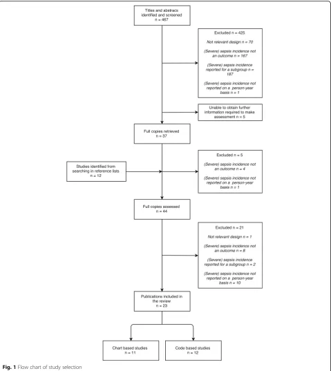
We included original studies with incidences of sepsis or severe sepsis in the general population (in person-years) as an outcome, published before 2016. Consequently, we excluded studies focusing on a specific subgroup of patients (e.g. neonatal sepsis, sepsis caused by a spe-cific microbial agent), as these studies would include only a fraction of the general population as their study population. The number of excluded studies and reasons for exclusion are described in Fig. 1.
We searched PubMed (search string
(((“Sepsis/epidemi-ology” [Mesh]) AND (“sepsis” [Title] OR “septicaemia” [Title])) AND “incidence” [Title/Abstract]) AND “english” [Language]), EMBASE (search string‘sepsis’/exp OR‘sepsis’
AND (‘epidemiology’/exp OR‘epidemiology’OR‘incidence’/ exp OR‘incidence’) AND [english]/lim) and Cochrane Li-brary (search string#1: MeSH descriptor: [Sepsis] explode all trees and with qualifier(s): [Epidemiology – EP] + #2: (“sepsis”:ti or“septicaemia:ti”) + #3:”incidence”:ti,ab).
The title and abstract of the resulting articles were screened and categorised according to predefined criteria if excluded (see section Availability of data and mate-rials). All included articles–along with additional articles found in reference lists –were retrieved, read in full and excluded according to the same criteria (see Fig. 1). Two authors (SEM and AHE) performed all rounds independ-ently; the final decision to exclude an article was reached by consensus.
Data were extracted from each study according to a predetermined list of variables (see section Availabil-ity of data and materials). If a study reported several incidences –e.g. for different years or applying different methodologies – each incidence measure was registered as an observation. We adapted a widely used terminology to categorise the studies according to method used to identify sepsis or severe sepsis: 1.“chart-based” including studies that identified patients by review of patient charts and 2. “code-based” including studies that identified patients using diagnostic codes [3, 14–16]. To examine regional differences in incidence of sepsis and severe sepsis each study was categorised according to World Bank region [17].
Data management and descriptive statistics were per-formed using R [18]. In order to examine the heterogen-eity that gives rise to the differences in incidence as well as possible interactions, we produced a number of box-plots based on crude data to allow for a visual evaluation of some of the factors that influence the reported inci-dence. Further, we present detailed tables that allow the reader to compare the included studies. The data set, along with the R-code and codebook, are freely available (see sectionAvailability of data and materials).
International Classification of Diseases (ICD)
In the code-based studies, ICD codes were used to identify cases from discharge databases without specific informa-tion on physiological parameters. Implementainforma-tion of the tenth revision of the ICD coding system (ICD-10) started in 1994 [19], but actual implementation dates vary among countries and was finally completed in the US as of October 1, 2015 [20]. Consequently, ICD-10 data was used in only two studies [21, 22]. A table with the full lists of specific sepsis codes in the ICD-9 and ICD-10 coding systems are provided as an additional file (see Additional file 1).
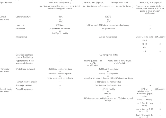
Below is a brief summary of the development of the guidelines used; Table 1 offers a detailed comparison of sepsis, severe sepsis, septic shock and multiple organ dysfunction syndrome.
The 1991 ACCP/SCCM Consensus Conference guidelines In 1992 Bone et al. proposed a standardised definition of sepsis [5]. This included an introduction of the four SIRS criteria: 1. Temperature >38 °C or <36 °C; 2. heart rate >90 beats per minute; 3. respiratory rate >20 breaths per minute or PaCO2 < 32 mmHg; and 4. white blood cell count >12,000/cu mm, <4,000/cu mm, or >10 % im-mature (band) forms. According to this, systemic inflam-matory response syndrome (SIRS) was defined as at least two SIRS criteria, and sepsis was defined as (suspected) infection and at least two SIRS criteria. In addition it was suggested that use of the term“septicaemia”should
Table 1Criteria proposed to define sepsis and severe sepsis; comparison of guidelines
Sepsis definition Boneet al., 1992 (Sepsis-1) Levyet al., 2003 (Sepsis-2) Dellingeret al., 2013 Singeret al., 2016 (Sepsis-3)
Infection, documented or suspected, and at least 2 of the following (SIRS criteria):
Infection, documented or suspected, and some of the following: Suspected or documented infection and an acute increase of≥2 SOFA
points (a proxy for organ dysfunction)
General parameters
Core temperature >38°C
or <36°C
>38.3°C or <36°C
–
Heart rate >90 bpm >90 bpm or >2 SD above the normal value for age –
Tachypnea >20 breaths per minute
or PaCO2<32 mmHg
No specification –
Mental status – Altered mental status Glasgow coma scale: SOFA score:
13-14 1
10-12 2
6-9 3
<6 4
Significant edema or
positive fluid balance –
>20 mL/kg over 24 hrs –
Hyperglycemia in the absence of diabetes
– Plasma glucose >120
mg/dL or >7.7 mM/L
Plasma glucose >140 mg/dL or >7.7 mM/L
–
Inflammatory parameters
White blood cell count >12,000/cu mm (leukocytosis) or
<4,000/cu mm (leukopenia) or
>10% immature (bands) forms
>12,000/μL (leukocytosis) or
<4,000/μL (leukopenia) or
Normal white blood cell count with >10% immature forms
–
Plasma C reactive protein – >2 SD above the normal value –
Plasma procalcitonin – >2 SD above the normal value –
Hemodynamic parameters
Arterial hypotension – SBP <90 mmHg
or MAP <70
or
SBP decrease >40 mmHg in adults or <2 SD below normal for age
MAP or administration of vasopressors (μg/kg/
min):
SOFA score:
MAP < 70 mm/Hg 1
dop≤5 or dob (any dose)
2
dop > 5 or epi≤0.1 or nor≤0.1
3
dop > 15 or epi > 0.1 or nor > 0.1
4
Mariansda
tter
et
al.
BMC
Medical
Research
Methodolog
y
(2016) 16:137
Page
3
of
Table 1Criteria proposed to define sepsis and severe sepsis; comparison of guidelines(Continued)
Mixed venous oxygen saturation
– >70% – –
Cardiac index – >3.5 L/min/m2 – –
Organ dysfunction parameters
Arterial hypoxemia – PaO2/FIO2<300 PaO2/FIO2: SOFA score:
<400 1
<300 2
<200 and mechanically
ventilated
3
<100 and mechanically
ventilated
4
Acute oliguria – Urine output <0.5 mL/
kg/hr or 45 mmol/L for at least 2 hrs
Urine output <0.5 mL/kg/hr for at least 2 hrs despite adequate fluid
resuscitation
Creatinine (mg/dl) [μmol/L] (or urine output):
SOFA score:
1.2–1.9 [110-170] 1
2.0–3.4 [171-299] 2
3.5–4.9 [300-440] (or < 500 mL/d)
3
> 5.0 [> 440] (or < 200 mL/d)
4
Creatinine increase – >0.5 mg/dL >0.5 mg/dL or 44.2μmol/L
Coagulation abnormalities – INR >1.5 or aPTT >60 s –
Ileus – Absent bowel sounds –
Thrombocytopenia – Platelet count <100 x 109/L Platelets x 103/μL: SOFA
score:
< 150 1
< 100 2
< 50 3
< 20 4
Hyperbilirubinemia – Plasma total bilirubin >4 mg/dL or 70 mmol/L Bilirubin (mg/dl)
[μmol/L]:
SOFA score:
1.2–1.9 [> 20-32] 1
2.0–5.9 [33-101] 2
6.0–11.9 [102-204] 3
> 12.0 [> 204] 4
Tissue perfusion parameters
Hyperlactatemia – >1 mmol/L –
Mariansda
tter
et
al.
BMC
Medical
Research
Methodolog
y
(2016) 16:137
Page
4
of
Table 1Criteria proposed to define sepsis and severe sepsis; comparison of guidelines(Continued)
Capillary refill – Decreased capillary refill or mottling –
Severe sepsis definition Boneet al., 1992 Dellingeret al., 2013 Singeret al., 2016
Sepsis associated with but not limited to Any of the below thought to be due to the infection –
Hypo-perfusion
Hypotension (sepsis-induced), in the absence of other causes
Systolic blood pressure < 90 mmHg or
A reduction of≥40 mmHg from baseline.
As defined for sepsis –
Lactate Lactic acidosis Lactate above upper limit of laboratory normal –
Organ failure
Kidney injury Oliguria As defined for sepsis
but
Creatinine > 2 mg/dL (176.8μmol/L)
–
Acute lung injury – Pneumonia not the infectious source: PaO2/FIO2< 250
or
Pneumonia the infectious source: PaO2/FIO2< 200
–
Liver injury – As defined for sepsis
but
Bilirubin > 2 mg/dL (34.2μmol/L)
–
Mental status Acute alteration As defined for sepsis –
Septic shock Hypotension despite adequate fluid resuscitation along with the presence of perfusion
abnormalities, as listed above.
Hypotension not reversed with fluid resuscitation. Sepsis with persisting hypotension requiring vasopressors to maintain MAP≥65 mmHg and having a serum lactate level >2 mmol/L (18mg/dL) despite adequate
volume resuscitation.
Multiple organ dysfunction syndrome (MODS) Altered organ dysfunction in an acutely ill patient such that homeostasis cannot be maintained
without intervention.
– –
Mariansda
tter
et
al.
BMC
Medical
Research
Methodolog
y
(2016) 16:137
Page
5
of
be avoided. We will refer to this definition as the“Bone criteria”.
International Sepsis Definitions Conference modifications In 2003, the first Surviving Sepsis Campaign was pub-lished [6]. In an effort to increase the clinical utility, the diagnostic criteria were expanded to include other parame-ters, among these inflammatory, hemodynamic and tissue
perfusion. It was emphasised that none of these new cri-teria were specific for sepsis. The latest campaign edition published in 2012 contained only minor revisions, and thus these expanded criteria have remained the recommended clinical standard [3]. However, a revised international definition of sepsis criteria has recently been published [13], in which the SIRS criteria are replaced by the sepsis-related organ failure assessment (SOFA) score [23]. Fig. 1Flow chart of study selection
Results
Our search identified 467 articles of which 430 were excluded after screening (see Fig. 1). An additional 12 articles were identified from the reference lists of the included articles, of which five were excluded after going through the abstracts. Of 44 articles read in full 21 were excluded: 10 articles did not provide sepsis or severe sepsis incidence on a person-year basis [15, 24–32], eight articles did not report sepsis or severe sepsis incidence as an out-come [33–40], two articles reported sepsis or severe sepsis incidence for a subgroup of patients [41, 42] and one article did not use a relevant design to compute sepsis and severe sepsis incidences [43]. Thus, we included a total of 23 articles: 11 chart-based and 12 code-based studies. Summaries of the included studies can be found in Tables 2 and 3.
Chart-based studies
Nine studies [44–52] screened patients according to pre-defined criteria for sepsis and/or severe sepsis; two studies [53, 54] analysed previously collected data. One chart-based study on severe sepsis reported incidences for several years. Most chart-based studies used the Bone criteria (or a modification hereof) and Protein C Worldwide Evaluation in Severe Sepsis (PROWESS) study criteria to identify cases of sepsis and severe sepsis (Table 2). For organ dysfunction definitions, adaptations of the PROWESS study criteria [55] were the most frequently used (see Additional file 2 for a detailed description).
Code-based studies
Three code-based studies applied different algorithms to the same data set [16, 22, 56] while three and six code-based studies reported several years' observations of sep-sis and severe sepsep-sis incidences, respectively [22, 56–63] (Table 3).
Most code-based studies used ICD-9, though there was great diversity in what and how many codes were used, ranging from 1 to more than 1200 (see Additional file 3).
Three code-based studies used the Bone criteria for validation: Angus et al. and Shen et al. [14, 56] used the combination of ICD codes defined in their methods ap-plied to an alternate cohort and a randomly selected database sample, respectively, while Martin et al. [63] compared only the ICD-9 codes specific for septicaemia to a chart-based method. In general, there was a high degree of agreement between patients identified using ICD codes and patients identified by the Bone criteria, respectively. However, Angus et al. did find that their ICD codes generated higher incidences than what was found for the reference cohort using clinical and physio-logic data [14].
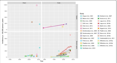
Sepsis and severe sepsis incidence in the general population Overall, we found great variation in incidence both between and across methods used to identify sepsis and severe sepsis, ranging from 74 to 1180 per 100,000 person-years and 3 to 1074 per 100,000 person-years, respectively. The incidence of both sepsis and severe sepsis increased over time (Fig. 2). When stratifying on method used to identify sepsis, we found that chart-based studies in general reported a higher incidence of sepsis than the code-based studies, whereas the opposite was the case for severe sepsis. There was a great diversity in the data source used: studies including patients from all wards in the hospital (”Hospital wide”) found the highest sepsis incidence whereas studies only including patients from intensive care units (ICUs) found a relatively low severe sepsis incidence (see Additional files 4 and 5). Stratifying on World Bank region, we found the lowest sepsis incidence in North America and the lowest severe sepsis incidence in the Europe & Central Asia region; in both cases the incidence was highest in the East Asia & Pacific region (Fig. 3). In addition, we examined for inter-action between calendar year, World Bank region and method (plots not shown). While we did find interaction with calendar year for both World Bank region and chart/ code based studies, there was a consistent trend in the rise of incidence. The interaction of method and World Bank region can be seen in Fig. 3.
Discussion
In this literature review, we found that the reported in-cidence of sepsis and severe sepsis in the general popu-lation varied greatly between the included studies. We compared the methods used and the demographic char-acteristics of the studied populations. We found that the variation may in part be attributable to whether a chart-based or a code-chart-based method was used, differences in the criteria used for identifying cases of sepsis or severe sepsis within these groups, year of incidence measure, and the World Bank region in which the study was conducted.
In most chart-based studies on severe sepsis incidence, cases were identified in ICUs only. Such selection might introduce bias towards a lower incidence because patients that fulfil the criteria for severe sepsis but did not need ICU care were excluded. Indeed, these studies did on aver-age find a lower incidence of severe sepsis than studies with other inclusion criteria. However, the chart-based study by Karlsson et al. [50] included admissions to both ICUs and other hospital wards, and still found an inci-dence of severe sepsis in adults much lower than what was found within a similar time period in the code-based studies of Dombrovskiy et al. [60] and Kumar et al. [62]. This indicates that other factors play an important role for the observed differences in incidence between chart- and code-based studies, and the question is whether these very
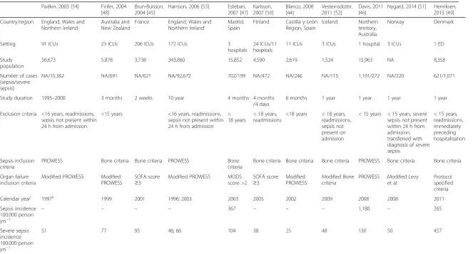
Table 2Chart-based studies of sepsis and severe sepsis incidence in the general population
Padkin, 2003 [54] Finfer, 2004 [48]
Brun-Buisson, 2004 [45]
Harrison, 2006 [53] Esteban, 2007 [47]
Karlsson, 2007 [50]
Blanco, 2008 [44]
Vesteinsdottir, 2011 [52]
Davis, 2011 [46]
Nygard, 2014 [51] Henriksen, 2015 [49]
Country/region England, Wales and Northern Ireland
Australia and New Zealand
France England, Wales and Northern Ireland
Madrid, Spain
Finland Castilla y Leòn Region, Spain
Iceland Northern territory, Australia
Norway Denmark
Setting 91 ICUs 23 ICUs 206 ICUs 172 ICUs 3
hospitals
24 ICUs/11 hospitals
11 ICUs 3 ICUs 1 hospital 3 ICUs 1 ED
Study population
56,673 5,878 3,738 343,860 15,852 4,500 2,619 1,524 15,963 NA 8,358
Number of cases (sepsis/severe sepsis)
NA/15,362 NA/691 NA/621 NA/92,672 702/199 NA/472 NA/246 NA/115 1,191/272 NA/220 621/1,071
Study duration 1995–2000 3 months 2 weeks 10 year 4 months 4 months
/4 days
6 months 1 year 1 year 1 year 1 year
Exclusion criteria <16 years, readmissions, sepsis not present within 24 h from admission
<15 years <16 years, readmissions, sepsis not present within 24 h from admission
< 18 years
< 18 years, readmissions
<18 years < 18 years, readmissions, sepsis not present on admission
< 15 years < 15 years, severe sepsis not present within 24 h from admission, transferred with diagnosis of severe sepsis
< 15 years, readmissions, immediately preceding hospitalisation
Sepsis inclusion criteria
PROWESS Bone criteria Bone criteria PROWESS Bone
criteria
Bone criteria Bone criteria Bone criteria PROWESS Bone criteria Bone criteria
Organ failure inclusion criteria
Modified PROWESS Modified PROWESS
SOFA score
≥3
Modified PROWESS MODS score >2
SOFA score
≥3
Modified PROWESS
Modified Bone criteria
PROWESS Modified Levy et al.
Protocol specified criteria
Calendar yeari 1997ii 1999 2001 1996; 2003 2003 2005 2002 2009 2008 2008 2011
Sepsis incidence 100.000 person yrs−1
– – – – 367 – – – 1,180 – 265
Severe sepsis incidence 100.000 person yrs−1
51 77 95 46; 66 104 38 25 48 130 50 457
Characteristics of chart based studies of sepsis and severe sepsis incidence extrapolated to the general population. i) If study is conducted in two consecutive calendar years the last year is reported. ii) If full data were not available for 1997, the closest full year’s data were used.Abbreviations:−, not calculated;EDemergency department,hrshours,ICUintensive care unit,MODSmultiple organ dysfunction syndrome,NAnot available, PROWESSProtein C Worldwide Evaluation in Severe Sepsis,SOFA, sequential organ failure assessment,yrsyears old
Mariansda
tter
et
al.
BMC
Medical
Research
Methodolog
y
(2016) 16:137
Page
8
of
Table 3Code-based studies of sepsis and severe sepsis incidence in the general population
CDC, 1990 [57]
Angus, 2001 [14]
Martin, 2003 [63]
Flaatten, 2004 [21]
Dombrovskiy, 2005 [60]
Esper, 2006 [61]
Dombrovskiy, 2007 [59]
Shen, 2010 [56] Wilhelms, 2010 [22]
Kumar, 2011 [62]
Lagu, 2012 [16]
Chen, 2013 [58]
Country/Region USA USA (7
states)
USA Norway New Jersey, USA
USA USA Taiwan Sweden USA USA Taiwan
Coding system ICD-9 ICD-9 ICD-9 ICD-10 ICD-9 ICD-9 ICD-9 ICD-9 ICD-9/10vi ICD-9 ICD9 ICD9
Data source NHDS Constructed
database
NHDS NPR New Jersey
SID
NHDS NIS NHIRD SHDR NIS NIS NHIRD
Study population NA 6,621,559 NA 700,107 7,364,550 NA NA 201,657iv
200,000v
2,024,793 NA NA NA
Number of cases (sepsis/severe sepsis)
NA NA/192,980 NA 6665/NA 24,765 - 30,081/ 8096 - 13,453
NA NA NA/7531iv
NA/5258v NA/37,990 vii
NA/27,655vii NA/12,512vii
NA NA NA/40,856
-116,749
Exclusion criteria <1 year Neonate
sepsis
<18 years Previous episode
of severe sepsis
Neonate sepsis, previous episode of severe sepsis
<18 years <18 years
Internal validation No Yes Yes No No (Yes)ii No Yes No No No No
Calendar year 1979; 1987
1995 1979; 2000 1999 1995–2002 1979; 2003 1993–2003 1997; 2006 1987; 2005 2000; 2007
2007 1997-2008
Sepsis incidence 100.000 person yrs−1
74; 176 – 83; 240i 149 – 83; 275i – – – – –
-Severe sepsis incidence
100.000 person yrs−1
– 300 – – 135–208 – 65–135 153; 359iii,iv
135; 217iii,v 10; 35 vii
25; 43vii 3; 13vii
143; 343 1074viii
303viii 188 - 507
Characteristics of code based studies of sepsis and severe sepsis incidence extrapolated to the general population. i) Age-standardized to fit the population distribution in the 2000 U.S. consensus. ii) Method validated by Martin et al. iii) Age-standardized using 2000 world population reported by WHO as standard. iv) No exclusion criteria. v) Exclusion criteria as stated. vi) Discharge diagnoses were classified according to ICD-9 until the end of 1996. These were translated into ICD-10 for the methods of Angus et al. and Martin et al. vii) Using the method proposed in Angus et al., Flaaten et al. (time of incidence measure: 1997; 2005) and Martin et al., respectively. viii) Using the method proposed in Angus et al. and Dombrovskiy et al., respectively.Abbreviations:−, not calculated;SHDRSwedish hospital discharge register,NAnot available,NHDSnational hospital discharge survey (USA),NHIRDnational health insurance research (Taiwan),NISnationwide inpatient sample (USA),NPRNorwegian patient register; yrs, years
Mariansda
tter
et
al.
BMC
Medical
Research
Methodolog
y
(2016) 16:137
Page
9
of
different approaches are even comparable. Wilhelms et al. [22] addressed this by applying the methods of Angus et al.,Flaatten, and Martin et al. [14, 21, 63] to the same database. Notably, Wilhelms found that the methods identified very different patient cohorts with little overlap, questioning whether the ICD codes correspond to the clinical definition of severe sepsis. As mentioned previ-ously, Angus et al. did indeed find that their criteria
generated higher incidences than the Bone criteria, but most of the code-based studies did not explore the clinical characteristics of identified cases, even though many codes not specific for sepsis were used. In a US study by Gaieski et al. [15], the methods of Angus et al., Wang et al., Dombrovskiy et al., and Martin et al. [2, 14, 59, 63], were all applied to a cohort identified using the Nationwide In-patient Sample (NIS) database, which was also used in
Chart Code
0 300 600 900 1200
0 300 600 900 1200
Sepsis
Severe sepsis
1980 1990 2000 2010 1980 1990 2000 2010
Year
Incidence pr. 100,000 person−years
Study
Angus et al., 2001
Blanco et al., 2008
Brun et al., 2004
CDC et al., 1990
Chen et al., 2013
Davis et al., 2011
Dombrovskiy et al., 2005
Dombrovskiy et al., 2007
Esper et al., 2006
Esteban et al., 2007
Finfer et al., 2004
Flaatten et al., 2004
Harrison et al., 2006
Henriksen et al., 2015
Karlsson et al., 2007
Kumar et al., 2011
Lagu et al., 2012a
Lagu et al., 2012b
Martin et al., 2003
Nygard et al., 2014
Padkin et al., 2003
Shen et al., 2010a
Shen et al., 2010b
Vesteinsdottir et al., 2011
Wilhelms et al., 2010a
Wilhelms et al., 2010b
Wilhelms et al., 2010c
Fig. 2Incidence over time. Each study is identified by colour and symbol
Sepsis Severe sepsis
0 300 600 900 1200
East Asia & Pacific Europe & Central Asia North America East Asia & Pacific Europe & Central Asia North America World Bank region
Incidence pr. 100,000 person−years
Method to identify (severe) sepsis
Chart
Code
Fig. 3Boxplot of the incidence of sepsis and severe sepsis stratified on World Bank region. The figure gives a crude estimate of the median, the interquartile range (IQR), and the highest and lowest value within 1.5 × IQR. Data beyond the end of the whiskers are plotted as black points. Points represent single observations that contribute data to the estimate; colours indicate whether the study is chart- or code-based
some of the included code-based studies [16, 59, 62]. The incidences found using each of these methods were com-pared to the incidence found using the specific ICD-9 sep-sis codes only. Apart from finding that these methods led to very different estimates of severe sepsis, the authors also found that only between 14 % (Wang, Angus) and 48 % (Dombrovskiy) of severe sepsis cases had been assigned the ICD-9 severe sepsis code (995.92).
The increase found in both sepsis and severe sepsis in-cidence over the years could be due to an actual increase caused by factors such as increasing prevalences of co-morbidities in the general population, a change in the population demographics with more elderly, use of intra-venous accesses or other predisposing factors for sepsis. However, an increased clinical and political awareness of sepsis, as pursued by the Surviving Sepsis campaigns, or perhaps a change in coding practice could also lead to higher estimates [64]. Probably, the increase in reported incidences is caused by a combination of several or all of these. As recently suggested, an automatic epidemiological surveillance system based on electronic health records for patients with sepsis, may give better estimates for both sepsis incidence and mortality [65].
When stratifying on World Bank region, we found a variation in incidences of both sepsis and severe sepsis. Remarkably, the incidence of sepsis was generally lower in the North America region compared to Europe & Central Asia, whereas the opposite was the case for se-vere sepsis. These differences may arise from differences in coding practice and the related economic incentive, and access to hospital and ICU care. The study by Wilhelms et al. [22] supports this observation: When reproducing the studies by Angus et al. [14] and Martin et al. [63] on a Swedish cohort they find remarkably lower incidences than was reported for the studies set in North America.
The relatively low number of studies on sepsis and severe sepsis incidence after stratifying on code-based or chart-based studies limits our review. Also, the great heterogeneity of the included studies, such as the number and type of codes used to define sepsis and severe sepsis in the code-based studies, may not only give rise to major differences in outcome but also impedes direct com-parison, as the studies differs from each other by several variables.
The importance of reaching a greater consistency in the definition of sepsis and severe sepsis used in epi-demiological studies has been commented by Singer et al. [13], following the third international sepsis definition consensus conference, and recommendations are given for both clinical identification of sepsis as well as ICD coding. If these recommendations are successfully im-plemented worldwide, this may offer a more simple and intuitive approach to diagnosis of sepsis and septic
shock. This approach, together with the proposed rec-ommendations for registration of the condition, may not only lead to a more prompt recognition of sepsis, but also enable a higher consistency for epidemiological studies reporting sepsis incidence.
Conclusion
The reported incidence of sepsis and severe sepsis in the general population varies greatly between studies. In this literature review, we present a detailed systematic exam-ination of all original studies reporting the incidence of sepsis or severe sepsis in the general population as a main outcome. We find that the methods used differ between the studies to a degree that greatly hampers the inference about any variable's impact on the incidence. This high-lights the importance of standardised definitions and acquisition of data regarding sepsis and severe sepsis.
Additional files
Additional file 1:The specific ICD codes for sepsis in the 9th and 10th revision. (PDF 69 kb)
Additional file 2:Comparison of the different criteria used to define organ dysfunction in the chart-based studies. (PDF 120 kb)
Additional file 3:The ICD-9 codes used in the included studies (except study by Flaaten in which ICD-10 codes were used). (PDF 139 kb)
Additional file 4:Boxplot of the incidence of sepsis stratified by protocol used to identify cases and on data source. (PDF 6 kb)
Additional file 5:Boxplot of the incidence of severe sepsis stratified by protocol used to identify cases and on data source. (PDF 10 kb)
Abbreviations
ACCP/SCCM:American College of Chest Physicians/Society of Critical Care Medicine; ICD: International Classification of Diseases; ICU: Intensive care unit; PROWESS: Protein C Worldwide Evaluation in Severe Sepsis; SIRS: Systemic inflammatory response syndrome
Availability of data and materials
The dataset supporting the conclusions of this article together with codebook and the R-code is available in the GitHub repository https://github.com/eiset/ SepsisIncidence.git.
Authors’contributions
SEM and AHE contributed equally to all parts of the research project and in drafting the manuscript as co-first authors. All authors made substantial contributions to conception and design, or acquisition of data, or analysis and interpretation of data. KKS and CFC contributed in revising the manuscript; given final approval of the version to be published. All authors take public responsibility for appropriate portions of the content; and agree to be accountable for all aspects of the work in ensuring that questions related to the accuracy or integrity of any part of the work are appropriately investigated and resolved. All authors read and approved the final manuscripts.
Competing interests
The authors declare that they have no competing interests.
Consent for publications Not applicable.
Ethics approval and consent to participate Not applicable.
Received: 27 April 2016 Accepted: 29 September 2016
References
1. Liu V, Escobar GJ, Greene JD, et al. Hospital deaths in patients with sepsis from 2 independent cohorts. JAMA. 2014;312:90–2.
2. Wang HE, Devereaux RS, Yealy DM, Safford MM, Howard G. National variation in United States sepsis mortality: a descriptive study. Int J Health Geogr. 2010;9:9.
3. Dellinger RP, Levy MM, Rhodes A, Annane D, Gerlach H, Opal SM, et al. Surviving sepsis campaign: international guidelines for management of severe sepsis and septic shock, 2012. Intensive Care Med. 2013;39:165–228. 4. Bone RC. Sepsis, the sepsis syndrome, multi-organ failure: a plea for
comparable definitions. Ann Intern Med. 1991;114:332–3. 5. Bone RC, Balk RA, Cerra FB, Dellinger RP, Fein AM, Knaus WA, et al.
Definitions for sepsis and organ failure and guidelines for the use of innovative therapies in sepsis. The ACCP/SCCM consensus conference committee. American college of chest physicians/society of critical care medicine. Chest. 1992;101:1644–55.
6. Levy MM, Fink MP, Marshall JC, Abraham E, Angus D, Cook D, et al. 2001 SCCM/ESICM/ACCP/ATS/SIS International Sepsis Definitions Conference. Intensive Care Med. 2003;29:530–8.
7. Dear VJL, SIRS. I’m sorry to say that I don’t like you. Crit Care Med. 1997;25:372–4. 8. Pittet D, Rangel-Frausto S, Li N, Tarara D, Costigan M, Rempe L, et al.
Systemic inflammatory response syndrome, sepsis, severe sepsis and septic shock: incidence, morbidities and outcomes in surgical ICU patients. Intensive Care Med. 1995;21:302–9.
9. Salvo I, de Cian W, Musicco M, Langer M, Piadena R, Wolfler A, et al. The Italian SEPSIS study: preliminary results on the incidence and evolution of SIRS, sepsis, severe sepsis and septic shock. Intensive Care Med. 1995;21 Suppl 2:S244–9. 10. Shapiro N, Howell MD, Bates DW, Angus DC, Ngo L, Talmor D. The association
of sepsis syndrome and organ dysfunction with mortality in emergency department patients with suspected infection. Ann Emerg Med. 2006;48:583–90. 590.e1.
11. Mayo Clinic Staff. Sepsis Symptoms - Mayo Clinic [Internet]. 2014 [cited 2015 Sep 5]. Available from: http://www.mayoclinic.org/diseases-conditions/ sepsis/basics/symptoms/con-20031900
12. UCLA Medical Center. Sepsis_Nurse_Driven_Protocol [Internet]. 2013 [cited 2015 Sep 5]. Available from: http://sepsis.mednet.ucla.edu/files/view/ downloads/NurseDrivenProtocol.pdf
13. Singer M, Deutschman CS, Seymour C, et al. The third international consensus definitions for sepsis and septic shock (sepsis-3). JAMA. 2016;315:801–10. 14. Angus DC, Linde-Zwirble WT, Lidicker J, Clermont G, Carcillo J, Pinsky MR.
Epidemiology of severe sepsis in the United States: analysis of incidence, outcome, and associated costs of care. Crit Care Med. 2001;29:1303–10. 15. Gaieski DF, Edwards JM, Kallan MJ, Carr BG. Benchmarking the incidence and
mortality of severe sepsis in the United States. Crit Care Med. 2013;41:1167–74. 16. Lagu T, Rothberg MB, Shieh MS, Pekow PS, Steingrub JS, Lindenauer PK.
What is the best method for estimating the burden of severe sepsis in the United States? J Crit Care. 2012;27:414.e1–9.
17. The World Bank. The World Bank, Country and Lending Groups | Data [Internet]. Ctry. Lend. Groups. 2013 [cited 2015 Sep 23]. Available from: http://data.worldbank.org/about/country-and-lending-groups
18. R Core Team. R. A language and environment for statistical computing. Vienna: R Foundation for Statistical Computing; 2015. Available from: http://www.R-project.org/.
19. World Health Organization. International Classification of Diseases (ICD) [Internet]. Int. Classif. Dis. ICD. [cited 2015 Sep 5]. Available from: http://www.who. int/classifications/icd/en/
20. Centers for Medicare. CMS_ICD10_Blog [Internet]. 2015 [cited 2016 Jan 29]. Available from: https://www.cms.gov/medicare/coding/icd10/index.html 21. Flaatten H. Epidemiology of sepsis in Norway in 1999. Crit Care Lond Engl.
2004;8:R180–4.
22. Wilhelms SB, Huss FR, Granath G, Sjoberg F. Assessment of incidence of severe sepsis in Sweden using different ways of abstracting International Classification of Diseases codes: difficulties with methods and interpretation of results. Crit Care Med. 2010;38:1442–9.
23. Moreno R, Vincent JL, Matos R, Mendonca A, Cantraine F, Thijs L, et al. The use of maximum SOFA score to quantify organ dysfunction/failure in intensive care. Results of a prospective, multicentre study. Working Group on Sepsis related Problems of the ESICM. Intensive Care Med. 1999;25:686–96.
24. Ayala-Ramirez OH, Dominguez-Berjon MF, Esteban-Vasallo MD. Trends in hospitalizations of patients with sepsis and factors associated with inpatient mortality in the Region of Madrid, 2003–2011. Eur J Clin Microbiol Infect Dis. 2014;33:411–21.
25. Ballester JCA, Ballester F, Gonzalez Sanchez A, Almela Quilis A, Colomer Rubio E, Penarroja OC. Epidemiology of sepsis in the Valencian Community (Spain), 1995–2004. Infect Control Hosp Epidemiol. 2008;29:630–4.
26. Braun L, Riedel AA, Cooper LM. Severe sepsis in managed care: analysis of incidence, one-year mortality, and associated costs of care. J Manag Care Pharm. 2004;10:521–30.
27. Danai PA, Sinha S, Moss M, Haber MJ, Martin GS. Seasonal variation in the epidemiology of sepsis. Crit Care Med. 2007;35:410–5.
28. Scheckler WE. Septicemia in a Community Hospital 1970 through 1973. JAMA. 1977;237:1938–41.
29. Seymour CW, Rea TD, Kahn JM, Walkey AJ, Yealy DM, Angus DC. Severe sepsis in pre-hospital emergency care: analysis of incidence, care, and outcome. Am J Respir Crit Care Med. 2012;186:1264–71.
30. Seymour CW, Iwashyna TJ, Cooke CR, Hough CL, Martin GS. Marital status and the epidemiology and outcomes of sepsis. Chest. 2010;137:1289–96. 31. Shen H-N, Lu C-L, Li C-Y. Do physicians have lower risk of severe sepsis and
associated mortality? A matched cohort study*. Crit Care Med. 2014;42:816–23. 32. Stiermaier T, Herkner H, Tobudic S, Burgmann K, Staudinger T, Schellongowski P, et al. Incidence and long-term outcome of sepsis on general wards and in an ICU at the General Hospital of Vienna: an observational cohort study. Wien Klin Wochenschr. 2013;125:302–8.
33. Barnato AE, Alexander SL, Linde-Zwirble WT, Angus DC. Racial variation in the incidence, care, and outcomes of severe sepsis: analysis of population, patient, and hospital characteristics. Am J Respir Crit Care Med. 2008;177:279–84. 34. Geerdes HF, Ziegler D, Lode H, Hund M, Loehr A, Fangmann W, et al. Septicemia in 980 patients at a university hospital in Berlin: prospective studies during 4 selected years between 1979 and 1989. Clin. Infect. Dis. Off. Publ. Infect. Dis. Soc. Am. 1992;15.
35. Klein Klouwenberg PMC, Ong DSY, Bonten MJM, Cremer OL. Classification of sepsis, severe sepsis and septic shock: the impact of minor variations in data capture and definition of SIRS criteria. Intensive Care Med. 2012;38:811–9. 36. Laupland KB, Davies HD, Church DL, Louie TJ, Dool JS, Zygun DA, et al.
Bloodstream infection-associated sepsis and septic shock in critically ill adults: a population-based study. Infection. 2004;32:59–64.
37. Majuran M, Clancy M. Determination of the size of the different sepsis categories presenting to a UK teaching hospital emergency department. Emerg Med J. 2008;25:11–4.
38. Martin GS, Mannino DM, Moss M. The effect of age on the development and outcome of adult sepsis. Crit. Care Med. 2006;34
39. Mayr FB, Yende S, Linde-Zwirble WT, Peck-Palmer OM, Barnato AE, Weissfeld LA, et al. Infection rate and acute organ dysfunction risk as explanations for racial differences in severe sepsis. JAMA. 2010;303:2495–503.
40. Sundararajan V, Macisaac CM, Presneill JJ, Cade JF, Visvanathan K. Epidemiology of sepsis in Victoria, Australia. Crit. Care Med. 2005;33. 41. Gasparovic V, Gornik I, Ivanovic D. Sepsis syndrome in Croatian intensive
care units: piloting a national comparative clinical database. Croat Med J. 2006;47:404–9.
42. Guidet B, Aegerter P, Gauzit R, Meshaka P, Dreyfuss D. Incidence and impact of organ dysfunctions associated with sepsis. Chest. 2005;127:942–51. 43. van Gestel A, Bakker J, Veraart CPWM, van Hout BA. Prevalence and incidence
of severe sepsis in Dutch intensive care units. Crit Care Lond Engl. 2004;8:R153–62.
44. Blanco J, Muriel-Bombin A, Sagredo V, Taboada F, Gandia F, Tamayo L, et al. Incidence, organ dysfunction and mortality in severe sepsis: a Spanish multicentre study. Crit. Care Lond. Engl. 2008;12.
45. Brun-Buisson C, Meshaka P, Pinton P. EPISEPSIS: a reappraisal of the epidemiology and outcome of severe sepsis in French intensive care units. Intensive Care Med. 2004;30:580–8.
46. Davis JS, Cheng AC, McMillan M, Humphrey AB, Stephens DP, Anstey NM. Sepsis in the tropical Top End of Australia’s Northern Territory: disease burden and impact on Indigenous Australians. Med J Aust. 2011;194:519–24. 47. Esteban A, Frutos-Vivar F, Ferguson ND, Penuelas O, Lorente JA, Gordo F,
et al. Sepsis incidence and outcome: contrasting the intensive care unit with the hospital ward. Crit Care Med. 2007;35:1284–9.
48. Finfer S, Bellomo R, Lipman J, French C, Dobb G, Myburgh J. Adult-population incidence of severe sepsis in Australian and New Zealand intensive care units. Intensive Care Med. 2004;30:589–96.
49. Henriksen DP, Laursen CB, Jensen TG, Hallas J, Pedersen C, Lassen AT. Incidence rate of community-acquired sepsis among hospitalized acute medical patients-a population-based survey. Crit. Care Med. 2015;43 50. Karlsson S, Varpula M, Ruokonen E, Pettila V, Parviainen I, Ala-Kokko TI, et al.
Incidence, treatment, and outcome of severe sepsis in ICU-treated adults in Finland: the Finnsepsis study. Intensive Care Med. 2007;33:435–43. 51. Nygard ST, Langeland N, Flaatten HK, Fanebust R, Haugen O, Skrede S.
Aetiology, antimicrobial therapy and outcome of patients with community acquired severe sepsis: a prospective study in a Norwegian university hospital. BMC Infect. Dis. 2014;14.
52. Vesteinsdottir E, Karason S, Sigurdsson SE, Gottfredsson M, Sigurdsson GH. Severe sepsis and septic shock: a prospective population-based study in Icelandic intensive care units. Acta Anaesthesiol Scand. 2011;55:722–31. 53. Harrison DA, Welch CA, Eddleston JM. The epidemiology of severe sepsis in
England, Wales and Northern Ireland, 1996 to 2004: secondary analysis of a high quality clinical database, the ICNARC Case Mix Programme Database. Crit. Care Lond. Engl. 2006;10.
54. Padkin A, Goldfrad C, Brady AR, Young D, Black N, Rowan K. Epidemiology of severe sepsis occurring in the first 24 h in intensive care units in England, Wales, and Northern Ireland. Crit Care Med. 2003;31:2332–8.
55. Bernard GR, Vincent J-L, Laterre P-F, LaRosa SP, Dhainaut J-F, Lopez-Rodriguez A, et al. Efficacy and safety of recombinant human activated protein C for severe sepsis. N Engl J Med. 2001;344:699–709.
56. Shen H-N, Lu C-L, Yang H-H. Epidemiologic trend of severe sepsis in Taiwan from 1997 through 2006. Chest. 2010;138.
57. CDC C for DC. From the Centers for Disease Control. Increase in National Hospital Discharge Survey rates for septicemia–United States, 1979–1987. JAMA. 1990;263:937–8.
58. Chen Y-C, Chang S-C, Pu C, Tang G-J. The impact of nationwide education program on clinical practice in sepsis care and mortality of severe sepsis: a population-based study in Taiwan. PloS One. 2013;8.
59. Dombrovskiy VY, Martin AA, Sunderram J, Paz HL. Rapid increase in hospitalization and mortality rates for severe sepsis in the United States: a trend analysis from 1993 to 2003. Crit Care Med. 2007;35:1244–50. 60. Dombrovskiy VY, Martin AA, Sunderram J, Paz HL. Facing the challenge:
decreasing case fatality rates in severe sepsis despite increasing hospitalizations. Crit Care Med. 2005;33:2555–62.
61. Esper AM, Moss M, Lewis CA, Nisbet R, Mannino DM, Martin GS. The role of infection and comorbidity: Factors that influence disparities in sepsis. Crit Care Med. 2006;34:2576–82.
62. Kumar G, Kumar N, Taneja A, Kaleekal T, Tarima S, McGinley E, et al. Nationwide trends of severe sepsis in the 21st century (2000–2007). Chest. 2011;140:1223–31.
63. Martin GS, Mannino DM, Eaton S, Moss M. The epidemiology of sepsis in the United States from 1979 through 2000. N Engl J Med. 2003;348:1546–54. 64. Cluzet VC, Lautenbach E. Editorial Commentary: We Are Seeing More
Sepsis…But Are We Seeing the Whole Picture? Clin Infect Dis. 2016;62:704–6. 65. Klompas M, Rhee C. We Need Better Tools for Sepsis Surveillance*. Crit Care
Med. 2016;44:1441–2.
• We accept pre-submission inquiries
• Our selector tool helps you to find the most relevant journal • We provide round the clock customer support
• Convenient online submission • Thorough peer review
• Inclusion in PubMed and all major indexing services • Maximum visibility for your research
Submit your manuscript at www.biomedcentral.com/submit