R E S E A R C H
Open Access
Transcriptional analysis of the effect of
exogenous decanoic acid stress on
Streptomyces
roseosporus
Guojian Liao
†, Qing Liu
†and Jianping Xie
*Abstract
Backgroud:Daptomycin is an important antibiotic against infections caused by drug-resistant pathogens. Its production critically depends on the addition of decanoic acid during fermentation. Unfortunately, decanoic acid (>2.5 mM) is toxic to daptomycin producer,Streptomyces roseosporus.
Results:To understand the mechanism underlying decanoic tolerance or toxicity, the responses ofS. roseosporus was determined by a combination of phospholipid fatty acid analysis, reactive oxygen species (ROS) measurement and RNA sequencing. Assays using fluorescent dyes indicated a sharp increase in reactive oxygen species during decanoic acid stress; fatty acid analysis revealed a marked increase in the composition of branched-chain fatty acids by approximately 10%, with a corresponding decrease in straight-chain fatty acids; functional analysis indicated decanoic acid stress has components common to other stress response, including perturbation of respiratory functions (nuoandcydoperons), oxidative stress, and heat shock. Interestingly, our transcriptomic analysis revealed that genes coding for components of proteasome and related to treholase synthesis were up-regulated in the decanoic acid–treated cells.
Conclusion:These findings represent an important first step in understanding mechanism of decanoic acid toxicity and provide a basis for engineering microbial tolerance.
Keywords:Daptomycin, Decanoic acid, Toxic, Tolerance,Streptomyces roseosporus
Background
Daptomycin, produced by Stretomyces roseosporus, is
a 10-membered cyclic lipopeptide showing excellent activity against Gram-positive pathogens, including
methicillin-resistant Staphylococcus aureus (MRSA) or
vancomycin-resistant Enterococci (VRE) [1]. Intensive
efforts to improve daptomycin yield are carried out, including strain improvement as well as optimization of process conditions and growth media [2-4]. Daptomycin is the minor component of A21978C factors isolated
from cultures of S. rosoesporus [5]. The mixture has a
common cyclic peptide nucleus with different fatty acid moieties attached to N-terminal Trp (Figure 1). The
addition of decanoic acid (DA) to the culture broth was shown to be essential for increasing daptomycin yield and productivity [6]. However, DA is highly toxic to S. rosoesporus and its feeding rate must be kept under strict control in large-scale industrial production [6]. As metabolic engineering efforts continue to increase dap-tomycin production titers, concomitant with addition of more DA during fermentation, it will be crucial to develop strategies for increasing DA tolerance.
The mechanism of toxicity of free fatty acids (FFA) varies with the length, branching and saturation status of the carbon backbone [7]. The degree of toxicity of a fatty acid also varies across bacteria, with some bacteria being more affected by the length of the carbon back-bone while others are more affected by saturation. Their antibacterial mode of action is poorly understood, but most toxicity studies have proposed the cell membrane as the most affected target of fatty acids. In yeast, it has been proposed that DA inserts itself into the lipid bilayer * Correspondence:jianpingxie@vip.sina.com
†Equal contributors 1
Institute of Modern Biopharmaceuticals, State Key Laboratory Breeding Base of Eco-Environment and Bio-Resource of the Three Gorges Area, School of life sciences, School of Pharmaceutical Sciences Southwest University, Chongqing 400715, China
of membrane and physically disturbs the membrane, resulting in increased fluidity of the membrane, leading to conformational changes in membrane proteins, the release of intracellular components [8]. It has been observed that increase of membrane fluidity induced by free fatty acid is accompanied by an increase of ROS production [9]. It can also be hypothesized that the same mechanism may be true for DA.
To elucidate the cytotoxicity mechanism of DA, we combine phospholipid fatty acid analysis, ROS measure-ment and RNA sequencing technologies to characterize the physiological response to DA and found that resis-tance to DA likely involves a functional shift of cell mem-brane composition, increase the gene expression involved in oxidative stress response and oxidative phospholytion. Our findings represent an important advance to under-stand the mechanism of DA and also provide a list of potential gene targets for further engineering DA tole-rance inS. roseosporus.
Results and discussion
The effect of decanoic acid on the growth ofS. roseosporus
DA was routinely added into the cell culture during the late exponential stage to direct the biosynthesis of daptomycin. In this study, cells were subsequently grown in the presence of a wide range of DA with the FFA added at the late exponential growth phase. Growth of SR was not influenced by 0.5 mM DA. However there was a sharp boundary between sub-inhibitory and growth inhibitory concentrations of DA. A concentration of 1 M caused an approximate 48-52 h lag, followed by normal exponential growth, but a concentration of 2.5 mM halted the growth (Figure 2). The concentration
of DA found to be inhibitory to S. roseosporus in this
study is consistent with that toS. coelicolor(2.5 mM).
The concentration of DA that caused stress but not significant cell death was found to be 1 mM and was used in the other growth assay and gene expression analysis.
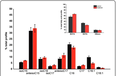
Effect of decanoic acid onS. roseosporusphospholipid fatty acid composition
Change in fatty acid profile is associated with FFA adap-tation in bacteria [10]. To investigate whether cell mem-brane reorganization is involved in the DA tolerance,
we compare the phospholipid fatty acid profile of S.
roseosporuscells during late exponential phase growth in either TSB (control), or 1 mM DA. The anteisopen-tadecanoic (anteisoC15), isopalmitic (isoC16), palmitic (C16), and pentedecanoic (C16:1) fatty acids made up the majority of total phospholipid composition (Figure 3). During DA stress, the branched-chain Fatty acids
Figure 1The chemical structure of daptomycin and other A21978C factors.
(BCFA) dramatically increased, with a corresponding decrease in straight-chain fatty acids (SCFA). Especially, a significant decrease in palmitoleic acid (C16:1) content from approximately 10% to 5% was detectable, implying
diminished membrane fluidity. Studies inListeria
mono-cytogenes have demonstrated that the increase of the ratio of BCFA/SCFA plays a significant role in tolerance to acid, temperature, and other stresses by reducing mem-brane fluidity and decreasing permeability [11]. Similarly, the switch to a fatty acid profile dominated by BCFA sug-gested a response mechanism leading to a more rigid membrane to mitigate the toxicity of DA.
Effect of decanoic on the generation of ROS inS. roseosporus
It has been observed that increase of membrane fluidity induced by FFA is accompanied by an increase of ROS production. To investigate whether ROS involved in the toxicity of DA, we compared the intracellular ROS levels of control and DA-exposed cultures. Cells were labeled with 5(and 6)-carboxy-2’,7’-dichlorodihydro-fluorescein
diacetate (carboxy-H2DCFDA), a known fluorogenic
marker for ROS in vivo. Cells exposed to DA showed
dramatically high level of fluorescence (Figure 4). Inte-restingly, cells exposed to different level of DA showed almost the same level of fluorescence, implying that 1 mM DA is enough to induce the ROS generation. These results suggested that exposure to DA may cause massive oxidative stress toS. roseosporus.
Effect of decanoic acid onS. roseosporustranscriptome
To elucidate molecular mechanisms underlying tole-rance, global gene expression changes during SR growth with DA were analyzed using Illumina RNA deep se-quencing (RNA-seq) technology. Tanscriptome libraries
were constructed using SR cells grown in the absence (control) or the presence of DA (1 mM).
RNA-seq data revealed a small subset of genes with differential transcription; 134 genes were up-regulated and 12 genes were down-regulated. The presence of DA at 1 mM resulted in transcriptional reprogramming of genes in three major discernible categories, including: energy production and conversion, posttranslational modification and protein turnover, and carbohydrate metabolism (Additional file 1).
Changes in energy metabolism
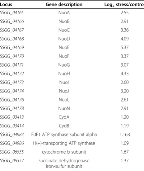
Exposure to DA resulted in the up-regulation of genes encoding enzymes proteins (enzymes) involved in energy
production and conversion (Table 1). nuo operon and
cyd operon were among the most significantly increased
transcripts during DA stress. The nuo operon encodes
the NADH ubiquinone oxidoreductase (complex I), which form core components of the electron transport chain [12]. The increase in the transcript levels of 12
member of nuo operon in response to DA stress
sug-gested that an increased requirement for energy or an impairment in the respiratory efficiency. Increases in the
transcripts in the cyd operon, which encodes a terminal
oxidase and gene (SSGG_06557) encoded the succinate dehydrogenase, both involved in oxidative phosphory-lation, also suggested a perturbation in respiratory
ba-lance. The up-regulated expression of nuo and cyo is
consistent with the recent discovery that nuo and cyo
played major roles in combating against oxidative stress in E. coli exposure to exogenous n-butanol and en-dogenous fatty acid production [10,13].
In addition, the increase in the transcripts in genes encoding the member of the ATP synthase complex
Figure 3Phospho-lipid fatty acid profiles ofS. roseosporusafter two hours of decanoic acid-stress.Control and stressed cell cultures were grown in triplicate. Stressed samples were exposed to1 mM decanoic acid for two hours before being harvested. Cell cultures (50 ml each) were harvested, centrifuged, washed in PBS.
Figure 4Measurement of intracellular reactive oxygen species using carboxy-H2DCFDA.Stressed samples(1, 2) were exposed to 1 mM decanoic acid for 30 min. control cells treated with tertbutyl hydroperoxide (TBHP), known to produce intracellular H2O2and
(SSGG_04986 and SSGG_04898) was observed. ATP syn-thase is responsible for generation of ATP through oxida-tive phosphorylation . It uses energy stored in the pH and potential gradients, created by pumping of protons across the membrance by enzymes of the respiratory chian, to synthesize ATP. Similar results were observed in yeast, where exposure of cells to octanoic acid (C8) led to the
activation of plasma membrane H +−ATPase [14]. These
results suggested that an increased requirement for energy was required to deal with the DA stress.
Induction of oxidative stress response
Consistent with results of ROS analysis, the
trans-criptomic response of S. roseosporus to DA showed a
massive induction of genes with a role in adaptation to oxidative stress. This response included activation of
clpB (proteases), sodF and several heat shock proteins
(Table 2).
In addition, expression levels of genes coding for
com-ponents of proteasome (pafA, pup, pfafA2, mpa) were
enhanced. Bacterial proteasomes could only be found in actinomycetes [15]. Mpa assembles into a hexameric ATPase and Pup, a prokaryotic ubiquitin-like protein, is ligated by PafA to substrate proteins. Subsequently, proteins tagged with Pup were targeted for degradation by the proteasome [16]. Recently, it was shown that proteasome is important for defense against reactive
nitrogen intermediates (RNI) inMycobacterium
tubercu-losis [17]. Increased expression of proteasome and clpB may degrade the misfolded proteins impaired by ROS.
Instreptomyces, the expression of genes coding for pro-teins involved in antioxidative defense systems was under the control of several key regulators, such as OxyR [18]. However, the homologue of OxyR, master regulator of
oxidative response, was absent in S. roseosporus. FurS
(SSGG_00190) is a zinc-containing redox regulator of S. reticuli which binds to an operator upstream of the furS-cepB [19]. Under oxidative stress conditions, an internal S-S bridge formed FurS abrogated its capability to
block the transcription of furS-cpeB [20]. SSGG_00191
shared 80% amino acid identity tocpeB ofS. reticuli and was induced during DA stress. Further studies are required to determine the roles of FurS in oxidative response.
Changes in carbohydrate transport and metabolism
Transcriptomic analysis suggested a metabolic adapta-tion dedicated to increase the producadapta-tion of trehalose (Table 3). Expression of genes coding for the puta-tive maltose ABC transporter (SSGG_01377), and Tres (SSGG_05057) which catalyzes the conversion of mal-tose into Trehalose was elevated. In addition, gene expression of alpha-amylase (SSGG_05058) was induced, which degrade starch to provide the maltose for synthe-sis of trehalose. The accumulation of this nonreducing sugar has been previously viewed as an important osmoprotectant and stress protectant [21]. It was recently reported that trehalose played an important protective role in the cellular response to oxidative stress
caused by hydrogen peroxide in Candida albicans[22].
Table 1 Selected genes involved in energy production and conversion with significant change in expression in DA-stress relative to control
Locus Gene description Log2stress/control
SSGG_04165 NuoA 2.55
SSGG_04166 NuoB 2.91
SSGG_04167 NuoC 3.36
SSGG_04168 NuoD 4.09
SSGG_04169 NuoE 5.37
SSGG_04170 NuoF 3.37
SSGG_04171 NuoG 3.07
SSGG_04172 NuoH 4.33
SSGG_04173 NuoI 2.60
SSGG_04174 NuoJ 3.20
SSGG_04176 NuoL 2.61
SSGG_04178 NuoN 2.91
SSGG_03413 CydA 1.20
SSGG_03414 CydB 1.19
SSGG_04984 F0F1 ATP synthase subunit alpha 1.168
SSGG_04986 H(+)-transporting ATP synthase 1.09
SSGG_06555 cytochrome b subunit 1.67
SSGG_06557 succinate dehydrogenase iron-sulfur subunit
1.37
Table 2 Selected genes involved in oxidative stress response with significant change in expression in DA-stress relative to control
Locus Gene description Log2stress/control
SSGG_00741 PafA 1.06
SSGG_00747 Pup 1.68
SSGG_00748 PafA2 1.52
SSGG_00750 Mpa 1.67
SSGG_01786 ClpB 1.59
SSGG_01803 SodF 1.18
SSGG_00191 CepB 2.58
SSGG_01307 phage shock protein A 1.45
SSGG_04333 Hsp 18 1.65
SSGG_04334 hsp60-like protein 1.81
SSGG_03360 bifunctional thioredoxin reductase/thioredoxin
1.59
SSGG_03361 thioredoxin reductase 2.02
Similarly, S. roseospous could counteract the DA stress by increasing trehalose production.
Genes encoding proteins involved in the TCA cycle were nearly unchanged. Interestingly, expression of genes in-volved in pyruvate production was up-regulated. Genes coding for fructose-bisphosphate aldolase (SSGG_03685), glyceraldehyde-3-phosphate dehydrogenase (SSGG_06343), phosphopyruvate hydratase (SSGG_02477) and pyruvate kinase (SSGG_01114) were upregulated after exposure to DA stress. Pyruvate is a key intermediate involved in a number of metabolic pathways. It was recently reported that pyruvate is involved into octanoic acid (C8) stress of
E. coli[23].Addition of pyruvate into media helps the cell partially recover from stress, but the exact mechanism was unclear. Similarly, enhanced production of pyruvate may helpS. roseosporusto recover from DA stress.
Preliminary model for the mechanism of DA toxicity and cell response
Based on several lines of evidence, we propose a prelimi-nary model for DA toxicity and cell’s response (Figure 5). DA incorporation into the cell membrane causes the dis-ruption of membrane fluidity. The membrane disdis-ruption results in destabililization of membrane-bound proteins, such as NADH dehydrogenase, H-ATPase. Furthermore, DA may directly interact with and partially inhibit of components of electron transport chain, resulting in facilitating electron leakage and generation of ROS. The malfunction of those enzymes will cause inhibition of electron transport chain and lead to less ATP
produc-tion. To counteract the impaired effect, S. roseosporus
utilizes multiple ways to protect the cells. Cells increase the ratio of BCFA/SCFA to reduce membrane fluidity and decrease permeability; to protect against the damage caused by oxidative stress, cells activate a number of antioxidant enzymes and repair activities, such as Su-peroxide dismutase, catalase, and heat shock proteins; furthermore, cells produce high level of trehalose and pyruvate to alleviate the toxicity of ROS. In addition, Table 3 Selected genes related to carbohydrate
metabolism and transport with significant change in expression in DA-stress relative to control
Locus Gene description Log2stress/
control
SSGG_01377 putative maltose ABC transporter permease
2.46
SSGG_05057 trehalose synthase 3.09
SSGG_05058 alpha-amylase 2.12
SSGG_03685 fructose-bisphosphate aldolase 1.37
SSGG_06343 phosphopyruvate hydratase 1.63
SSGG_01114 pyruvate kinase 1.30
SSGG_02477 phosphopyruvate hydratase 1.53
cells increase the expression of genes encoding proteins involved in oxidative phosphoralation to supply proteins to replace the impaired proteins.
Conclusions
Multiple mechanisms are involved in the mitigation of the toxicity of DA. The relative contribution of particular mechanism to the toxicity of fatty acid remains elusive. Taken together, our study provided insights into the toxi-city or tolerance mechanism underlying DA exposure and several candidates that may be targeted for further engi-neering to mitigate the toxicity of DA.
Methods Culture conditions
For toxicity assays,Streptomyces roseosporus(NRRL11579) was grown in TSB medium. To determine the effect of
DA on S. roseosporus growth, wide range of
concentra-tions was tested first (data not shown) and then narrowed to a range that caused stress but not significant cell death. Growth assays to test the effect of different concentrations
of DA onS. roseosporuswere performed in 250 ml shake
flasks with 25 ml of TSB medium with a 2% inoculation culturing at 200 rpm at 28°C. Unless specified otherwise, all subsequent DA assays were conducted at 1 mM DA.
Phospholipid fatty acid analysis
Cells were harvested in control culture and DA-stressed S. rosoeporuscultures (2 h after exposure to 1 mM DA) of growth by centrifugation at 3000 × g and 4 for 15 min, and the pellet was washed three times with distilled water. The fatty acids in the cells (40–50 mg in wet weight) were saponified and methylated. The methyl ester mixtures were separated using an Agilent 5890 dual tower gas chromatograph. Fatty acids were identified by the MIDI microbial identification system (Sherlock 4.5 microbial identification system) [24]. Minor fatty acids (<0.6% of the total) are not reported.
Reactive oxygen species assay
Control and DA-stressed S. rosoeporus cultures were
grown in TSB medium as described with various concentrations of DA. Positive controls for oxidative stressed cells were prepared by adding 10 ul of 7.78 M tert-butyl hydroperoxide (TBHP) (Invitrogen, USA) to one set of control cells before incubation. Ten
microli-ters of 25 mM carboxy-H2DCFDA was added to all cells.
Florescence at 535 nm was measured after 30 min.
RNA extraction
S. roseosprouswas cultured in TSB to exponential phase (48 h). DA was added to a final concentration of 1 mM, and biomass was collected after treatment of 30 min. the cultures were centrifuged at 3000 × g and 4 for 15 min,
and cell pellets were immediately frozen in liquid
nitro-gen and stored at −80 for subsequent RNA isolation.
Total RNA was extracted using Trizol (Invitrogen),
following manufacturer’s protocols. RNA preparations
were treated with RNase-free DNase (Promega) and the integrity of the RNA was determined using Bioanalyzer 2100 (Agilent Technologies). mRNA was enriched by removing the rRNAs using MICROBExpress kit. The mRNA remaining in the supernatant was recovered by ethanol precipitation and quantified by Bioanalyzer 2100. A cDNA library was constructed and sequenced by Illumina HiseqTM2000 [25].
Data processing and analysis
Raw sequencing reads were mapped against the S.
roseosprous genome. Reads that mapped to more than one region of the genome (5 to 8% of the total) failed to be unambiguously mapped were excluded for subsequent analyses. Analyses of differential expression including FDR calculations were performed using DESeq [26,27].
Only P values of <0.01, FDR≤0.01 were considered to
be significant.
Additional file
Additional file 1: Table S1.Statistically significant log2 gene expression values for S. roseosporus cultures responding to decanoic acid stress.
Competing interests
The authors declare that they have no competing interests.
Authors’contributions
GL and QL carried out the experiments and analyzed the primary data. GL wrote the draft manuscript. JX supervised the whole work and revised the manuscript. All authors read and approved the final manuscript.
Acknowledgements
This work was supported by grants from National Natural Science Foundation of China (Grant No. 31100069), the Natural Science Foundation Project of CQ CSTC (cstc2012jjA10149) and the Fundamental Research Funds for the Central Universities (XDJK2013A010), New Century Excellent Talents in Universities (NCET-11-0703).
Received: 26 December 2012 Accepted: 19 February 2013 Published: 21 February 2013
References
1. Baltz RH:Daptomycin: mechanisms of action and resistance, and biosynthetic engineering.Curr Opin Chem Biol2009,13:144–151. 2. Wang L, Zhao Y, Liu Q, Huang Y, Hu C, Liao G:Improvement of A21978C
production in Streptomyces roseosporus by reporter-guided rpsL mutation selection.J Appl Microbiol2012,112:1095–1101.
3. Yu G, Jia X, Wen J, Lu W, Wang G, Caiyin Q, Chen Y:Strain improvement of Streptomyces roseosporus for daptomycin production by rational screening of He-Ne laser and NTG induced mutants and kinetic modeling.Appl Biochem Biotechnol2011,163:729–743.
4. Lu W, Fan J, Wen J, Xia Z, Caiyin Q:Kinetic analysis and modeling of daptomycin batch fermentation by Streptomyces roseosporus.
Appl Biochem Biotechnol2011,163:453–462.
of lipopeptide antibiotic A21978C: the synthesis and evaluation of daptomycin (LY146032).J Antibiot (Tokyo)1988,41:1093–1105.
6. Huber FM, Pieper RL, Tietz AJ:The formation of daptomycin by supplying decanoic acid to Streptomyces roseosporus cultures producing the antibiotic complex A21978C.J Biotechnol1988,7:283–292.
7. Desbois AP, Smith VJ:Antibacterial free fatty acids: activities, mechanisms of action and biotechnological potential.Appl Microbiol Biotechnol2010,
85:1629–1642.
8. Alexandre H, Mathieu B, Charpentier C:Alteration in membrane fluidity and lipid composition, and modulation of H(+)-ATPase activity in Saccharomyces cerevisiae caused by decanoic acid.Microbiology1996,
142(Pt 3):469–475.
9. Heiskanen KM, Savolainen KM:Erucic acid and erucic acid anilide-induced oxidative burst in human polymorphonuclear leukocytes.Free Radic Res
1997,27:477–485.
10. Lennen RM, Kruziki MA, Kumar K, Zinkel RA, Burnum KE, Lipton MS, Hoover SW, Ranatunga DR, Wittkopp TM, Marner WD 2nd, Pfleger BF:Membrane stresses induced by overproduction of free fatty acids in Escherichia coli.
Appl Environ Microbiol2011,77:8114–8128.
11. Giotis ES, McDowell DA, Blair IS, Wilkinson BJ:Role of branched-chain fatty acids in pH stress tolerance in Listeria monocytogenes.Appl Environ Microbiol2007,73:997–1001.
12. Ingledew WJ, Poole RK:The respiratory chains of Escherichia coli.
Microbiol Rev1984,48:222–271.
13. Rutherford BJ, Dahl RH, Price RE, Szmidt HL, Benke PI, Mukhopadhyay A, Keasling JD:Functional genomic study of exogenous n-butanol stress in Escherichia coli.Appl Environ Microbiol2010,76:1935–1945.
14. Viegas CA, Almeida PF, Cavaco M, Sa-Correia I:The H(+)-ATPase in the plasma membrane of Saccharomyces cerevisiae is activated during growth latency in octanoic acid-supplemented medium accompanying the decrease in intracellular pH and cell viability.Appl Environ Microbiol
1998,64:779–783.
15. De Mot R, Nagy I, Walz J, Baumeister W:Proteasomes and other self-compartmentalizing proteases in prokaryotes.Trends Microbiol1999,7:88–92. 16. Darwin KH:Prokaryotic ubiquitin-like protein (Pup), proteasomes and
pathogenesis.Nat Rev Microbiol2009,7:485–491.
17. Darwin KH, Ehrt S, Gutierrez-Ramos JC, Weich N, Nathan CF:The proteasome of Mycobacterium tuberculosis is required for resistance to nitric oxide.Science2003,302:1963–1966.
18. Hahn JS, Oh SY, Roe JH:Role of OxyR as a peroxide-sensing positive regulator in Streptomyces coelicolor A3(2).J Bacteriol2002,
184:5214–5222.
19. Ortiz De Orue Lucana D, Schrempf H:The DNA-binding characteristics of the Streptomyces reticuli regulator FurS depend on the redox state of its cysteine residues.Mol Gen Genet2000,264:341–353.
20. Ortiz De Orue Lucana D, Troller M, Schrempf H:Amino acid residues involved in reversible thiol formation and zinc ion binding in the Streptomyces reticuli redox regulator FurS.Mol Genet Genomics2003,
268:618–627.
21. Benaroudj N, Lee DH, Goldberg AL:Trehalose accumulation during cellular stress protects cells and cellular proteins from damage by oxygen radicals.J Biol Chem2001,276:24261–24267.
22. Cao Y, Wang Y, Dai B, Wang B, Zhang H, Zhu Z, Xu Y, Jiang Y, Zhang G:
Trehalose is an important mediator of Cap1p oxidative stress response in Candida albicans.Biol Pharm Bull2008,31:421–425.
23. Fu Y:Metabolic flux analysis of Escherichia coli MG1655 under octanoic acid (C8) stress. Iowa State University: Department of Chemical and Biological Engineering; 2011.
24. Li Y, Florova G, Reynolds KA:Alteration of the fatty acid profile of
Streptomyces coelicolor by replacement of the initiation enzyme 3-ketoacyl acyl carrier protein synthase III (FabH).J Bacteriol2005,187:3795–3799. 25. Mandlik A, Livny J, Robins WP, Ritchie JM, Mekalanos JJ, Waldor MK:
RNA-Seq-based monitoring of infection-linked changes in Vibrio cholerae gene expression.Cell Host Microbe2011,10:165–174.
26. Gentleman RC, Carey VJ, Bates DM, Bolstad B, Dettling M, Dudoit S, Ellis B, Gautier L, Ge Y, Gentry J,et al:Bioconductor: open software development for computational biology and bioinformatics.Genome Biol2004,5:R80. 27. Khudyakov JI, D'Haeseleer P, Borglin SE, Deangelis KM, Woo H, Lindquist EA,
Hazen TC, Simmons BA, Thelen MP:Global transcriptome response to ionic liquid by a tropical rain forest soil bacterium, Enterobacter lignolyticus.Proc Natl Acad Sci USA2012,109:E2173–2182.
doi:10.1186/1475-2859-12-19
Cite this article as:Liaoet al.:Transcriptional analysis of the effect of exogenous decanoic acid stress onStreptomyces roseosporus.Microbial Cell Factories201312:19.
Submit your next manuscript to BioMed Central and take full advantage of:
• Convenient online submission
• Thorough peer review
• No space constraints or color figure charges
• Immediate publication on acceptance
• Inclusion in PubMed, CAS, Scopus and Google Scholar
• Research which is freely available for redistribution