R E S E A R C H
Open Access
Improved cell surface display of
Salmonella
enterica
serovar Enteritidis antigens in
Escherichia coli
Martin Gustavsson
1, Thi-Huyen Do
2, Petra Lüthje
3, Ngoc Tan Tran
2, Annelie Brauner
3, Patrik Samuelson
1,
Nam Hai Truong
2and Gen Larsson
1*Abstract
Background:Salmonella entericaserovar Enteritidis (SE) is one of the most potent pathogenicSalmonellaserotypes causing food-borne diseases in humans. We have previously reported the use of theβ-autotransporter AIDA-I to express theSalmonellaflagellar protein H:gm and the SE serotype-specific fimbrial protein SefA at the surface of E. colias live bacterial vaccine vehicles. While SefA was successfully displayed at the cell surface, virtually no full-length H:gm was exposed to the medium due to extensive proteolytic cleavage of the N-terminal region. In the present study, we addressed this issue by expressing a truncated H:gm variant (H:gmd) covering only the serotype-specific central region. This protein was also expressed in fusion to SefA (H:gmdSefA) to understand if the excellent translocation properties of SefA could be used to enhance the secretion and immunogenicity.
Results:H:gmd and H:gmdSefA were both successfully translocated to theE. coliouter membrane as full-length proteins using the AIDA-I system. Whole-cell flow cytometric analysis confirmed that both antigens were displayed and accessible from the extracellular environment. In contrast to H:gm, the H:gmd protein was not only expressed as full-length protein, but it also seemed to promote the display of the protein fusion H:gmdSefA. Moreover, the epitopes appeared to be recognized by HT-29 intestinal cells, as measured by induction of the pro-inflammatory interleukin 8.
Conclusions:We believe this study to be an important step towards a live bacterial vaccine againstSalmonelladue to the central role of the flagellar antigen H:gm and SefA inSalmonellainfections and the corresponding immune responses againstSalmonella.
Keywords:Autotransport, Surface expression,Escherichia coli, AIDA-I, Live vaccines,Salmonella enterica
Background
Bacterial surface display of recombinant proteins has been used for various applications in microbiology, mo-lecular biology, biotechnology and vaccinology [1,2]. The field is dominated by display inEscherichia coli, mainly due to the extensive knowledge concerning the genetics, the presence of transformation protocols, the rapid cell growth on simple media, and the multitude of produc-tion techniques and protocols for recombinant protein production that are associated with this organism.
Further, a range of specific mutant strains with desirable features is available for metabolic engineering.
An area of particular interest is the display of im-munogenic peptides at the cell surface for direct use as live vaccine delivery vehicles [2]. This technique offers some interesting advantages over conventional vaccines the first being that a surface display system provides the prerequisites for a more economic process [3]. The rea-son is that it is designed to circumvent the step of anti-gen purification and relies on a one-step combination of the production and purification processes. Secondly, the live vaccine systems may have in-built adjuvant effects from endogenous components of the host cell e.g. from the distinctive structure of theE. colilipopolysaccharides
* Correspondence:gen@kth.se
1
Royal Institute of Technology (KTH), Division of Industrial Biotechnology, AlbaNova University Center, SE 10691 Stockholm, Sweden
Full list of author information is available at the end of the article
(LPS) but also the lipoteichoic acids, peptidoglycans, lipoproteins etc., which are well recognized by the host’s immune system [4] and therefore elicit strong immune responses. Finally, these vehicles may have prolonged retention times due to colonization effects and they are probably also safer to use than inactivated or attenuated pathogen-based vaccines, which may revert to their pathogenic form.
Surface display relies today on a number of different techniques. We have on several occasions successfully used the autotransporter AIDA-I (Adhesin Involved in Diffuse Adherence of enteropathogenicE. coli) [5] to ex-press and display different peptides and proteins on the cell surface of a laboratoryE. coliK-12 strain into which the AIDA-inherent system of pathogenicE. coliwas trans-fered [6-8]. Our autotransporter vector pAIDA1 consists of a specific N-terminal signal peptide followed by a passenger protein, a linker region and a C-terminal trans-location unit, AIDAC, which forms a β-barrel-type outer membrane (OM) pore [9,10], as illustrated in Figure 1. The signal peptide is cleaved off after the Sec-mediated translocation over the inner membrane, followed by insertion and formation of the β-barrel pore through which the linker and passenger moiety eventually pass to become surface exposed [9,10]. Recently, we expressed the Salmonella entericaserovar Enteritidis (SE) proteins SefA and H:gm at theE. colicell surface [7,11,12] to investigate the potential of the surface expression technique as a process for live vaccine production. Although two differ-ent versions of the recombinant AIDA-system allowed us to express both the flagellar protein H:gm and the fimbrial protein SefA inE. coli, only SefA was successfully exposed
as a full-length protein at the surface [7,11]. Western blot analysis showed the presence of H:gm in the OM fraction but revealed that it was proteolytically cleaved.
It is well known that SefA and H:gm both play central roles inSalmonella infections and elicit a protective im-mune response against SE [13-17]. Hence, the inability to express and display H:gm as a full-length protein on the E. coli cell surface might reduce the efficacy of this strain as a live vaccine. We hypothesized that our previ-ous difficulties of surface expression of H:gm, and the partial proteolysis observed, was due either to the larger size of this protein compared to SefA or due to prema-ture folding of the quite complex strucprema-ture of the H:gm protein in the OM both leading to a longer exposure time to periplasmic and OM proteases. In the present investigation we addressed this issue by protein engin-eering reducing H:gm into a smaller derivative consist-ing of only the serotype-specific region [18], denoted H:gmd (Figure 2). This smaller unit was expressed by the AIDA-I vector but also in fusion to SefA with the idea to boost the production by use of a protein with known and excellent translocation properties, as well as with the aim to produce a vaccine targeting both epitopes using a single cell.
Results
Surface expression of H:gm, H:gmd, SefA, and H:gmdSefA inE. coli
E. coli O17, with the deletion of the outer membrane
protease OmpT, was used for production to avoid the cleavage of the protein from the cell surface that would otherwise take place [6]. This strain was transformed
A
B
Figure 1Schematic representation of the recombinant autodisplay system pAIDA1. A)The surface display plasmid pAIDA1 based on the AIDA-I autotransporter. Expression is under control of the lacUV5 promoter, and two detection/affinity tags (His6and c-Myc), in addition to two
with the surface expression plasmid pAIDA1, which was used to produce the engineered variant of the Salmon-ella flagellar protein H:gm, here denoted H:gmd. In addition, this protein was also fused to the fimbrial protein SefA to investigate a supposed positive effect on production by utilizing the known successful transloca-tion of this particular protein. For comparison, strains producing the full-length protein H:gm, as well as SefA, were cultivated and the expression compared to the new constructs. A comparison of the properties of these xproteins can be found in Table 1. As a zero-reference,
E. coli growth without the plasmid was used. All
pro-cesses were performed by batch technology and sampled during the logarithmic phase for subsequent analysis.
As seen in Figure 1, the pAIDA1 vector is constructed to harbor two distinct tags, His6and c-Myc that are flank-ing the recombinant passenger protein on the N- and C-terminal sides, respectively. Using fluorescently labeled antibodies specific for the tags allows us to determine the
presence and integrity of the different fusion proteins using flow cytometry.
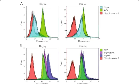
All recombinant fusion proteins showed whole-cell fluorescence intensities greater than the negative control when probed with the anti-c-Myc-antibody (Figure 3), which is located closest to the cell surface, i.e. on the in-side with respect to the surface exposed antigen (Figure 1). This confirms that all constructs were expressed at least as far as to the translocation unit (AIDAC) with the linker and c-Myc-tag that were inserted and accessible in the outer cell membrane to the binding of the fluorophore.
Figure 3 shows also the data for the second detection system based on the His6-tag that is situated outside the antigen facing the medium. All constructs, except H:gm (Additional file 1: Figure S1), stained positive for the presence of this tag at the cell surface. The fluorescence intensity of the H:gm-expressing cells was comparable to the negative control while the new constructs exhibited a significant increase in fluorescence. In contrast to H:gm, the novel construct containing the smaller flagel-lar antigen H:gmd was expressed very well at the cell surface. In fact, this construct resulted in the highest fluorescence of all constructs tested for both the anti-His6 and the anti-c-Myc antibody (Figure 3B).
Subcellular localization and size of the fusion proteins
To specify the localization and integrity of the different fusion proteins whole-cell lysates were fractioned and the OM fraction was collected for comparison of the different constructs. This fraction was analyzed by SDS-PAGE and the result is shown in Figure 4. As can be seen, the
Salmonella antigens were correctly expressed in the OM
and appeared to generate full-length fusion proteins of the expected sizes; 68.8 kDa (H:gmd-AIDAC), 83.3 kDa (H:gmdSefA-AIDAC), 72.6 kDa (SefA-AIDAC). As antici-pated, the OM fractions of the negative controls (wild type
E. coliO17ΔOmpT or non-induced O17ΔOmpT
contain-ing pAIDA1-SefA) did not generate any detectable protein bands corresponding to those of the relevant Salmonella antigen fusions. These results thus corroborate well with the whole-cell fluorescence data of Figure 3. Additional bands for the outer membrane proteins OmpF (39 kDa) and OmpA (35 kDa) [19] were observed in all analysed samples.
A maybe even more important finding was that H:gmd did not show any visible signs of proteolytic degradation, neither in the FACS fluorescence data (Figure 3) or by SDS-PAGE analysis (Figure 4), in contrast to the pre-viously used H:gm that gave rise to little full-length protein due to extensive proteolysis (Additional file 1: Figure S1) [7,11]. This is a significant improvement as H:gmd contains the serotype-specific specific amino acids, and thus might increase its potential as a live bacterial subunit vaccine againstSalmonella.
Figure 23D-structure of H:gm.Atomic model of theSalmonella
entericaflagellar antigen H:gm (507 aa) generated with Phyre2 (Protein Homology/Analogy Recognition Engine) [26] and drawn in PyMOL. The protein forms a tubular like structure with the N- and C-terminal ends in close proximity to each other. The region corresponding to H:gmd (aa 254–351) is highlighted in cyan.
Table 1 Description of the different passenger proteins expressed in this study
Protein Description Size (kDa) Length (aa) Reference
Functionality of the expressed proteins
Having verified their correct localization and surface exposure, we finally wanted to check if the engineered antigen H:gmd might still provoke an immunogenic response since a severe deletion was imposed on the full length H:gm protein. It has previously been reported that S. enterica flagellar and fimbrial proteins (i.e. SefA and H:gm), can induce an IL-8 response from gut epi-thelial cell line HT-29 [20]. Thus, an in vitro assay was devised by exposing this cell line to E. coli expressing the various epitope constructs, while measuring the re-lease of IL-8 as marker for a pro-inflammatory response. An elevated response was detected for the strains express-ing SefA, H:gm and H:gmd as compared to the strain carrying a surface expression vector without any of the SE epitopes (Figure 5). The highest response was seen from SefA followed by H:gmd and H:gm, where the two latter were of approximately the same magnitude. Interestingly, there was no significant response to the H:gmdSefA con-struct. Furthermore, it appears that the chosenE. colihost strain was unable to raise an IL-8 response by itself, as the control bearing the empty surface expression vector
showed similar or lower IL-8 levels as the uninfected negative control.
Discussion
The main goal with the present study was to successfully express and display H:gm-derived antigens at the surface
of E. coli by using the AIDA-I autotransporter system.
Our previous attempts to express and display H:gm were unsuccessful due to extensive proteolytic cleavage in the C-terminal region of that antigen [7,11]. To make H:gm less affected to proteolytic degradation could be achieved through several approaches. One option would be the usage of protease-deficient E. coli strains. However, we already used a mutant strain deficient for the protease OmpT (O17ΔOmpT) and a clear candidate protease could not be identified from our previous experiments. Instead, we chose to engineer the protein to get a H:gm-derivative that could potentially be efficiently displayed at the cell surface and be less sensitive to proteolytic cleavage. For this purpose, we here chose to include only a subpart, called H:gmd, comprising 98 aa out of the total 507 aa in the full-length H:gm (Figure 2). The rationale behind this
A
B
Figure 3Flow cytometry histograms of variousSalmonellaantigens expressed on the surface ofE. coli.The presence of the His6-tag and
c-Myc-tag in the displayed proteins were probed with THE™His Tag Antibody [FITC] and Anti-c-Myc [SureLight® Allophycocyanin] antibody, respectively. Negative controls (cells lacking surface display plasmids) are all shown in red.(A)Left panel: His-probedE. coli/pAIDA1-H:gm (light blue) andE. coli/pAIDA1-SefA (green). Right panel: c-Myc-probedE. coli/pAIDA1-H:gm (light blue) andE. coli/pAIDA1-SefA (green).(B)Left panel: His-probedE. coli/pAIDA1-H:gmd (dark blue),E. coli/pAIDA1-SefA (green), andE. coli/pAIDA1-H:gmdSefA (purple). Right panel: c-Myc-probed
was threefold. Firstly, the chosen region comprises the serotype-specific part of H:gm needed for development of immunity to SE [21,22]. Secondly, the large size of H:gm can be expected to lead to a longer time required for full translocation over the outer membrane, resulting in an extended exposure to periplasmic proteases. By expressing only a small part of H:gm this exposure should be reduced and consequently the protein should be less susceptible to proteolysis. Thirdly, it has been shown that passenger pro-teins that form stable folds in the periplasm, for instance due to disulphide formation, are difficult to display using autotransporters due to stalled translocation over the outer membrane [23,24]. As seen in Figure 2, H:gm folds with the N-and C-terminals in close proximity, which could be problematic given the autotransporter translocation mechanism. By expressing only the H:gmd domain this potentially problematic part of the protein is excluded.
This strategy proved to be very successful, as H:gmd generated whole-cell fluorescence intensities much greater than the negative control, both when the presence of the N-terminal His6-tag and the C-terminal c-Myc tag were analyzed (Figure 3B). This is in contrast to the original H:gm construct, which did not generate a signal above the negative control when probed with the His6-reactive anti-body, indicating proteolytic degradation (Figure 3A, left panel). Interestingly, not only was the smaller H:gmd pro-tein expressed to full-length at the cell surface, but it also appeared to promote the display and accessibility of the fused protein H:gmdSefA since H:gmd and H:gmdSefA both resulted in higher fluorescence intensities than SefA alone or the full-length H:gm. Although the true reasons behind the observed improvements in expression and cell surface display characteristics of H:gmd over H:gm at present are unclear, they can likely be attributed either to structural reasons as discussed previously, or that the protease sensitive amino acids in H:gm are not present in H:gmd.
With the ultimate goal to generate suitable tools for live vaccine development, we tested theE. colistrains ex-pressing the differentSalmonellaepitopes for their ability to induce an immune reaction in gut epithelial cells. The response in the in vitrosystem applied here was overall very low, probably related to the low immunogenicity of the non-pathogenicE. coliK-12 strain O17ΔOmpT, which was unable to stimulate IL-8 production on its own. How-ever, our results suggest that while SefA appeared the most potent immunogen, the truncated H:gmd variant was to some extent even superior to the full-length H:gm construct, in line with the flow cytometric resultss. In con-trast, the fusion construct H:gmdSefA did not perform well in this assay. A possible explanation might be that the epitopes of the single proteins are hidden by the structure of the fusion protein.
225 115 80 65
50
35
30
25
15 kDa
Figure 4SDS-PAGE analysis of recombinantSalmonella antigens recovered fromE. coliouter membrane fractions. Lane 1: Size marker (Spectra Multicolour Broadrange, Fermentas); Lane 2: O17ΔOmpT/pAIDA1-SefA (positive control; shake flask cultivation from a prior experiment [7]; Lane 3: empty O17ΔOmpT (negative control); Lane 4: non-induced O17ΔOmpT/pAIDA1-SefA (negative control); Lane 5: O17ΔOmpT/pAIDA1-H:gmd; Lane 6: O17ΔOmpT/ pAIDA1-H:gmdSefA; Lane 7 & 8: O17ΔOmpT/pAIDA1-SefA, replicates.
Conclusions
In this study we engineered a truncated variant of H:gm comprising only the serotype-specific amino acids. H: gmd was resistant to proteolytic cleavage and exhibited highly efficient surface display, in contrast to H:gm. This is a promising result from the perspective of creating a live vaccine against SE, though additional studies are needed to further confirm the immunogenicity of these constructs and to investigate their ability to raise SE-specific antibodies in an animal host.
Methods
Bacterial strains and plasmids
Escherichia coli K-12 strain O17ΔOmpT was used for
surface expression using the AIDA-I autotransporter, as previously reported [6]. The plasmid pAIDA1-SefA [7] was used to produce a recombinant fusion of SE fimbriae protein SefA and AIDA-I on the surface of the host cell. This plasmid encodes the C-terminal AIDA β-barrel (AIDAC) followed by a linker consisting of the first 54 amino acids (aa) of the native AIDA passenger, the recom-binant SefA passenger and finally the native AIDA signal peptide. Additionally, the plasmid contains two epitope tags (c-Myc and His6) located on the C- and N-terminal sides of SefA, respectively (Figure 1). pAIDA1-H:gm [6-8] encodes the SE flagellar antigen H:gm instead of SefA. For construction of pAIDA1-H:gmd and pAIDA1-H:gmdSefA, standard recombinant DNA techniques were used [25]. Restriction endonucleases, T4 DNA ligase, plasmid purifi-cation kits (miniprep), and DreamTaq DNA polymerase were purchased from Fermentas. Primers were supplied by Eurofins MWG Operon while GE Healthcare sup-plied the PCR DNA and Gel band Purification Kit. H: gmd_forw (5′ -acaggtaccactaaatctactgctggaaccgctgaag-3′) and H:gmd_rev (5′-actgagctcctggatccacgaccctccgcctcct gaacccccgcctccgtttttggttttatcatcaaaag-3′) were used to PCR amplify a H:gmd-encoding DNA fragment from pAIDA1-H:gm. The resulting amplicon was then purified, digested with KpnI and SacI, gel purified and finally ligated into KpnI- and SacI-digested pAIDA1 to generate pAIDA1-H: gmd. To create pAIDA1-H:gmdSefA, the purified H:gmd-amplicon was instead cleaved with KpnI and BamHI, gel purified and then ligated into pAIDA1-SefA previously digested with KpnI and BclI, thus yielding pAIDA1-H: gmdSefA. These novel constructs were all verified by DNA sequencing. Table 1 shows a summary of the properties of the different antigens.
Cultivation
All cultivations were performed in minimal salts medium, consisting of (per liter): 7.0 g (NH4)2SO4, 1.6 g KH2PO4, 6.6 g Na2HPO4.2H2O, and 0.5 g (NH4)2-H-Citrate. In addition, all media were supplemented with 1 ml l−1 of both 1 M MgSO4and trace element solution that were
sterile filtered into the different reactors. The trace elem-ent stock solution consisted of (per liter): 0.5 g CaCl2· 2H2O, 16.7 g FeCl3· 6H2O, 0.18 g ZnSO4· 7H2O, 0.16 g CuSO4· 5H2O, 0.15 g MnSO4· 4H2O, 0.18 g CoCl2· 6H2O, 20.1 g Na-EDTA.
All cultivations were performed in a batch format ini-tialized by shake flask growth in 1 L bottles containing 100 ml of minimal salts medium supplemented with 10 g l−1 glucose. These were inoculated from a frozen glycerol stock (−80°C) and placed inside a shaking incu-bator (37°C, 180 rpm) over night. In the following morn-ing, six samples were withdrawn and used to inoculate a six-parallel stirred-tank bioreactor unit (Greta, Belach Bioteknik AB, Sweden) where each reactor contained 800 ml of minimal medium to which 10 g l−1of separ-ately sterilized glucose had been added. After inocula-tion, the cultures were grown for two generations to an optical density of 0.4, before induction by addition of isopropyl β-D-1-thiogalactopyranoside (IPTG) to a con-centration of 200 μM. Zero-samples for surface expres-sion were withdrawn immediately before induction. The cultures were then grown for approximately four genera-tions, after which final samples were withdrawn for subsequent analysis and comparison.
The dissolved oxygen concentration (DOT) in the reactors was controlled at 40% by automatic adjustment of the stirrer speed. The airflow was initially set to 0.1 VVM, and then incrementally increased to 1.0 VVM. The temperature was automatically controlled at 37°C and pH was kept at 7.0 by titration with NH4OH (12.5% w/v). Foaming was kept at a minimum by manual addition of antifoaming agent when necessary.
Analyses Cell mass
Cell mass accumulation was followed by sampling at regular intervals. The optical density at 600 nm (OD600) was measured using a spectrophotometer (Genesys 20, Thermo Scientific). All samples were diluted in saline solution (0.9% NaCl) to an approximate OD600of 0.1 prior to measurement to compensate for the non-linearity of OD600 measurements, and actual OD600 values were de-rived by multiplication with the dilution factor.
Flow cytometric analyses of surface expression
were recorded. The excitation wavelength was 488 nm for the His6-tag (FITC) analysis and emission was detected at 525/40 nm, while the c-Myc-analysis (SureLight® Allophy-cocyanin) used 638 nm and 660/20 nm for the excitation and emission wavelengths, respectively.
Analysis of OM proteins
E. coliproteins were separated into soluble, inner mem-brane and OM fractions as previously reported [19]. The resulting outer membrane protein samples were separated using SDS-PAGE on 10 % Bis-Tris gels (NuPage, Invitrogen). The gels were then stained using PageBlue protein staining solution (Fermentas) according to the manufacturer’s protocol.
Cell stimulation assay
Colonrectal adenocarcinoma cell line HT-29 (ATCC, HTB-38) were grown in McCoy’s 5A medium supple-mented with 10% fetal bovine serum at 37°C in a hu-midified incubator with 5% CO2. For experiments, cells were seeded on 24-well plates and used when confluent. Bacteria were collected at the time point of optimal protein expression by centrifugation, washed once in phosphate-buffered saline (PBS) and adjusted to an OD600of 0.125 in PBS. Bacteria were diluted 1:100 in complete cell culture medium to reach a final concentration of approximately 106colony forming units (CFU)/ml. Medium was removed from the cells and 1 ml of fresh medium with bacteria was added. The plate was centrifuged at 600gfor 5 min to ac-celerate bacterial contact to the cells and correct for poten-tial differences in motility of the strains. The infected cells were incubated at 37°C in a humidified incubator with 5% CO2 for 4 hours. Then, the supernatants were collected, floating cells were removed by centrifugation at 300g for 5 min and the cleared supernatants were stored at−80°C until analysis. Secreted interleukin (IL) 8 was quantified by enzyme-linked immunosorbent assay (ELISA) according to the manufacturer’s protocol (R&D Systems).
Additional file
Additional file 1: Figure S1.Flow cytometric analysis ofE. coli O17ΔOmpT expressing pAIDA1-H:gm (green) and pAIDA1-SefA (blue). Significant N-terminal proteolysis of H:gm was evident from the low signal from the N-terminal His6-tag (A) relative to the C-terminal Myc-tag (B). Red: O17ΔOmpT without the surface display vectors (negative control). Reproduced from Jarmander et al. 2012 [7].
Competing interests
The authors declare that they have no competing interests.
Authors’contributions
MG designed and performed the expression study and finalized the manuscript draft. THD and NTT performed the cloning and initial expression studies. PL designed and performed the cell stimulation experiments. PS supervised the cloning and wrote the initial draft of the manuscript. AB designed and supervised the cell stimulation experiments. NTH and GL
were responsible for the original concept and supervised the work. All authors read and approved the final manuscript.
Acknowledgements
The Swedish International Development Agency (SIDA) is acknowledged for their financial support.
Author details
1Royal Institute of Technology (KTH), Division of Industrial Biotechnology,
AlbaNova University Center, SE 10691 Stockholm, Sweden.2Institute of
Biotechnology, Vietnam Academy of Science and Technology, 18-Hoang Quoc Viet, Cau Giay, Ha Noi, Vietnam.3Department of Microbiology, Tumor
and Cell Biology, Division of Clinical Microbiology, Karolinska Institute and Karolinska University Hospital, Stockholm, Sweden.
Received: 20 November 2014 Accepted: 12 March 2015
References
1. Benhar I. Biotechnological applications of phage and cell display. Biotechnol Adv. 2001;19:1–33.
2. Georgiou G, Stathopoulos C, Daugherty PS, Nayak AR, Iverson BL, Curtiss R. Display of heterologous proteins on the surface of microorganisms: from the screening of combinatorial libraries to live recombinant vaccines. Nat Biotechnol. 1997;15:29–34.
3. Dertzbaugh MT. Genetically engineered vaccines: an overview. Plasmid. 1998;39:100–13.
4. Weidemann B, Schletter J, Dziarski R, Kusumoto S, Stelter F, Rietschel ET, et al. Specific binding of soluble peptidoglycan and muramyldipeptide to CD14 on human monocytes. Infect Immun. 1997;65:858–64.
5. Benz I, Schmidt M. Adhesin (AIDA-I) Involved in Diffuse Adherence of Enteropathogenic Escherichia coli. Infect Immun. 1989;57:1506–11. 6. Gustavsson M, Bäcklund E, Larsson G. Optimisation of surface expression
using the AIDA autotransporter. Microb Cell Fact. 2011;10:72. 7. Jarmander J, Gustavsson M, Do T-H, Samuelson P, Larsson G. A dual tag
system for facilitated detection of surface expressed proteins in Escherichia coli. Microb Cell Fact. 2012;11:118.
8. Gustavsson M, Muraleedharan MN, Larsson G. Surface expression of ω-transaminase in Escherichia coli. Appl Environ Microb. 2014;80:2293–8. 9. Benz I, Schmidt MA. Structures and functions of autotransporter proteins in
microbial pathogens. Int J Med Microbiol. 2011;301:461–8.
10. Leyton DL, Rossiter AE, Henderson IR. From self sufficiency to dependence: mechanisms and factors important for autotransporter biogenesis. Nat Rev Microbiol. 2012;10:213–25.
11. Nhan NT, Gonzalez de Valdivia E, Gustavsson M, Hai T, Larsson G. Surface display of salmonella epitopes in Escherichia coli and Staphylococcus carnosus. Microb Cell Fact. 2011;10:22.
12. Jarmander J, Janoschek L, Lundh S, Larsson G, Gustavsson M. Process optimization for increased yield of surface-expressed protein in Escherichia coli. Bioproc Biosyst Eng. 2014;37:1685–93.
13. Strindelius L, Filler M, Sjöholm I. Mucosal immunization with purified flagellin from Salmonella induces systemic and mucosal immune responses in C3H/HeJ mice. Vaccine. 2004;22:3797–808.
14. Robertson JMC, McKenzie NH, Duncan M, Allen-Vercoe E, Woodward MJ, Flint HJ, et al. Lack of flagella disadvantages Salmonella enterica serovar Enteritidis during the early stages of infection in the rat. J Med Microbiol. 2003;52:91–9.
15. Edwards RA, Schifferli DM, Maloy SR. A role for Salmonella fimbriae in intraperitoneal infections. Proc Natl Acad Sci U S A. 2000;97:1258–62. 16. Clouthier SC, Müller KH, Doran JL, Collinson SK, Kay WW. Characterization of
three fimbrial genes, sefABC, of Salmonella enteritidis. J Bacteriol. 1993;175:2523–33.
17. Peralta RC, Yokoyama H, Ikemori Y, Kuroki M, Kodama Y. Passive immunisation against experimental salmonellosis in mice by orally administered hen egg-yolk antibodies specific for 14-kDa fimbriae of Salmonella enteritidis. J Med Microbiol. 1994;41:29–35.
19. Bäcklund E, Reeks D, Markland K, Weir N, Bowering L, Larsson G. Fedbatch design for periplasmic product retention in Escherichia coli. J Biotechnol. 2008;135:358–65.
20. Rochon M, Römling U. Flagellin in combination with curli fimbriae elicits an immune response in the gastrointestinal epithelial cell line HT-29. Microbes Infect. 2006;8:2027–33.
21. Mizumoto N, Toyota-Hanatani Y, Sasai K, Tani H, Ekawa T, Ohta H, et al. Detection of specific antibodies against deflagellated Salmonella Enteritidis and S. Enteritidis FliC-specific 9 kDa polypeptide. Vet Microbiol.
2004;99:113–20.
22. Li J, Nelson K, McWhorter AC, Whittam TS, Selander RK. Recombinational basis of serovar diversity in Salmonella enterica. Proc Natl Acad Sci U S A. 1994;91:2552–6.
23. Jong WSP, ten CM H-J, Blaauwen den T, Slotboom DJ, Tame JRH, Wickström D, et al. Limited tolerance towards folded elements during secretion of the autotransporter Hbp. Mol Microbiol. 2007;63:1524–36.
24. Leyton DL, Sevastsyanovich YR, Browning DF, Rossiter AE, Wells TJ, Fitzpatrick RE, et al. Size and conformation limits to secretion of disulfide-bonded loops in autotransporter proteins. J Biol Chem. 2011;286:42283–91.
25. Sambrook J, Fritsch EF, Maniatis T. Molecular cloning. 1989.
26. Kelley LA, Sternberg MJE. Protein structure prediction on the Web: a case study using the Phyre server. Nat Protoc. 2009;4:363–71.
Submit your next manuscript to BioMed Central and take full advantage of:
• Convenient online submission
• Thorough peer review
• No space constraints or color figure charges
• Immediate publication on acceptance
• Inclusion in PubMed, CAS, Scopus and Google Scholar
• Research which is freely available for redistribution


![Figure 4 SDS-PAGE analysis of recombinantpAIDA1-H:gmdSefA; Lane 7 & 8: O17from a prior experiment [7]; Lane 3: empty O17control); Lane 4: non-induced O17control); Lane 5: O17Lane 1: Size marker (Spectra Multicolour Broadrange, Fermentas); Lane2: O17antigen](https://thumb-us.123doks.com/thumbv2/123dok_us/9152143.1909940/5.595.57.292.410.644/figure-analysis-recombinantpaida-experiment-spectra-multicolour-broadrange-fermentas.webp)
