The pathophysiology of chronic
thromboembolic pulmonary hypertension
Gérald Simonneau
1, Adam Torbicki
2, Peter Dorfmüller
3and Nick Kim
4Affiliations: 1Assistance Publique–Hôpitaux de Paris, Service de Pneumologie, Hôpital Bicêtre, Le Kremlin-Bicêtre, Hôpital Marie Lannelongue, Paris Sud University, Paris, France. 2Dept of Pulmonary Circulation and Thromboembolic Diseases, Centre of Postgraduate Medical Education, Europejskie Centrum Zdrowia Otwock, Otwock, Poland. 3Dept of Pathology, Paris Sud University, Hôpital Marie Lannelongue, Le Plessis-Robinson, France.4Division of Pulmonary and Critical Care Medicine, University of California San Diego, La Jolla, CA, USA.
Correspondence: Gérald Simonneau, Assistance Publique–Hôpitaux de Paris, Service de Pneumologie, Hôpital Bicêtre, Le Kremlin-Bicêtre, Hôpital Marie Lannelongue, Paris Sud University, 94275 Paris, France. E-mail: gerald.simonneau@gmail.com
@ERSpublications
CTEPH has complex pathophysiology including persistent organised thrombus and extensive small-vessel diseasehttp://ow.ly/IgMH309hwmq
Cite this article as: Simonneau G, Torbicki A, Dorfmüller P, et al. The pathophysiology of chronic thromboembolic pulmonary hypertension. Eur Respir Rev 2017; 26: 160112 [https://doi.org/10.1183/ 16000617.0112-2016].
ABSTRACT Chronic thromboembolic pulmonary hypertension (CTEPH) is a rare, progressive pulmonary vascular disease that is usually a consequence of prior acute pulmonary embolism. CTEPH usually begins with persistent obstruction of large and/or middle-sized pulmonary arteries by organised thrombi. Failure of thrombi to resolve may be related to abnormal fibrinolysis or underlying haematological or autoimmune disorders. It is now known that small-vessel abnormalities also contribute to haemodynamic compromise, functional impairment and disease progression in CTEPH. Small-vessel disease can occur in obstructed areas, possibly triggered by unresolved thrombotic material, and downstream from occlusions, possibly because of excessive collateral blood supply from high-pressure bronchial and systemic arteries. The molecular processes underlying small-vessel disease are not completely understood and further research is needed in this area. The degree of small-vessel disease has a substantial impact on the severity of CTEPH and postsurgical outcomes. Interventional and medical treatment of CTEPH should aim to restore normal flow distribution within the pulmonary vasculature, unload the right ventricle and prevent or treat small-vessel disease. It requires early, reliable identification of patients with CTEPH and use of optimal treatment modalities in expert centres.
Introduction
Chronic thromboembolic pulmonary hypertension (CTEPH) is classed as group 4 in the present clinical classification of pulmonary hypertension [1]. It is a rare, progressive pulmonary vascular disease that has a poor outcome if left untreated [2]. For many years it has been clear that CTEPH can occur as a complication of acute pulmonary embolism (PE) following venous thromboembolism (VTE) [3]. The mechanism of pulmonary hypertension in CTEPH is multifactorial. Recent insights have revealed that CTEPH involves not only persistent organised thrombi in proximal pulmonary arteries (main, lobar and
Copyright ©ERS 2017. ERR articles are open access and distributed under the terms of the Creative Commons Attribution Non-Commercial Licence 4.0.
This article has supplementary material available from err.ersjournals.com
Received: Dec 02 2016 | Accepted after revision: Feb 17 2017
Conflict of interest: Disclosures can be found alongside this article at err.ersjournals.com
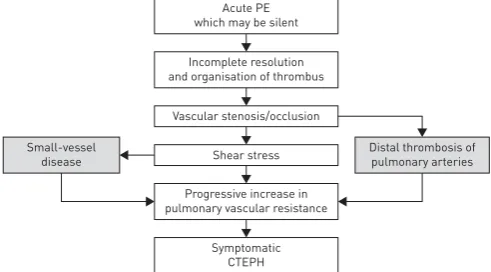
segmental), but also small-vessel disease, which plays an important role in the development and progression of the disease (figure 1) [4]. In this article we review recent advances in our understanding of the pathophysiology of CTEPH.
Relationship between PE and CTEPH
CTEPH is generally considered to be a rare and late complication of one or multiple episodes of acute PE that have not resolved despite ⩾3 months of curative anticoagulation. A large prospective international CTEPH registry has reported that 75% of included patients had a history of acute PE [5]. Nevertheless, this frequency is probably overestimated, because the diagnosis of acute PE was not well documented in a substantial number of cases and it is possible that the condition previously recorded as PE may have been the first manifestation of CTEPH. In fact, incomplete resolution of acute PE is not rare: some studies report that persistent lung perfusion defects are observed on scintigraphy in >50% of cases after 3 months of anticoagulation [6]. Fortunately, most of these patients do not present with symptomatic pulmonary hypertension.
CTEPH can develop several months or years after an acute PE (which may be silent), despite continuing anticoagulation, and in the absence of new symptoms or any new acute event [7–10].
Data from German and French pulmonary hypertension registries suggest an annual incidence of CTEPH of four and more than six per million adults per year, respectively [11] (G. Simonneau, Service de Pneumologie, Hôpital Bicêtre, Paris Sud University, Paris, France; unpublished data); this corresponds to
∼300 patients with newly diagnosed CTEPH per year in France. The incidence of CTEPH after acute PE has not yet been clearly established. In published prospective studies with the diagnosis confirmed by right heart catheterisation (RHC) the incidence of CTEPH after symptomatic acute PE is reported to range from 0.4% to 6.2% (online supplementary table S1) [7, 10, 12–22], giving a pooled incidence of 3.4% (95% CI 2.1−4.4%). Considering that ∼30 000 acute PE cases are diagnosed each year in France [23], a CTEPH incidence of 3.4% would lead to 1000 new CTEPH cases a year; far more than is actually observed. Most of these studies probably overestimated the incidence of CTEPH after an acute PE. One reason for this overestimation is that many patients had pre-existing, undiagnosed CTEPH at the time of the index PE, as we observe very frequently in our daily practice. Therefore, in reality, the incidences reported in these studies are a mix of incident and prevalent cases. In most studies, CTEPH was diagnosed a few months after the index PE, which is surprising because generally CTEPH develops after a“honeymoon period”of several years without any symptoms. Only one study, by GUERIN et al. [19], has addressed this issue
properly. 146 patients with acute PE were treated with curative anticoagulation. During a median follow-up of 26 months, eight of the 146 patients had suspected CTEPH because of persistent dyspnoea and abnormal echocardiographic findings, and CTEPH was confirmed by RHC in seven patients (4.8%, 95% CI 2.3−9.6%). However, at the time of the index acute PE, only two patients had systolic pulmonary arterial pressure (sPAP) <50 mmHg. In the remaining five patients, sPAP ranged from 62 to 102 mmHg;
Small-vessel disease
Residual PH arteriopathy Venous/capillary
disease Chronic
obstruction
Right heart function Inflammation
Infection
Blood (thrombosis) CTEPH
pathophysiology
this level of sPAP is not compatible with a first acute PE because the nonadapted right ventricle (RV) cannot generate such high pressures. It is therefore likely that CTEPH was present at the time of the index acute PE in these five patients. This suggestion was confirmed by review of their initial multidetector computed tomography scans by a senior radiologist, as all patients with confirmed CTEPH during follow-up had at least two signs of the condition at initial presentation. Thus, the cumulative incidence of CTEPH after acute PE in the GUERINet al.study was not 4.8%, but at most∼1.5% (two out of 146). This
would give an estimated rate of 450 new CTEPH cases per year in France, which is comparable with the 300 cases per year reported in the French registry. In view of the low incidence of CTEPH after acute PE, systematic ventilation/perfusion lung scanning to detect the presence of CTEPH in the follow-up of acute PE is not recommended.
Thrombus nonresolution in CTEPH
In most patients with PE, significant resolution of the embolus occurs, with subsequent restoration of blood flow and normalisation of haemodynamic parameters [24]. However, in a small subset of patients, a residual organised clot remains attached to pulmonary vessel walls. Why only a minority of patients fail to resolve fresh thrombi and develop CTEPH after an acute PE remains a mystery. Pathological specimens in acute PE and CTEPH are completely different: in acute PE, the fresh clots are red, easily detached from the pulmonary artery wall and consist mainly of red cells and platelets in a fibrin mesh. In CTEPH, the chronic clots are yellow, highly adherent to the pulmonary vascular wall, and contain collagen, elastin, inflammatory cells, re-canalisation vessels and, more rarely, calcification [25]. Organisation and fibrosis of this residual thrombotic material (described as “bands and webs” on pulmonary angiography) impairs blood flow, and ultimately leads to the development of CTEPH (figure 2) [2, 24]. Various factors have been suspected to underlie the failure of thrombus resolution; some of these factors are discussed below.
Clinical conditions predisposing to CTEPH
Several features of VTE appear to predispose individuals to poor thrombus resolution and subsequent development of CTEPH. For example, large pulmonary emboli appear to carry a higher risk of progression to CTEPH [2, 24], perhaps because the lytic system lacks the capacity to deal with the clot or is prevented from reaching and dissolving a large embolus sufficiently. Other features that appear to increase the risk of progression to CTEPH include recurrent pulmonary emboli and insufficient anticoagulation [2, 10]. However, these factors cannot explain the development of CTEPH in most patients, and other mechanisms must be involved.
An increased risk of CTEPH has been linked with numerous other factors, such as underlying autoimmune and haematological disorders [26] and comorbidities, as multiple comorbidities are present more frequently in patients with CTEPH than in those with pulmonary arterial hypertension (PAH) [27]. In a study comparing 433 patients with CTEPH against 254 patients with other nonthromboembolic forms of pulmonary hypertension, ventriculo-atrial shunts and infected pacemaker leads, splenectomy, prior VTE ( particularly recurrent VTE), non-O blood group, presence of lupus anticoagulant/ antiphospholipid antibodies, thyroid replacement therapy and a history of malignancy were all identified as carrying an increased risk of CTEPH [27].
Cancer
Patients with cancer have an increased risk of thromboembolic events, resulting from various mechanisms including activation of the fibrinolytic and coagulation systems, acute-phase reactions, inflammation and cytokine production [28]. Findings from a European database involving 687 patients with CTEPH (n=433)
FIGURE 2Natural history of chronic thromboembolic pulmonary hyper-tension (CTEPH). PE: pulmonary em-bolism. Reproduced and modified from [2] with permission.
Acute PE which may be silent
Incomplete resolution and organisation of thrombus
Progressive increase in pulmonary vascular resistance Small-vessel
disease
Distal thrombosis of pulmonary arteries Shear stress
and non-thromboembolic pulmonary hypertension (n=254) support an association between a history of malignancy and CTEPH (OR 3.76, 95% CI 1.47–10.43; p=0.005) [27]. The authors suggested that the evidence is sufficiently robust to warrant investigation for CTEPH in patients with a history of cancer who develop pulmonary hypertension.
Inflammation and infection
There appears to be an inflammatory component to CTEPH development, with higher levels of C-reactive protein (CRP) seen in patients compared with healthy controls, as well as a significant reduction in CRP after pulmonary endarterectomy (PEA) [29]. However, elevated CRP is not specific to CTEPH, as levels were also elevated in patients with PAH. More recent results confirmed that CRP, as well as interleukin
(IL)-10, monocyte chemotactic protein-1, macrophage inflammatory protein-1α and matrix
metalloproteinase (MMP)-9 were significantly elevated in patients with CTEPH [30]. Furthermore, surgical samples from patients who had undergone PEA contained numerous macrophages, lymphocytes and neutrophils, with correlations between CRP and neutrophil accumulation, and between MMP-9 and macrophage accumulation. In a prospective analysis of serum from eight patients with CTEPH, levels of IL-6, IL-8, interferon-γ-induced protein (IP)-10, monokine induced by interferon-γ and macrophage inflammatory protein-1αwere significantly elevated compared with age- and sex-matched healthy controls [31]. In patients with CTEPH, but not those with idiopathic PAH, levels of IP-10 (associated with fibroblast migration and activation) were negatively correlated with exercise capacity, cardiac output and cardiac index, while levels of IL-6 were positively correlated with pulmonary vascular resistance (PVR), right atrial pressure and levels of N-terminal prohormone of brain natriuretic peptide. Another inflammatory marker investigated for a connection to CTEPH is tumour necrosis factor-α: levels are elevated in patients with CTEPH compared with controls, and are reduced after PEA [32].
The presence of chronic infection (e.g. Staphylococcus aureus) has been identified in patients with CTEPH [33], although its relevance is unclear [34]. One study found staphylococcal DNA in six out of seven thromboemboli harvested during PEA from CTEPH patients with ventriculo-atrial shunts [33]. The authors suggested that thrombus infection was a trigger for the development of CTEPH. In a mouse model of intravenous thrombosis, staphylococcal infection delayed thrombus resolution in parallel with upregulation of transforming growth factor-βand connective tissue growth factor [33]. In a retrospective study of patients with CTEPH (n=433) and non-thromboembolic pulmonary hypertension (n=254), the presence of a ventriculo-atrial shunt or infected pacemaker was a significant risk factor for CTEPH development (OR 76.40, 95% CI 7.67–10 351; p<0.001) [27].
Biological and genetic risk factors for thrombus nonresolution
It has been suspected that patients with thrombus nonresolution could have a hypercoagulability state due to biological abnormalities. Interestingly, the classical hereditary thrombotic risk factors, e.g. protein C, protein S and antithrombin deficiencies, and mutations of factor V and II, are no more frequent in patients with CTEPH than in healthy control populations [35]. In this prospective study, only the frequency of antiphospholipid antibodies and lupus anticoagulant was higher in patients with CTEPH than in patients with idiopathic PAH. In a prospective case–control study, increased levels of clotting factor VIII were identified in 41% of patients with CTEPH, which was significantly higher than seen in both healthy controls and patients with non-thromboembolic PAH [36]. In the same study, levels of von Willebrand factor, an adhesive glycoprotein that stabilises and activates factor VIII, were significantly increased in patients with CTEPH compared with healthy controls and patients with PAH, with the increase persisting in patients who had undergone PEA.
ADAMTS13 (a disintegrin and metalloproteinase with thrombospondin type 1 motif, member 13), also known as von Willebrand factor-cleaving protease, regulates the size of von Willebrand factor and plays a fundamental role in haemostasis. Severe deficiency of ADAMTS13 causes thrombotic thrombocytopenic purpura [37]. In a case–control study, an excess of rare and low-frequency coding single-nucleotide variants of ADAMTS13 were found in patients with deep vein thrombosis compared with matched controls; in addition, these patients showed relatively lower plasma levels of ADAMTS13 activity [38].
Other studies have described increased tissue factor gene expression [46], an increased frequency of mutations known to be associated with PAH [47] and differentially expressed microRNAs [48, 49].
Blood groups
CTEPH is more common in patients with blood groups A, B and AB. In one study, 77% of patients with CTEPH had non-O blood group compared with 58% of patients with PAH ( p=0.003) [36]. A European registry suggested that non-O blood group was a significant predictor for the diagnosis of CTEPH (OR 2.09, 95% CI 1.12–3.94; p=0.019) [27]. The ABO locus is a susceptibility locus for VTE and non-O carriers have a higher risk for VTE than O carriers [50].
Fibrinogen and fibrinolytic abnormalities in CTEPH
Patients with CTEPH appear to have a high prevalence of abnormal fibrinogen molecules in the blood [51], such as fibrinogen Aα-Thr312Ala [52, 53]. This mutation leads to a modified fibrin structure in clots, including increased cross-linking ofα-chains [34]. Other heterozygous polymorphisms identified in patients with CTEPH include theβ-chain mutations P235L/γR375W, P235L/γY114H and P235L, and the α-chain mutations L69H and R554H [51]. More recently, theβ15–42fragment of the fibrinogen E chain
has been shown to delay thrombus resolutionin vivo[2]. The common feature of each fibrin abnormality so far detected in patients with CTEPH is that they are able to resist physiological thrombolysis, and thus affect thrombus resolution [2, 54]. For example, a study comparing fibrinogen from patients with CTEPH and healthy controls found that fibrin from patients was more resistant to plasmin-mediated lysis compared with controls [55]. The authors suggest that this is a result of alterations in the structure of fibrin and/or fibrinogen that affect accessibility to plasmin cleavage sites. In addition, there was a persistence of fibrin structural motifs (e.g. the N-terminus of the β-chain) within the pulmonary vasculature, which the authors speculate may be involved in progression from acute PE to CTEPH [55]. In the study by OLMANet al. [56] neither an increase in type 1 plasminogen activator inhibitor nor a blunted
response of tissue type plasminogen activator were observed, indicating that the plasma fibrinolytic system is intact in CTEPH. A recent study investigated the potential role of thrombin-activatable fibrinolysis inhibitor (TAFI), a plasma carboxypeptidase inhibitor produced by the liver that inhibits fibrinolysis, in the pathology of CTEPH [57]. Plasma TAFI levels and the release of TAFI from platelets were significantly higher in patients with CTEPH than in patients with PAH or controls. Moreover, TAFI levels were significantly correlated with resistance to clot lysis in a whole-blood assay and they remained unchanged after balloon pulmonary angioplasty. These observations suggest a significant role for TAFI in the pathophysiology of CTEPH.
Platelet function in CTEPH
The observation that platelet-activating conditions such as thyroid hormone replacement therapy and splenectomy are risk factors for CTEPH suggests a role for platelets in its genesis [5, 27]. Studies in a mouse model of impaired thrombus resolution suggested that the initial increase in thrombus volume after splenectomy is due to platelet activation [58]. The same study reported an increase in platelet microparticles in splenectomised versus non-splenectomised CTEPH patients. Compared with controls, patients with CTEPH have a decreased platelet count, higher mean platelet volume, increased spontaneous platelet aggregation and decreased platelet aggregation in response to agonists [59]. These observations suggest a prothrombotic state with higher platelet turnover in patients with CTEPH. YAOITA et al. [60]
reported that platelets from patients with CTEPH or PAH were activated compared with non-pulmonary hypertension controls when measured by surface expression on P-selectin, PAC-1 binding and the GTP-bound GTPase RhoA, which is involved in platelet aggregation. Surgical materials extracted by PEA from patients with CTEPH contains increased levels of platelet factor 4, which is released by platelets at sites of injury [61]. These observations suggest a role for platelet dysfunction in the pathology of CTEPH.
Impaired angiogenesis
Platelet endothelial cell adhesion molecule-1 and thrombosis
Platelet endothelial cell adhesion molecule (PECAM)-1 is a glycopeptide receptor expressed on platelets, endothelial cells and many other cell types. It is involved in leukocyte transmigration and responses to inflammatory stimuli, key components of venous thrombus resolution [63]. In a mouse model mimicking human deep vein thrombosis, PECAM-1 deficiency led to significantly larger thrombi and misguided thrombus resolution [64]. Furthermore, human unresolved deep vein thrombosis specimens showed accumulation of the cleaved form of PECAM-1, and patients with delayed thrombus resolution had significantly increased plasma levels of soluble cleaved PECAM-1 compared with those whose thrombi resolved [64]. White and red thrombi from patients with CTEPH show reduced PECAM-1 expression compared with unthrombosed vessels, implicating PECAM-1 deficiency in the pathology of CTEPH [62].
Small-vessel disease in CTEPH
Histological and mechanistic aspectsThe occlusion of proximal (main, lobar and segmental) pulmonary arteries by organised fibrotic clots is the initial trigger for developing CTEPH. However, it is not the only pulmonary hypertension mechanism in this setting. There is growing evidence that in addition to mechanical obstruction of proximal arteries, some patients develop a more or less severe pulmonary microvasculopathy (small-vessel disease), first described by MOSERand BLOOR[65] in lung tissue obtained by biopsy or at autopsy. Pathological studies by
MOSERand BLOORand other authors have disclosed a full range of pulmonary hypertensive lesions similar
to those observed in idiopathic PAH, including intimal thickening and remodelling of pulmonary resistance vessels, eccentric intimal fibrosis, intimal fibromuscular proliferation and plexiform lesions [2, 65, 66]. This vascular remodelling affects the wall of distal muscular pulmonary arteries (0.1−0.5 mm in diameter), and may even reach arterioles and venules of <0.1 mm in diameter. These changes are classically explained by redistribution of the pulmonary flow in nonoccluded pulmonary arteries exposed to high pressure and shear stress, leading to endothelial dysfunction, a progressive increase in PVR and ultimately to symptomatic CTEPH. However, this microvasculopathy is observed not only in lung areas served by nonoccluded proximal pulmonary arteries, but also distally to pulmonary arteries occluded by fibrotic material. This makes it unlikely that redistribution of the flow in the pulmonary arterial bed alone can explain the remodelling. DORFMÜLLER et al. [67] detected large anastomoses between the systemic
Distal thrombosis of PA with partial
recanalisation
PAH-like lesions of muscularised
arterioles
Venular fibrosis and muscularisation-
like PVOD
Capillary lesion-like haemangiomatosis Thromboembolic
material
Hypertrophic bronchial artery Bronchus
Pulmonary vein
Hypertrophic vasa vasorum
Pulmonary artery
circulation and pulmonary arterial circulation (via hypertrophic bronchial arteries and vasa vasorum) in patients with CTEPH. It has been speculated that pre-existing anastomoses are opened by the pressure gradient between bronchial arteries and postobstruction pulmonary arteries. This mechanism may help to maintain perfusion and support ischaemic tissue downstream of a proximal obstruction, but the exposure of the pulmonary artery circulation to the high-pressure systemic circulation may induce pulmonary arterial vascular remodelling in some patients, especially distal to chronic thromboembolic obstruction. In the aforementioned study, DORFMÜLLERet al. observed important reactive, nonthrombotic microvascular
remodelling in obstructed territories. Ink injection experiments in humans with CTEPH and an experimental piglet model of CTEPH revealed that small-vessel disease was not confined to precapillary arterioles, but additionally concerned postcapillary venules and small veins. In fact, anastomoses are known to exist between the systemic circulation and both pulmonary capillaries and pulmonary veins, probably leading to lesions that may be similar to capillary haemangiomatosis and pulmonary veno-occlusive disease (figure 3) [67]. Work is still needed to explain fully how small-vessel disease develops and progresses.
Small-vessel disease in CTEPH may also consist of distal thrombosis, in rare cases. The lesions can be diffuse, probably when small pulmonary arterioles distal to more proximal complete obstructions are not maintained open because bronchial arteries and anastomoses fail to develop. In addition to typical findings of CTEPH, pulmonary angiography in these patients reveals a special aspect of diffuse, poor subpleural perfusion in the capillary phase (P. Dartevelle, French National Reference Centre of Pulmonary Hypertension, Hôpital Bicêtre, Le Kremlin-Bicêtre, France; personal communication) (figure 4).
Molecular mechanisms of small-vessel disease
Recent evidence suggests the involvement of the nitric oxide (NO)–soluble guanylate cyclase (sGC)–cyclic guanosine monophosphate (cGMP) pathway in the pathophysiology of CTEPH. NO produced by vascular endothelium inhibits platelet aggregation and growth of smooth muscle cells [68, 69]. NO also activates sGC to synthesise cGMP, a second messenger with many actions including smooth muscle relaxation. Plasma levels of asymmetric dimethylarginine, an inhibitor of NO synthase, are increased in patients with CTEPHversuscontrols [70], and reduced endogenous NO levels have been found in patients with PAH and CTEPH [71]. The vascular remodelling in CTEPH may indicate dysfunction in antiproliferative mechanisms, including the NO pathway. The clinical and haemodynamic improvements seen with the sGC stimulator riociguat in patients with CTEPH ineligible for PEA or with persistent recurrent pulmonary hypertension after surgery also suggest an important role for the NO–sGC–cGMP pathway in the pathology of the condition [72–74].
Levels of endothelin (ET)-1 are elevated in patients with CTEPH and in animal models of the condition [75–79]. Recent evidence suggests a potential role for ET-1 in smooth muscle cell proliferation within the chronic clot in CTEPH and in small-vessel disease [80]. Levels of vascular endothelial growth factor A are significantly decreased after PEA in patients with CTEPH [81]. Increased levels of angiopoietin-1, a signalling molecule involved in angiogenesis and smooth muscle cell proliferation, have been identified in the lungs of patients with CTEPH [82] and high pre-operative levels of angiopoietin-2 are correlated with worse outcomes for PEA [83]. An initial study in a model of CTEPH in piglets [84] has suggested that small-vessel disease is associated with changes in ET-1 and IL-6 gene expression [85].
Clinical implications of small-vessel disease
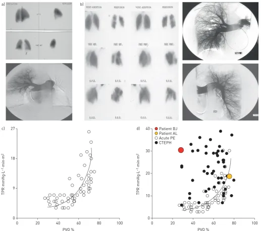
The progressive development of these microvascular changes could explain why some patients with CTEPH deteriorate even in the absence of recurrent pulmonary emboli. The severity of this small-vessel disease is highly variable between individual patients. The presence of extensive small-vessel disease in a patient should be suspected when the extent of the mechanical obstruction by proximal fibrotic clots (e.g.
modest obstruction based on assessment using perfusion lung scan and/or pulmonary angiography) does not correlate with the dramatic increase in PVR [86] (examples are shown in figure 5). These patients are at higher risk of postoperative mortality [87, 88], as small-vessel disease is not amenable to PEA and it contributes to persistent pulmonary hypertension after PEA, as does incomplete removal of proximal thrombotic material after surgery [89–92]. Thus, for optimal disease management it is essential to detect and treat CTEPH before small-vessel remodelling occurs. The adverse effect of extensive small-vessel disease on patient prognosis after surgery was confirmed in a prospective study of 26 patients with CTEPH [91]. Upstream resistance was assessed pre-operatively by analysis of the pulmonary occlusion waveform on RHC. Upstream resistance correlated significantly with postoperative total pulmonary resistance index and mean pulmonary arterial pressure (PAP), and all four postoperative deaths during the study were in patients with pre-operative upstream resistance <60%.
Small-vessel disease arising from distal thrombosis (detected as described earlier) also tends to be severe and associated with a poor outcome after PEA [93]. Interestingly, the presence of dilatation of the bronchial arteries and anastomosis within the pulmonary arterial circulation, frequently seen in CTEPH and preventing this distal thrombosis, is associated with good survival and major haemodynamic improvement after PEA [94].
As well as being not amenable to PEA, small-vessel disease in patients with CTEPH is not accessible to balloon pulmonary angioplasty, which may explain the persistence of pulmonary hypertension after this interventional procedure in these patients. Organised fibrotic clots narrowing distal pulmonary arteries can be treated by balloon pulmonary angioplasty using balloons 1–10 mm in diameter.
RV dysfunction and failure in CTEPH
CTEPH is characterised by a chronic increase in RV afterload and wall stress. Initially, these burdens result in RV hypertrophy characterised by increases in RV wall thickness and cell size through the addition of sarcomeres [95, 96]. This process is the result of the intrinsic ability of cardiac muscle cells to sense and respond to mechanical load [96]. At first, the increase in RV wall thickness results in decreased wall stress and improved pumping effectiveness by unloading of the individual muscle fibres [96], and RV function more closely resembles the left ventricle [97]. These changes are referred to as “adaptive” remodelling. This process is not limited to patients with CTEPH, but also occurs in patients with PAP <25 mmHg and chronic thromboembolic disease (CTED) [97]. This was demonstrated in a study using a conductance catheter, in which differences between patients with CTED, CTEPH and controls were observed in pressure–volume loop morphology, most notably during systolic ejection [97]. In patients with CTED, these changes resulted from the elevated RV afterload that develops exclusive of haemodynamic definition of pulmonary hypertension. In addition, the RV relaxed more slowly in patients with CTED and CTEPH compared with controls. The authors attributed this characteristic to a chronic elevation in RV afterload and lower arterial compliance. In a comparison of patients with idiopathic PAH, proximal CTEPH ( pre-and postsurgery) pre-and distal (inoperable) CTEPH, those with CTEPH had a significantly shorter time constant of the pulmonary circulation than those with PAH [98]. This is consistent with the suggestion from a small study that patients with CTEPH have lower RV contractility and fractional area change than patients with idiopathic PAH or Eisenmenger’s syndrome (although the patients with CTEPH (mean age 50.8 years) were older than the comparator groups (mean age 42.2 years and 41.2 years for idiopathic PAH and Einsenmenger’s syndrome, respectively)) [99].
RV dilatation, reduced RV contractile force, diastolic dysfunction and myocardial fibrosis [96]. RV dilatation increases wall tension, which increases oxygen demand and decreases perfusion, leading to a cycle of further compromised contractility and dilatation. Decreased RV stroke volume, reduced pulmonary flow and underfilling of the left ventricle lead to systemic hypotension and worsening of RV coronary perfusion. Progressive dysfunction ensues, ultimately resulting in RV failure, which is the main cause of death in CTEPH [95, 96]. The time course of RV failure in response to a pressure overload varies greatly between patients, possibly because of variations in pressure, load, phenotype and neurohumoral overdrive [95, 96]. While the progression to RV failure is clearly much slower, the sequence of events
a) b)
c) 27
18
TPR mmHg·L
–1·min·m
2
9
0
0 20 40
PVO %
60 80 100
d)
20 30
40 Patient BJ Patient AL
TPR mmHg·L
–1·min·m
2
10
0
0 20 40
PVO %
60 80 100
CTEPH Acute PE
resembles that occurring in acute PE in many aspects. The two most important differences are the progressive increase in RV afterload induced by ongoing remodelling of pulmonary arterioles and the possibility of adaptive hypertrophy of RV myocardium, both absent in acute PE. Whether increased RV mass is an independent risk factor for the subsequent development of progressive RV failure in CTEPH is unclear [96].
RV diastolic dysfunction, including increased stiffness, impaired filling and prolonged isovolumic relaxation, diffuse myocardial fibrosis and sarcomeric stiffening, may occur relatively early in the disease as patients with pulmonary hypertension can exhibit impaired RV diastolic function while RV systolic function is relatively preserved [96, 100, 101].
In animal models of pulmonary hypertension, disturbed angiogenesis and capillary rarefaction occur in dysfunctional RV hypertrophic tissue, and RV myocardial metabolism switches from mitochondria-based fatty acid oxidation to glycolysis [96]. Compared with controls, patients with pulmonary hypertension exhibit reduced RV contractility in response to exercise and a loss of pulmonary vascular distensibility [96, 102].
Animal models of pulmonary hypertension have indicated a significant increase in tissue fibrosis in maladaptive RV hypertrophy, relative to levels seen in adaptive hypertrophy [96, 103, 104]. The fibrosis seen in maladaptive hypertrophy is a pathological process resulting from upregulation and interaction between growth factors (including transforming growth factor-β and connective tissue growth factor), hormones and matrix metalloproteinases [96].
PEA improves RV function in patients with CTEPH, indicating that pathological remodelling can be reversed [105–108]. The functional improvements take time to develop, in contrast to the haemodynamic changes, which are immediate [105–108]. The reduction in systolic RV wall stress after PEA is a key factor in achieving resynchronisation of left ventricle and RV peak strains [106]. Balloon pulmonary angioplasty may also improve RV function [109–111], but it is less well established than PEA [112].
The status of the RV in patients who have undergone successful PEA or balloon pulmonary angioplasty for the treatment of CTEPH is unclear. Should such an unloaded RV be considered as“preconditioned”and therefore be expected to better sustain a potential increase in afterload, or, conversely, is it irreversibly damaged and dysfunctional? Is there a“point of no return”for the RV somewhere along the sequence of events induced by CTEPH? Clarification of those issues could affect our strategy and timing of therapeutic interventions.
Summary and conclusions
The current understanding of CTEPH has moved beyond a chronic obstruction caused by unresolved thrombotic material and the resultant RV dysfunction to encompass a complex disease comprising proximal chronic obstruction by fibrotic clots, small-vessel disease and remodelling throughout the pulmonary vascular bed [4]. The processes by which residual thrombosis persists in patients with PE, and by which such residual thrombi lead to CTEPH, are not fully understood, but inflammation and infection are believed to play a part. The degree of small-vessel disease has a substantial impact on the severity of CTEPH and postsurgical outcomes, and thus assessment of small-vessel disease can assist in ensuring optimal management for patients. The molecular processes underlying small-vessel disease are not completely understood, but insights gained to date have given clues about the potential mechanisms of benefit of medical therapies. Further research into small-vessel disease in patients with CTEPH could help to identify those who will benefit most from the three management strategies now available (PEA, medical therapy and balloon pulmonary angioplasty) and lead to the discovery of new treatment modalities preventing the progression to irreversible RV failure. Clarification of those issues could improve our management strategies and timing of therapeutic interventions.
Acknowledgements
Editorial assistance was provided by Adelphi Communications Ltd (Bollington, UK), supported by Bayer AG (Berlin, Germany).
References
1 Simonneau G, Gatzoulis MA, Adatia I,et al.Updated clinical classification of pulmonary hypertension.J Am Coll Cardiol2013; 62: D34–D41.
2 Lang IM, Dorfmüller P, Vonk Noordegraaf A. The pathobiology of chronic thromboembolic pulmonary hypertension.Ann Am Thorac Soc2016; 13: Suppl. 3, S215–S221.
3 Fedullo P, Kerr KM, Kim NH,et al.Chronic thromboembolic pulmonary hypertension.Am J Respir Crit Care Med2011; 183: 1605–1613.
4 Kim NH. Group 4 pulmonary hypertension: chronic thromboembolic pulmonary hypertension: epidemiology, pathophysiology, and treatment.Cardiol Clin2016; 34: 435–441.
6 Wartski M, Collignon MA. Incomplete recovery of lung perfusion after 3 months in patients with acute pulmonary embolism treated with antithrombotic agents. THESEE Study Group. Tinzaparin ou Heparin Standard: Evaluation dans l’Embolie Pulmonaire Study.J Nucl Med2000; 41: 1043–1048.
7 Klok FA, van Kralingen KW, van Dijk AP,et al. Prospective cardiopulmonary screening program to detect chronic thromboembolic pulmonary hypertension in patients after acute pulmonary embolism.Haematologica 2010; 95: 970–975.
8 Lang I. Chronic thromboembolic pulmonary hypertension: a distinct disease entity.Eur Respir Rev 2015; 24: 246–252.
9 Olsson KM, Meyer B, Hinrichs J,et al. Chronic thromboembolic pulmonary hypertension.Dtsch Arztebl Int 2014; 111: 856–862.
10 Pengo V, Lensing AW, Prins MH,et al. Incidence of chronic thromboembolic pulmonary hypertension after pulmonary embolism.N Engl J Med2004; 350: 2257–2264.
11 Hoeper MM, Humbert M, Souza R,et al.A global view of pulmonary hypertension.Lancet Respir Med2016; 4: 306–322.
12 Becattini C, Agnelli G, Pesavento R,et al.Incidence of chronic thromboembolic pulmonary hypertension after a first episode of pulmonary embolism.Chest2006; 130: 172–175.
13 Martí D, Gómez V, Escobar C,et al.Incidencia de hipertensión pulmonar tromboembólica crónica sintomática y asintomática. [Incidence of symptomatic and asymptomatic chronic thromboembolic pulmonary hypertension.] Arch Bronconeumol2010; 46: 628–633.
14 Poli D, Grifoni E, Antonucci E, et al. Incidence of recurrent venous thromboembolism and of chronic thromboembolic pulmonary hypertension in patients after a first episode of pulmonary embolism.J Thromb Thrombolysis2010; 30: 294–299.
15 Surie S, Gibson NS, Gerdes VE,et al.Active search for chronic thromboembolic pulmonary hypertension does not appear indicated after acute pulmonary embolism.Thromb Res2010; 125: e202–e205.
16 Berghaus TM, Barac M, von Scheidt W,et al.Echocardiographic evaluation for pulmonary hypertension after recurrent pulmonary embolism.Thromb Res2011; 128: e144–e147.
17 Held M, Hesse A, Gött F,et al.A symptom-related monitoring program following pulmonary embolism for the early detection of CTEPH: a prospective observational registry study.BMC Pulm Med2014; 14: 141.
18 Giuliani L, Piccinino C, D’Armini MA,et al.Prevalence of undiagnosed chronic thromboembolic pulmonary hypertension after pulmonary embolism.Blood Coagul Fibrinolysis2014; 25: 649–653.
19 Guérin L, Couturaud F, Parent F,et al.Prevalence of chronic thromboembolic pulmonary hypertension after acute pulmonary embolism.Thromb Haemost2014; 112: 598–605.
20 Kayaalp I, Varol Y, Çimen P,et al.The incidence of chronic thromboembolic pulmonary hypertension secondary to acute pulmonary thromboembolism.Tuberk Toraks2014; 62: 199–206.
21 Vavera Z, Vojacek J, Pudil R,et al.Chronic thromboembolic pulmonary hypertension after the first episode of pulmonary embolism? How often? Biomed Pap Med Fac Univ Palacky Olomouc Czech Repub 2016; 160: 125–129.
22 Klok FA, Tesche C, Rappold L,et al.External validation of a simple non-invasive algorithm to rule out chronic thromboembolic pulmonary hypertension after acute pulmonary embolism.Thromb Res2015; 135: 796–801. 23 Oger E. Incidence of venous thromboembolism: a community-based study in Western France.Thromb Haemost
2000; 83: 657–660.
24 Banks DA, Pretorius GV, Kerr KM, et al. Pulmonary endarterectomy: part I. Pathophysiology, clinical manifestations, and diagnostic evaluation of chronic thromboembolic pulmonary hypertension. Semin Cardiothorac Vasc Anesth2014; 18: 319–330.
25 Wagenvoort CA. Pathology of pulmonary thromboembolism.Chest1995; 107: 10S–17S.
26 Blauwet LA, Edwards WD, Tazelaar HD,et al.Surgical pathology of pulmonary thromboendarterectomy: a study of 54 cases from 1990 to 2001.Hum Pathol2003; 34: 1290–1298.
27 Bonderman D, Wilkens H, Wakounig S,et al.Risk factors for chronic thromboembolic pulmonary hypertension. Eur Respir J2009; 33: 325–331.
28 Karimi M, Cohan N. Cancer-associated thrombosis.Open Cardiovasc Med J2010; 4: 78–82.
29 Quarck R, Nawrot T, Meyns B,et al. C-reactive protein: a new predictor of adverse outcome in pulmonary arterial hypertension.J Am Coll Cardiol2009; 53: 1211–1218.
30 Quarck R, Wynants M, Verbeken E, et al. Contribution of inflammation and impaired angiogenesis to the pathobiology of chronic thromboembolic pulmonary hypertension.Eur Respir J2015; 46: 431–443.
31 Zabini D, Heinemann A, Foris V, et al. Comprehensive analysis of inflammatory markers in chronic thromboembolic pulmonary hypertension patients.Eur Respir J2014; 44: 951–962.
32 Langer F, Schramm R, Bauer M,et al.Cytokine response to pulmonary thromboendarterectomy.Chest2004; 126: 135–141.
33 Bonderman D, Jakowitsch J, Redwan B,et al.Role for staphylococci in misguided thrombus resolution of chronic thromboembolic pulmonary hypertension.Arterioscler Thromb Vasc Biol2008; 28: 678–684.
34 Toshner M, Pepke-Zaba J. Chronic thromboembolic pulmonary hypertension: time for research in pathophysiology to catch up with developments in treatment.F1000 Prime Rep2014; 6: 38.
35 Wolf M, Boyer-Neumann C, Parent F,et al.Thrombotic risk factors in pulmonary hypertension.Eur Respir J 2000; 15: 395–399.
36 Bonderman D, Turecek PL, Jakowitsch J,et al. High prevalence of elevated clotting factor VIII in chronic thromboembolic pulmonary hypertension.Thromb Haemost2003; 90: 372–376.
37 Sadler JE. Von Willebrand factor, ADAMTS13, and thrombotic thrombocytopenic purpura.Blood 2008; 112: 11–18.
38 Lotta LA, Tuana G, Yu J,et al.Next-generation sequencing study finds an excess of rare, coding single-nucleotide variants of ADAMTS13 in patients with deep vein thrombosis.J Thromb Haemost2013; 11: 1228–1239. 39 Gu S, Su P, Yan J,et al.Comparison of gene expression profiles and related pathways in chronic thromboembolic
40 Lindner J, Maruna P, Kunstyr J,et al. Hemodynamic instability after pulmonary endarterectomy for chronic thromboembolic pulmonary hypertension correlates with cytokine network hyperstimulation.Eur Surg Res2009; 43: 39–46.
41 Tanabe N, Amano S, Tatsumi K,et al.Angiotensin-converting enzyme gene polymorphisms and prognosis in chronic thromboembolic pulmonary hypertension.Circ J2006; 70: 1174–1179.
42 Chen Z, Nakajima T, Tanabe N,et al.Susceptibility to chronic thromboembolic pulmonary hypertension may be conferred bymiR-759 viaits targeted interaction with polymorphic fibrinogen alpha gene.Hum Genet2010; 128: 443–452.
43 Feng YX, Liu D, Sun ML,et al.BMPR2 germline mutation in chronic thromboembolic pulmonary hypertension. Lung2014; 192: 625–627.
44 Suntharalingam J, Machado RD, Sharples LD,et al.Demographic features, BMPR2 status and outcomes in distal chronic thromboembolic pulmonary hypertension.Thorax2007; 62: 617–622.
45 Ulrich S, Szamalek-Hoegel J, Hersberger M,et al. Sequence variants in BMPR2 and genes involved in the serotonin and nitric oxide pathways in idiopathic pulmonary arterial hypertension and chronic thromboembolic pulmonary hypertension: relation to clinical parameters and comparison with left heart disease.Respiration2010; 79: 279–287.
46 Yang M, Deng C, Wu D,et al.The role of mononuclear cell tissue factor and inflammatory cytokines in patients with chronic thromboembolic pulmonary hypertension.J Thromb Thrombolysis2016; 42: 38–45.
47 Xi Q, Liu Z, Zhao Z,et al. High frequency of pulmonary hypertension-causing gene mutation in Chinese patients with chronic thromboembolic pulmonary hypertension.PLoS One2016; 11: e0147396.
48 Guo L, Yang Y, Liu J,et al.Differentially expressed plasma microRNAs and the potential regulatory function of Let-7b in chronic thromboembolic pulmonary hypertension.PLoS One2014; 9: e101055.
49 Wang L, Guo LJ, Liu J,et al.MicroRNA expression profile of pulmonary artery smooth muscle cells and the effect of let-7d in chronic thromboembolic pulmonary hypertension.Pulm Circ2013; 3: 654–664.
50 Wu O, Bayoumi N, Vickers MA,et al. ABO(H) blood groups and vascular disease: a systematic review and meta-analysis.J Thromb Haemost2008; 6: 62–69.
51 Morris TA, Marsh JJ, Chiles PG,et al. High prevalence of dysfibrinogenemia among patients with chronic thromboembolic pulmonary hypertension.Blood2009; 114: 1929–1936.
52 Le Gal G, Delahousse B, Lacut K,et al.Fibrinogen Aα-Thr312Ala and factor XIII-A Val34Leu polymorphisms in idiopathic venous thromboembolism.Thromb Res2007; 121: 333–338.
53 Suntharalingam J, Goldsmith K, van Marion V,et al.Fibrinogen AαThr312Ala polymorphism is associated with chronic thromboembolic pulmonary hypertension.Eur Respir J2008; 31: 736–741.
54 Marsh JJ, Chiles PG, Liang NC, et al. Chronic thromboembolic pulmonary hypertension-associated dysfibrinogenemias exhibit disorganized fibrin structure.Thromb Res2013; 132: 729–734.
55 Morris TA, Marsh JJ, Chiles PG,et al.Fibrin derived from patients with chronic thromboembolic pulmonary hypertension is resistant to lysis.Am J Respir Crit Care Med2006; 173: 1270–1275.
56 Olman MA, Marsh JJ, Lang IM,et al.Endogenous fibrinolytic system in chronic large-vessel thromboembolic pulmonary hypertension.Circulation1992; 86: 1241–1248.
57 Yaoita N, Satoh K, Satoh T, et al. Thrombin-activatable fibrinolysis inhibitor in chronic thromboembolic pulmonary hypertension.Arterioscler Thromb Vasc Biol2016; 36: 1293–1301.
58 Frey MK, Alias S, Winter MP,et al.Splenectomy is modifying the vascular remodeling of thrombosis.J Am Heart Assoc2014; 3: e000772.
59 Remková A, Šimková I, Valkovičová T. Platelet abnormalities in chronic thromboembolic pulmonary hypertension.Int J Clin Exp Med2015; 8: 9700–9707.
60 Yaoita N, Shirakawa R, Fukumoto Y,et al.Platelets are highly activated in patients of chronic thromboembolic pulmonary hypertension.Arterioscler Thromb Vasc Biol2014; 34: 2486–2494.
61 Zabini D, Nagaraj C, Stacher E,et al.Angiostatic factors in the pulmonary endarterectomy material from chronic thromboembolic pulmonary hypertension patients cause endothelial dysfunction.PLoS One2012; 7: e43793. 62 Alias S, Redwan B, Panzenböck A, et al. Defective angiogenesis delays thrombus resolution: a potential
pathogenetic mechanism underlying chronic thromboembolic pulmonary hypertension.Arterioscler Thromb Vasc Biol2014; 34: 810–819.
63 Privratsky JR, Newman PJ. PECAM-1: regulator of endothelial junctional integrity.Cell Tissue Res2014; 355: 607–619. 64 Kellermair J, Redwan B, Alias S,et al.Platelet endothelial cell adhesion molecule 1 deficiency misguides venous
thrombus resolution.Blood2013; 122: 3376–3384.
65 Moser KM, Bloor CM. Pulmonary vascular lesions occurring in patients with chronic major vessel thromboembolic pulmonary hypertension.Chest1993; 103: 685–692.
66 Pietra GG, Capron F, Stewart S,et al.Pathologic assessment of vasculopathies in pulmonary hypertension.J Am Coll Cardiol2004; 43: 25S–32S.
67 Dorfmüller P, Günther S, Ghigna MR, et al. Microvascular disease in chronic thromboembolic pulmonary hypertension: a role for pulmonary veins and systemic vasculature.Eur Respir J2014; 44: 1275–1288.
68 Klinger JR, Abman SH, Gladwin MT. Nitric oxide deficiency and endothelial dysfunction in pulmonary arterial hypertension.Am J Respir Crit Care Med2013; 188: 639–646.
69 Tonelli AR, Haserodt S, Aytekin M,et al.Nitric oxide deficiency in pulmonary hypertension: pathobiology and implications for therapy.Pulm Circ2013; 3: 20–30.
70 Skoro-Sajer N, Mittermayer F, Panzenboeck A, et al. Asymmetric dimethylarginine is increased in chronic thromboembolic pulmonary hypertension.Am J Respir Crit Care Med2007; 176: 1154–1160.
71 Stasch JP, Evgenov OV. Soluble guanylate cyclase stimulators in pulmonary hypertension.Handb Exp Pharmacol 2013; 218: 279–313.
72 Ghofrani HA, D’Armini AM, Grimminger F, et al. Riociguat for the treatment of chronic thromboembolic pulmonary hypertension.N Engl J Med2013; 369: 319–329.
74 Simonneau G, D’Armini AM, Ghofrani HA,et al.Predictors of long-term outcomes in patients treated with riociguat for chronic thromboembolic pulmonary hypertension: data from the CHEST-2 open-label, randomised, long-term extension trial.Lancet Respir Med2016; 4: 372–380.
75 Reesink HJ, Meijer RC, Lutter R, et al. Hemodynamic and clinical correlates of endothelin-1 in chronic thromboembolic pulmonary hypertension.Circ J2006; 70: 1058–1063.
76 Kim H, Yung GL, Marsh JJ, et al. Pulmonary vascular remodeling distal to pulmonary artery ligation is accompanied by upregulation of endothelin receptors and nitric oxide synthase.Exp Lung Res2000; 26: 287–301. 77 Kim H, Yung GL, Marsh JJ,et al.Endothelin mediates pulmonary vascular remodelling in a canine model of
chronic embolic pulmonary hypertension.Eur Respir J2000; 15: 640–648.
78 Langer F, Bauer M, Tscholl D, et al. Circulating big endothelin-1: an active role in pulmonary thromboendarterectomy?J Thorac Cardiovasc Surg2005; 130: 1342–1347.
79 Bauer M, Wilkens HC, Langer F,et al.Selective upregulation of endothelin B receptor gene expression in severe pulmonary hypertension.Circulation2002; 105: 1034–1036.
80 Southwood M, MacKenzie Ross RV, Kuc RE, et al. Endothelin ETA receptors predominate in chronic
thromboembolic pulmonary hypertension.Life Sci2016; 159: 104–110.
81 Southwood M, Hadinnapola C, Mosely E,et al.Vascular endothelial cell growth factor-a (VEGF-a) signalling and neovascularisation of pulmonary endarterectomy material in chronic thromboembolic pulmonary hypertension (CTEPH).Thorax2014; 69: Suppl. 2, Abstract S37.
82 Hoeper MM, Mayer E, Simonneau G,et al.Chronic thromboembolic pulmonary hypertension.Circulation2006; 113: 2011–2020.
83 Bates DM, Fernandes TM, Chiles PG, et al. Preoperative plasma angiopoietin-2 levels may predict adverse outcomes following pulmonary thromboendarterectomy.Am J Respir Crit Care Med2015; 191: A5411.
84 Noly PE, Guihaire J, Coblence M,et al.Chronic thromboembolic pulmonary hypertension and assessment of right ventricular function in the piglet.J Vis Exp2015; 34: e53133.
85 Boulate D, Perros F, Dorfmüller P, et al. Pulmonary microvascular lesions regress in reperfused chronic thromboembolic pulmonary hypertension.J Heart Lung Transplant2015; 34: 457–467.
86 Azarian R, Wartski M, Collignon MA,et al. Lung perfusion scans and hemodynamics in acute and chronic pulmonary embolism.J Nucl Med1997; 38: 980–983.
87 Thistlethwaite PA, Mo M, Madani MM,et al. Operative classification of thromboembolic disease determines outcome after pulmonary endarterectomy.J Thorac Cardiovasc Surg2002; 124: 1203–1211.
88 Dartevelle P, Fadel E, Mussot S,et al.Chronic thromboembolic pulmonary hypertension.Eur Respir J2004; 23: 637–648.
89 Moser KM, Auger WR, Fedullo PF,et al.Chronic thromboembolic pulmonary hypertension: clinical picture and surgical treatment.Eur Respir J1992; 5: 334–342.
90 Jenkins D. Pulmonary endarterectomy: the potentially curative treatment for patients with chronic thromboembolic pulmonary hypertension.Eur Respir Rev2015; 24: 263–271.
91 Kim NH, Fesler P, Channick RN,et al.Preoperative partitioning of pulmonary vascular resistance correlates with early outcome after thromboendarterectomy for chronic thromboembolic pulmonary hypertension.Circulation 2004; 109: 18–22.
92 Jamieson SW, Kapelanski DP, Sakakibara N,et al.Pulmonary endarterectomy: experience and lessons learned in 1,500 cases.Ann Thorac Surg2003; 76: 1457–1462.
93 Tanabe N, Sugiura T, Jujo T,et al.Subpleural perfusion as a predictor for a poor surgical outcome in chronic thromboembolic pulmonary hypertension.Chest2012; 141: 929–934.
94 Heinrich M, Uder M, Tscholl D,et al.CT scan findings in chronic thromboembolic pulmonary hypertension: predictors of hemodynamic improvement after pulmonary thromboendarterectomy.Chest2005; 127: 1606–1613. 95 Delcroix M, Vonk Noordegraaf A, Fadel E, et al. Vascular and right ventricular remodelling in chronic
thromboembolic pulmonary hypertension.Eur Respir J2013; 41: 224–232.
96 van de Veerdonk MC, Bogaard HJ, Voelkel NF. The right ventricle and pulmonary hypertension.Heart Fail Rev 2016; 21: 259–271.
97 McCabe C, White PA, Hoole SP,et al.Right ventricular dysfunction in chronic thromboembolic obstruction of the pulmonary artery: a pressure-volume study using the conductance catheter. J Appl Physiol 2014; 116: 355–363.
98 MacKenzie Ross RV, Toshner MR, Soon E, et al.Decreased time constant of the pulmonary circulation in chronic thromboembolic pulmonary hypertension.Am J Physiol Heart Circ Physiol2013; 305: H259–H264. 99 Giusca S, Popa E, Amzulescu MS,et al.Is right ventricular remodeling in pulmonary hypertension dependent on
etiology? An echocardiographic study.Echocardiography2016; 33: 546–554.
100 Murch SD, La Gerche A, Roberts TJ,et al.Abnormal right ventricular relaxation in pulmonary hypertension. Pulm Circ2015; 5: 370–375.
101 Trip P, Rain S, Handoko ML, et al. Clinical relevance of right ventricular diastolic stiffness in pulmonary hypertension.Eur Respir J2015; 45: 1603–1612.
102 Lau EM, Chemla D, Godinas L,et al.Loss of vascular distensibility during exercise is an early hemodynamic marker of pulmonary vascular disease.Chest2016; 149: 353–361.
103 Bogaard HJ, Abe K, Vonk Noordegraaf A,et al. The right ventricle under pressure: cellular and molecular mechanisms of right-heart failure in pulmonary hypertension.Chest2009; 135: 794–804.
104 Drake JI, Bogaard HJ, Mizuno S,et al.Molecular signature of a right heart failure program in chronic severe pulmonary hypertension.Am J Respir Cell Mol Biol2011; 45: 1239–1247.
105 D’Armini AM, Zanotti G, Ghio S,et al.Reverse right ventricular remodeling after pulmonary endarterectomy. J Thorac Cardiovasc Surg2007; 133: 162–168.
106 Mauritz GJ, Vonk-Noordegraaf A, Kind T, et al. Pulmonary endarterectomy normalizes interventricular dyssynchrony and right ventricular systolic wall stress.J Cardiovasc Magn Reson2012; 14: 5.
108 Reesink HJ, Marcus JT, Tulevski II,et al.Reverse right ventricular remodeling after pulmonary endarterectomy in patients with chronic thromboembolic pulmonary hypertension: utility of magnetic resonance imaging to demonstrate restoration of the right ventricle.J Thorac Cardiovasc Surg2007; 133: 58–64.
109 Sato H, Ota H, Sugimura K, et al. Balloon pulmonary angioplasty improves biventricular functions and pulmonary flow in chronic thromboembolic pulmonary hypertension.Circ J2016; 80: 1470–1477.
110 Tsugu T, Murata M, Kawakami T, et al. Significance of echocardiographic assessment for right ventricular function after balloon pulmonary angioplasty in patients with chronic thromboembolic induced pulmonary hypertension.Am J Cardiol2015; 115: 256–261.
111 Yamasaki Y, Nagao M, Abe K,et al.Balloon pulmonary angioplasty improves interventricular dyssynchrony in patients with inoperable chronic thromboembolic pulmonary hypertension: a cardiac MR imaging study.Int J Cardiovasc Imaging2017; 3: 229–239.