Is bronchiectasis really a disease?
Michal Shteinberg
1,2, Patrick A. Flume
3and James D. Chalmers
4Number 2 in the Series
“
Controversies in bronchiectasis
”
Edited by James Chalmers and Michal Shteinberg
Affiliations:1Pulmonology Institute and CF Center, Carmel Medical Center, Haifa, Israel.2Faculty of Medicine, Technion Israel Institute of Technology, Haifa, Israel. 3Dept of Medicine and Dept of Pediatrics, Medical University of South Carolina, Charleston, SC, USA.4Scottish Centre for Respiratory Research, University of Dundee, Ninewells Hospital and Medical School, Dundee, UK.
Correspondence: Michal Shteinberg, Pulmonology Institute and CF center, Carmel Medical Center, 7 Michal St, Haifa 3436212, Israel. E-mail: [email protected]
@ERSpublications
Despite the heterogeneity in phenotype and response to treatment, bronchiectasis should be defined as a disease, and the large heterogeneity and overlap in pathogenesis, clinical features and response to treatments should be regarded as treatable traitshttp://bit.ly/2YL02lQ
Cite this article as:Shteinberg M, Flume PA, Chalmers JD. Is bronchiectasis really a disease?Eur Respir Rev2020; 29: 190051 [https://doi.org/10.1183/16000617.0051-2019].
ABSTRACT The definition of a disease requires that distinguishing signs and symptoms are present that are common, and that the constellation of signs and symptoms differentiate the condition from other causes. In bronchiectasis, anatomical changes, airways inflammation and airway infection are the distinguishing features that are common to this disease. However, bronchiectasis is a heterogenous disease: signs and symptoms are shared with other airway diseases, there are multiple aetiologies and certain phenotypes of bronchiectasis have distinct clinical and laboratory features that are not common to all people with bronchiectasis. Furthermore, response to therapeutic interventions in clinical trials is not uniform. The concept of bronchiectasis as a treatable trait has been suggested, but this may be too restrictive in view of the heterogeneity of bronchiectasis. It is our opinion that bronchiectasis should be defined as a disease in its own right, but one that shares several pathophysiological features and“treatable traits” with other airway diseases. These traits define the large heterogeneity in the pathogenesis and clinical features and suggest a more targeted approach to therapy.
Introduction
What is a disease? According to the Merriam–Webster dictionary, the definition of disease is“a condition of the living animal […] or of one of its parts that impairs normal functioning and is typically manifested by distinguishing signs and symptoms”. However, what counts as a disease may evolve over time. As an example, age-related dementia is an example of a process with dire consequences for the inflicted individuals and their family, but previously dementia was viewed as a normal phenomenon of ageing and was not classified as a disease until 1906 [1].
Development of diagnostic technologies may also change what we classify as disease [2]. “Asthma” has been described for centuries, the term thought to have originated from an ancient Greek word meaning
“panting” [3]. Therefore, the term “asthma” was used to describe all causes of breathlessness. With the
Copyright ©ERS 2020. This article is open access and distributed under the terms of the Creative Commons Attribution Non-Commercial Licence 4.0.
Previous articles in this series:No. 1:Amati F, Simonetta E, Gramegna A,et al. The biology of pulmonary exacerbations in bronchiectasis.Eur Respir Rev2019; 28: 190055.
Provenance: Commissioned article, peer reviewed.
development of diagnostic tests that could measure airway calibre, the term was narrowed to the process that causes reversible bronchial constriction. Further development in definitions of bronchial inflammation allowed us to refine and differentiate between different phenotypes of asthma. Thus, improvements in diagnostic technology allow us to define“new”diseases and refine definitions of old ones.
The invention of the stethoscope by Laennec enabled him to describe the clinical features of bronchiectasis for the first time and correlate with anatomical findings [4]. However, bronchiectasis was not frequently acknowledged until the last two decades when it was appreciated that there was a greater prevalence of bronchiectasis and consequently greater attention and research [5, 6]. High-resolution computed tomography (HRCT) scanning allowed detection of bronchiectasis in people with symptoms that may have been previously diagnosed as other diseases based on clinical features (e.g.asthma or chronic bronchitis). Recent advances in phenotyping patients with bronchiectasis (e.g. biomarkers or genetics) may further change the way we classify bronchiectasis.
In this review we will attempt to outline the arguments in favour and against labelling bronchiectasis as a disease, looking at pathogenesis, aetiology, clinical features, patterns of inflammation and infection and efficacy of treatment modalities. Lastly, we will focus on implications of the different approaches to labelling bronchiectasis as a disease or otherwise.
Thesis: bronchiectasis is a disease
Bronchiectasis fulfils the dictionary definition of a disease. It is unquestionably the result of impaired function of the airways and is characterised by distinguishing signs (i.e. radiographic features) [7, 8]. Although there are a number of aetiologies or associated conditions, a common pathophysiological mechanism has been suggested, in which impaired mucociliary clearance and retention of airway phlegm, consequent bacterial infection and inflammation contribute to a destructive process, leading to one another and propelling bronchiectasis [9, 10].
A central feature of most cases of bronchiectasis is airway inflammation. GAGA et al. [11] found increased inflammatory infiltrates in bronchial biopsies performed in patients with bronchiectasis compared with healthy controls. Studies have further characterised inflammatory cells to include lymphocytes ( predominantly CD4 positive, with a smaller proportion of CD8 positive), abundant neutrophilic infiltrate and increased presence of interleukin (IL)-8 [11–15]. Elevated neutrophil count has been strongly correlated with impaired lung function, bronchiectasis severity and disease duration [11]. Neutrophil elastase, a serine protease produced by neutrophils and excreted in response to infectious stimuli [16], has been found to increase in quantity during exacerbations of airways disease and decrease after treatment of the exacerbation and with resolution of symptoms. In bronchiectasis due to cystic fibrosis (CF), neutrophil elastase level was significantly correlated with the rate of lung function decline [17]. In different research settings, neutrophil elastase has been proven to contribute to disease severity being associated with an elevated risk of exacerbations, lung function decline and mortality [18].
Another common finding in bronchiectasis is persistent infection of the airways by bacterial pathogens, most notably Pseudomonas aeruginosa, Haemophilus influenza, Staphylococcus aureus and non-tuberculous mycobacteria (NTM) as well as fungi [13, 19–22]. Infection with P. aeruginosa is associated with worse outcomes including more frequent exacerbations, worse quality of life and elevated mortality [22–25]. Inflammation is increased in people chronically infected with P. aeruginosa and decreased in response to antimicrobial treatment, both short- and long-term [26].
Antithesis: bronchiectasis is not a disease
Fundamentally, the word bronchiectasis means dilation of the bronchi; as such it describes an anatomical abnormality. There are few other examples of anatomical, pathological or radiological terms which are also used to describe diseases. This is because most such abnormalities usually can be caused by multiple diseases or pathological processes. One example in the respiratory field of an anatomical abnormality also used to describe a disease is pulmonary fibrosis. This term might be said to meet the criteria set out in the introduction for diseases, since it has a defined pathophysiology and set of associated symptoms. Nevertheless, we know clearly that pulmonary fibrosis caused by hypersensitivity pneumonitis is a very different disease with a different treatment to usual interstitial pneumonia or sarcoidosis. Thus, pulmonary fibrosis is a common anatomical phenomenon derived from a number of possible pathologies. It may not always be possible to determine the initiating process (e.g.drug induced or hypersensitivity) solely on the basis of the radiological morphology, but it is nonetheless important in order to treat correctly.
Similar to pulmonary fibrosis, typical symptoms of bronchiectasis are not exclusive: productive cough may also occur in COPD (chronic bronchitis) without bronchiectasis, and dry cough (which is classically described in pulmonary fibrosis) may be the main complaint in up to 27% of people with bronchiectasis [47]. Auscultatory findings of wheeze and rales are likewise far from specific. Physiologically, bronchiectasis is classically characterised by obstructive air flow limitation. However, in a prospective multicentre study of 187 patients with bronchiectasis, only 41% had obstruction on spirometry, while 58% had a normal spirogram [48]. Another study of 277 patients found an obstructive pattern in 43%, and 28.7% had a restrictive or mixed pattern [49].
Radiological bronchiectasis is not specific or sensitive
The diagnosis of bronchiectasis is based upon the demonstration of airway dilatation on imaging. However, defining this feature as the hallmark of bronchiectasis is problematic for several reasons. First, radiological bronchiectasis is evident in other airway diseases. In asthma, the prevalence of bronchiectasis is highly variable (more prevalent in severe cases) and ranges between 25 and 68% [50–55]. Likewise, there are several studies performing HRCT chest scans that have found a prevalence of bronchiectasis >57% in patients with COPD [50, 56–64].
One of the criteria for the diagnosis of bronchiectasis on HRCT chest scan is that the ratio of the bronchus to an accompanying blood vessel is >1. However, chest CT scans in elderly people without pulmonary symptoms have found increased bronchial diameter and an increase in bronchial wall thickness in 40–60% of healthy elderly subjects [65–68]. Similar findings have been described in people with rheumatoid arthritis (RA) with no pulmonary symptoms [69]. Therefore, mild radiological airway dilatation, or cylindrical bronchiectasis, may be typical of normal ageing and be clinically unimportant.
Radiological features of bronchiectasis are not specific to clinical features of bronchiectasis. As an example, bronchiectasis can be a feature of idiopathic pulmonary fibrosis (IPF), which behaves differently from other cases of bronchiectasis as it is not typically associated with neutrophilic inflammation and is uniformly excluded from the clinical definition of bronchiectasis in clinical practice and in registries [70, 71] and clinical trials.
The finding of airway dilatation on HRCT may not be sensitive for the detection of early airway disease that may eventually lead to overt bronchiectasis. In CF, it has been demonstrated that early structural airway changes are dependent on the scanning protocol, and expiratory CT chest scans may result in a low sensitivity for detection of airway abnormalities [72]. Likewise, in children, protracted bacterial bronchitis (PBB) [73] and chronic suppurative lung disease (CSLD) [74] are clinically similar to bronchiectasis (e.g.persistent symptoms, airway neutrophilia and chronic bacterial airway infection) without evidence of bronchiectasis on imaging [75]. There is also evidence of “chronic wet cough” in adults without bronchiectasis [76], which is hypothesised to be the adult equivalent of PBB/CSLD [77]. It may be hypothesised that these two entities represent early stages of bronchiectasis, which may be reversible with treatment [78].
consequence of the underlying disease rather than being the disease itself, and may be prevented with early treatment directed at the underlying disease as has been suggested in PBB/CSLD.
Bronchiectasis is heterogeneous and comprised of many different clinical entities
Bronchiectasis has many phenotypes that are distinct from each other [10]. Some of these phenotypes are considered aetiologies (e.g. bronchiectasis associated with inflammatory bowel disease or with COPD) while others are a description of distinctive clinical features (e.g. “dry bronchiectasis”) [47]. One of the best studied but not the most common, is the phenotype of CF-associated bronchiectasis. CF is thought of as a clinical syndrome of single gene disorder resulting in multiorgan involvement, with bronchiectasis as the pulmonary manifestation. Coordinated centre-based CF care developed early on and separately from that of bronchiectasis, and it is probably for historic reasons that CF is excluded from bronchiectasis registries, clinical trials and guidelines [27, 80]. However, in recent years it has been increasingly acknowledged that cystic fibrosis transmembrane regulator (CFTR) dysfunction is increasingly common among people with bronchiectasis [81–86]: There is increased frequency of CF-causing mutations [81, 82] and other CFTR variants [84, 86] than the frequency in the general population, as well as physiological abnormalities that are related to CFTR deficiency in people with bronchiectasis [86, 87]. It is therefore more useful to acknowledge that CFTR dysfunction is contributing to many people with bronchiectasis, rather than to make the simple distinction of CF and non-CF bronchiectasis.
Similar to CF, other phenotypes of bronchiectasis exist that have distinct aetiology, clinical features and therapeutic modalities. Perhaps the best example is primary ciliary dyskinesia (PCD). Like CF, PCD has a distinct genetic aetiology, albeit more complex and not always identified [88]. Like CF, PCD shares airway pathology with bronchiectasis but has distinct clinical features and extrapulmonary involvement (i.e. middle ear infections, situs abnormalities, sperm dysmotility in males) [89]. Ongoing clinical trials (e.g. inhibition of epithelial sodium channel, ClinicalTrials.gov Identifier: NCT02871778) test therapeutic modalities in people with PCD and may lead to the first registered therapy for this indication. However, PCD is currently considered as one of the aetiologies of bronchiectasis and people with PCD-bronchiectasis are included in bronchiectasis registries and some clinical trials [90]. The convention that CF-bronchiectasis is considered a different entity from bronchiectasis, while PCD-bronchiectasis is not, is therefore methodologically inconsistent. For this reason, use of the term“non-CF bronchiectasis”is now discouraged [91].
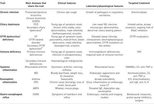
Other examples of bronchiectasis with distinct clinical features and specific treatment modalities also exist. These are immune deficiencies, genetic and acquired (haematological malignancies), that may be treated with immunoglobulin replacement therapy, autoimmune-related (such as RA and inflammatory bowel disease (IBD) and allergic bronchopulmonary aspergillosis (ABPA), for which treatment with systemic corticosteroids and anti-IgE antibodies are indicated. Asthma, COPD and chronic rhinosinusitis (CRS) that are frequently causes and/or associated with bronchiectasis [50], have distinct treatment modalities, as summarised in table 1.
Airway inflammation in bronchiectasis may also not be uniform. In the study by DENTEet al.[15] most patients with bronchiectasis had elevated sputum neutrophils. However, 20% of patients had more than 3% eosinophils in sputum, with total sputum eosinophil count ranging from 0 to 70%. A subgroup of eosinophilic predominant inflammation was also found in 17.5% of 40 patients in another study [92]. Similarly, in a previous single-centre study of people with bronchiectasis, presence of CRS was associated with elevated peripheral blood eosinophils and IgE [93]. It may be that some people with bronchiectasis have a type 2 inflammation, perhaps associated with asthma, CRS and ABPA, while the majority of people with bronchiectasis have a neutrophilic inflammation. These differences may be important in developing treatments that target inflammation in bronchiectasis.
Chronic infection is common in bronchiectasis, with P. aeruginosa being the most common organism isolated [22, 47, 71]. However, presence of chronic infection is not uniform and ranges from 16% to 50% in different series. While infection with P. aeruginosa is associated with worse outcomes [24, 25], The impact ofP. aeruginosais also heterogeneous in relation to exacerbation frequency [22]. It is hypothesised that the immune response to infection may impact on disease severity [94, 95].
Response to therapeutic interventions is not uniform and not specific
Single-centre studies demonstrated significant reductions in sputum production and improvement in symptoms in patients with bronchiectasis [98, 99]. However, later studies did not show a similar benefit although cough was significantly improved [100]. Currently, treatment with ICS is not recommended in people with bronchiectasis, unless indicated for concomitant asthma or COPD [27]. It remains to be seen whether future studies will be successful in defining subgroups of patients, perhaps according to patterns of inflammation, which will be responsive to previously tested modalities, such as ICS, inhaled antibiotics and rhDNase.
Therapeutic modalities with established efficacy in bronchiectasis are also far from specific and beneficial in other airway diseases. Macrolides have proven efficacy in reducing exacerbations in CF [101], but also in diffuse panbronchiolitis [102, 103], bronchiolitis obliterans in lung transplant recipients [104–106], COPD [107–109], CRS [110] and, recently, asthma [111]. Airway clearance and inhaled mucolytic agents, such as hypertonic saline, are the first-line treatment for CF [112]. Rehabilitation has established benefits in COPD care [113], only later to be utilised in bronchiectasis.
Is bronchiectasis a
“
treatable trait
”
?
The concept of“treatable traits”in airway diseases was recently introduced [114]. It was suggested that, in the age of precision medicine, a pragmatic, therapy-focused approach to airway disorders would be more successful in treatment than classifying diseases and treating accordingly. Focusing on asthma and COPD, classifying people into “diseases” may lead to suboptimal management because diseases with different TABLE 1“Treatable traits”of bronchiectasis
Trait Main diseases that
share the trait
Clinical features
Laboratory/physiological features
Targeted treatment
Chronic infection Protracted bacterial bronchitis
Chronic wet cough Growth of pathogens in respiratory secretions
Antimicrobials
Chronic bronchitis (COPD)
Ciliary dyskinesia PCD Young age of symptom onset, chronic otitis media, situs abnormalities, male infertility
(asthenospermia), sinusitis
Low nasal NO, electron microscopic abnormalities, abnormal ciliary beating pattern
Inhaled saline, airway clearance, ongoing trial of
ENaC inhibition Secondary ciliary
dyskinesia#
CFTR dysfunction/ deficiency
CF Young age of symptom onset, pancreatitis, malnutrition, bowel
obstruction, male infertility (azoospermia), sinusitis
Elevated sweat chloride, characteristic electrophysiological abnormalities, CFTR mutations on
two alleles CFTR modulators CFTR-related disorder Secondary CFTR dysfunction# Immune deficiency Primary immune
deficiencies
Young age of symptom onset, infection in extrapulmonary
sites
Immunoglobulin deficiencies, impaired tests of immune function
IVIG Secondary immune deficiencies Haematological malignancies Systemic inflammation
Rheumatoid arthritis Symmetric arthritis, morning stiffness
Rheumatoid factor DMARDs, CS, anti-TNF-α
IBD Bloody diarrhoea, weight loss, GI ulceration
Endoscopic appearance and histology
Aminosalicylates, CS, anti-TNF-α Eosinophilic
inflammation
Asthma Seasonality Airway reversibility ICS, bronchodilators,
anti-IgE, anti-IL-5 CRS Chronic nasal discharge, loss of
smell, facial pain
Sinus inflammation on endoscopy, sinus CT
ABPA Wheeze, mucus plugs Elevated IgE,Aspergillusspp. sensitisation Gastro-oesophageal
reflux
GORD Symptoms of heartburn and reflux
Endoscopic, motility and imaging studies
Behavioural measures, proton-pump inhibitors,
surgery
endotypes require different therapeutic strategies. The classical approach limits our investigation of the causes of morbidity of people whose symptoms do not meet the definition of one airway disease. This may result in limited development of drugs that target one endotype and not another. Lastly, this approach may also limit the generalisability of clinical trials, because many patients with a “disease” targeted in clinical trials may have features of another“disease”and therefore are excluded from those trials, obvious examples being the exclusion of people with airway reversibility from COPD trials and smokers from asthma trials. The list of traits of airway diseases that was included in that paper contained, among others, emphysema, eosinophilic inflammation and bronchiectasis.
Emphysema is considered to be a trait of people with the “classic” definition of COPD. Similar to bronchiectasis, radiological emphysema may be asymptomatic or be associated with severe symptoms (e.g. exercise intolerance). It is therefore not considered a disease by itself, but a clinical feature, or trait, of people with COPD, especially smokers and, in the presence of α1-antitrypsin deficiency. Lung volume reduction is a therapeutic option that targets emphysema, making the trait a treatable one [115].
Bronchiectasis was suggested to be one of the treatable traits of airways diseases [114]. However, the diversity of bronchiectasis would make it very difficult to narrow into a treatable trait as the treatable aspect suggests there is a narrow indicated treatment such as inhaled corticosteroids for the treatable trait of eosinophilic inflammation. Rather, we suggest that bronchiectasis be viewed as a disease that is heterogeneous and shares treatable traits with other diseases. Examples of treatable traits of bronchiectasis may be airway infection (targeted by antimicrobials), failure of mucociliary clearance (treatable with airway clearance and pharmacological adjuncts) and CFTR dysfunction (treatment with CFTR modulators). Table 1 shows several examples of such treatable traits that are shared by subsets of people with bronchiectasis and other airway and systemic diseases. Determining an aetiology or a treatable trait has important implications for future care since identification of the underlying abnormality, which may be inflammatory, ciliary or epithelial in origin, and early treatment may prevent the development of bronchiectasis.
The treatable traits model is powerful specifically because it does not require a clinician to identify a disease or to decide which disease is predominant in a specific circumstance. If your patient has airway infection and frequent exacerbations, they may benefit from antimicrobial and anti-inflammatory treatment with a macrolide regardless of whether the primary disease label is bronchiectasis, COPD or asthma. Likewise, if the patient has exercise limitation and deconditioning, they are likely to benefit from pulmonary rehabilitation regardless of the underlying disease label. Thus, moving from thinking of airway diseases as labels and instead as complex systems composed of multiple traits may lead to more holistic treatment.
Does it really matter?
AGUSTI et al. [114] suggest that the pulmonology community move away from “Oslerian” diagnoses towards treatable traits definitions, but this approach does have implications to be considered. For health authorities, labelling patients with distinct diagnoses allows planning and distributing health services, drug and treatment registration, and more. For patients, naming their symptoms as a diagnosis may be reassuring, apart from the hope of improving with treatment [116]. A repeated complaint from our patients with bronchiectasis is that their condition was misdiagnosed and neglected for decades. Moving away from labelling bronchiectasis as a disease may wipe away the important achievements of bronchiectasis research and organisation of the past years. In asthma and COPD, heterogeneity and the importance of phenotyping is increasingly recognised. However, the medical community has not moved away from the definition of these diagnoses, and international guidelines on diagnosis and management are still available and regularly updated [113, 117], while important advancement in“precision medicine” directed towards specific phenotypes continue [118, 119]. The same should be the case for bronchiectasis: maintaining the“disease” label while acknowledging its limitations, the heterogeneity of the disease and the importance of identifying treatable traits to appropriately target new therapies.
Conclusion
Conflict of interest: M. Shteinberg reports grants, personal fees and other funding from GSK, grants and other funding from Novartis, other funding from Actelion, grants from Trudell pharma, personal fees from Astra Zeneca and Horizon Pharma, speakers fees from Teva, other from Rafa, and personal fees and other funding from Boehringer Ingelheim, outside the submitted work. P.A. Flume reports grants and personal fees from Bayer Healthcare AG and Insmed, grants from Novoteris, and personal fees from Eloxx Pharmaceuticals and Horizon Pharma, outside the submitted work. J.D. Chalmers reports grants and personal fees from GlaxoSmithKline, Boehringer Ingelheim, AstraZeneca, Grifols/Aradigm and Insmed, personal fees from Zambon, and personal fees from Novartis, Bayer, Napp and Savara, outside the submitted work.
References
1 Bondi MW, Edmonds EC, Salmon DP. Alzheimer’s disease: past, present, and future.J Int Neuropsychol Soc 2017; 23: 818–831.
2 Scully JL. What is a disease?EMBO Rep2004; 5: 650–653.
3 Marketos SG, Ballas CN. Bronchial asthma in the medical literature of Greek antiquity. J Asthma1982; 19: 263–269.
4 Sakula A. R T H Laënnec 1781–1826 his life and work: a bicentenary appreciation.Thorax1981; 36: 81–90. 5 Quint JK, Millett ERC, Joshi M,et al.Changes in the incidence, prevalence and mortality of bronchiectasis in the
UK from 2004 to 2013: a population-based cohort study.Eur Respir J2016; 47: 186–193.
6 Roberts HJ, Hubbard R. Trends in bronchiectasis mortality in England and Wales. Respir Med 2010; 104: 981–985.
7 Hill AT, Haworth CS, Aliberti S, et al. Pulmonary exacerbation in adults with bronchiectasis: a consensus definition for clinical research.Eur Respir J2017; 49: 1700051.
8 Kapur N, Masters IB, Chang AB. Exacerbations in noncystic fibrosis bronchiectasis: clinical features and investigations.Respir Med2009; 103: 1681–1687.
9 Cole PJ. Inflammation: a two-edged sword–the model of bronchiectasis.Eur J Respir Dis Suppl1986; 147: 6–15. 10 Flume PA, Chalmers JD, Olivier KN. Advances in bronchiectasis: endotyping, genetics, microbiome, and disease
heterogeneity.Lancet2018; 392: 880–890.
11 Gaga M, Bentley AM, Humbert M,et al.Increases in CD4+ T lymphocytes, macrophages, neutrophils and interleukin 8 positive cells in the airways of patients with bronchiectasis.Thorax1998; 53: 685–691.
12 Schaaf B, Wieghorst A, Aries SP,et al.Neutrophil inflammation and activation in bronchiectasis: comparison with pneumonia and idiopathic pulmonary fibrosis.Respiration2000; 67: 52–59.
13 Angrill J, Agustí C, De Celis R,et al.Bronchial inflammation and colonization in patients with clinically stable bronchiectasis.Am J Respir Crit Care Med2001; 164: 1628–1632.
14 Chalmers JD, Moffitt KL, Suarez-Cuartin G,et al.Neutrophil elastase activity is associated with exacerbations and lung function decline in bronchiectasis.Am J Respir Crit Care Med2017; 195: 1384–1393.
15 Dente FL, Bilotta M, Bartoli ML, et al. Neutrophilic bronchial inflammation correlates with clinical and functional findings in patients with noncystic fibrosis bronchiectasis.Mediators Inflamm2015; 2015: 642503. 16 Papayannopoulos V, Metzler KD, Hakkim A, et al. Neutrophil elastase and myeloperoxidase regulate the
formation of neutrophil extracellular traps.J Cell Biol2010; 191: 677–691.
17 Sagel SD, Wagner BD, Anthony MM,et al.Sputum biomarkers of inflammation and lung function decline in children with cystic fibrosis.Am J Respir Crit Care Med2012; 186: 857–865.
18 Gramegna A, Amati F, Terranova L,et al.Neutrophil elastase in bronchiectasis.Respir Res2017; 18: 211. 19 Cabello H, Torres A, Celis R,et al.Bacterial colonization of distal airways in healthy subjects and chronic lung
disease: a bronchoscopic study.Eur Respir J1997; 10: 1137–1144.
20 Tunney MM, Einarsson GG, Wei L, et al. Lung microbiota and bacterial abundance in patients with bronchiectasis when clinically stable and during exacerbation.Am J Respir Crit Care Med2013; 187: 1118–1126. 21 Moss RB. Fungi in cystic fibrosis and non-cystic fibrosis bronchiectasis.Semin Respir Crit Care Med2015; 36:
207–216.
22 Araújo D, Shteinberg M, Aliberti S,et al.The independent contribution ofPseudomonas aeruginosainfection to long-term clinical outcomes in bronchiectasis.Eur Respir J2018; 51: 1701953.
23 Finch S, McDonnell MJ, Abo-Leyah H,et al.A comprehensive analysis of the impact ofPseudomonas aeruginosa colonization on prognosis in adult bronchiectasis.Ann Am Thorac Soc2015; 12: 1602–1611.
24 Chalmers JD, Goeminne P, Aliberti S,et al.The bronchiectasis severity index. An international derivation and validation study.Am J Respir Crit Care Med2014; 189: 576–585.
25 Martínez-García MÁ, de Gracia J, Vendrell Relat M,et al.Multidimensional approach to non-cystic fibrosis bronchiectasis: the FACED score.Eur Respir J2014; 43: 1357–1367.
26 Chalmers JD, Smith MP, McHugh BJ, et al. Short- and long-term antibiotic treatment reduces airway and systemic inflammation in non-cystic fibrosis bronchiectasis.Am J Respir Crit Care Med2012; 186: 657–665. 27 Polverino E, Goeminne PC, McDonnell MJ,et al.European Respiratory Society guidelines for the management
of adult bronchiectasis.Eur Respir J2017; 50: 1700629.
28 Pasteur MC, Bilton D, Hill AT,et al.British Thoracic Society guideline for non-CF bronchiectasis.Thorax2010; 65: Suppl. 1, i1–58.
29 Chalmers JD, Aliberti S, Blasi F. State of the art review: management of bronchiectasis in adults.Eur Respir J 2015; 45: 1446–1462.
30 Muñoz G, de Gracia J, Buxó M,et al.Long-term benefits of airway clearance in bronchiectasis: a randomised placebo-controlled trial.Eur Respir J2018; 51: 1701926.
31 Herrero-Cortina B, Vilaró J, Martí D, et al. Short-term effects of three slow expiratory airway clearance techniques in patients with bronchiectasis: a randomised crossover trial.Physiotherapy2016; 102: 357–364. 32 Guimarães FS, Moço VJR, Menezes SLS,et al.Effects of ELTGOL and Flutter VRP1 on the dynamic and static
pulmonary volumes and on the secretion clearance of patients with bronchiectasis.Rev Bras Fisioter2012; 16: 108–113.
34 Herrero-Cortina B, Alcaraz V, Vilaró J,et al.Impact of hypertonic saline solutions on sputum expectoration and their safety profile in patients with bronchiectasis: a randomized crossover trial.J Aerosol Med Pulm Drug Deliv 2018; 31: 281–289.
35 Nicolini A, Cardini F, Landucci N,et al.Effectiveness of treatment with high-frequency chest wall oscillation in patients with bronchiectasis.BMC Pulm Med2013; 13: 21.
36 Tsang KW, Chan WM, Ho PL,et al.A comparative study on the efficacy of levofloxacin and ceftazidime in acute exacerbation of bronchiectasis.Eur Respir J1999; 14: 1206–1209.
37 Ip M, Shum D, Lauder I,et al.Effect of antibiotics on sputum inflammatory contents in acute exacerbations of bronchiectasis.Respir Med1993; 87: 449–454.
38 Hill SL, Stockley RA. Effect of short and long term antibiotic response on lung function in bronchiectasis. Thorax1986; 41: 798–800.
39 Altenburg J, de Graaff CS, Stienstra Y, et al. Effect of azithromycin maintenance treatment on infectious exacerbations among patients with non-cystic fibrosis bronchiectasis: the BAT randomized controlled trial.JAMA 2013; 309: 1251–1259.
40 Serisier DJ, Martin ML, McGuckin MA, et al. Effect of long-term, low-dose erythromycin on pulmonary exacerbations among patients with non-cystic fibrosis bronchiectasis: the BLESS randomized controlled trial. JAMA2013; 309: 1260–1267.
41 Wong C, Jayaram L, Karalus N, et al. Azithromycin for prevention of exacerbations in non-cystic fibrosis bronchiectasis (EMBRACE): a randomised, double-blind, placebo-controlled trial.Lancet2012; 380: 660–667. 42 Haworth CS, Foweraker JE, Wilkinson P,et al. Inhaled colistin in patients with bronchiectasis and chronic
Pseudomonas aeruginosainfection.Am J Respir Crit Care Med2014; 189: 975–982.
43 Rubin BK, Williams RW. Aerosolized antibiotics for non-cystic fibrosis bronchiectasis. Respiration 2014; 88: 177–184.
44 Shteinberg M, Johnston C, Haworth C. Long-term inhaled antibiotictreatment in bronchiectasis.In:Chalmers J, Polverino E, Aliberti S, eds. Bronchiectasis: The EMBARC Manual. Berlin, Springer, 2017; pp: 223–239. 45 Nadig TR, Flume PA. Aerosolized antibiotics for patients with bronchiectasis.Am J Respir Crit Care Med2016;
193: 808–810.
46 Chalmers JD, Aliberti S, Filonenko A, et al. Characterization of the “frequent exacerbator phenotype” in bronchiectasis.Am J Respir Crit Care Med2018; 197: 1410–1420.
47 Aliberti S, Lonni S, Dore S,et al.Clinical phenotypes in adult patients with bronchiectasis.Eur Respir J2016; 47: 1113–1122.
48 Radovanovic D, Santus P, Blasi F,et al.A comprehensive approach to lung function in bronchiectasis. Respir Med2018; 145: 120–129.
49 Dimakou K, Triantafillidou C, Toumbis M, et al. Non CF-bronchiectasis: aetiologic approach, clinical, radiological, microbiological and functional profile in 277 patients.Respir Med2016; 116: 1–7.
50 Polverino E, Dimakou K, Hurst J,et al.The overlap between bronchiectasis and chronic airways diseases: state of the art and future directions.Eur Respir J2018; 52: 1800328.
51 Gupta S, Siddiqui S, Haldar P,et al.Qualitative analysis of high-resolution CT scans in severe asthma.Chest 2009; 136: 1521–1528.
52 Gupta S, Siddiqui S, Haldar P,et al.Quantitative analysis of high-resolution computed tomography scans in severe asthma subphenotypes.Thorax2010; 65: 775–781.
53 Machado D, Pereira C, Teixeira L,et al.Thoracic high resolution computed tomography (HRCT) in asthma.Eur Ann Allergy Clin Immunol2009; 41: 139–145.
54 Bisaccioni C, Aun MV, Cajuela E,et al.Comorbidities in severe asthma: frequency of rhinitis, nasal polyposis, gastroesophageal reflux disease, vocal cord dysfunction and bronchiectasis.Clinics2009; 64: 769–773.
55 Dimakou K, Gousiou A, Toumbis M,et al.Investigation of bronchiectasis in severe uncontrolled asthma.Clin Respir J2018; 12: 1212–1218.
56 Martínez-García MÁ, Soler-Cataluña JJ, Donat Sanz Y,et al.Factors associated with bronchiectasis in patients with COPD.Chest2011; 140: 1130–1137.
57 O’Donnell AE. Bronchiectasis in patients with COPD: a distinct COPD phenotype?Chest2011; 140: 1107–1108. 58 Martínez-García M-A, de la Rosa Carrillo D, Soler-Cataluña J-J,et al.Prognostic value of bronchiectasis in patients
with moderate-to-severe chronic obstructive pulmonary disease.Am J Respir Crit Care Med2013; 187: 823–831. 59 Ni Y, Shi G, Yu Y,et al. Clinical characteristics of patients with chronic obstructive pulmonary disease with
comorbid bronchiectasis: a systemic review and meta-analysis. Int J Chron Obstruct Pulmon Dis 2015; 10: 1465–1475.
60 Ringshausen FC, de Roux A, Pletz MW,et al.Bronchiectasis-associated hospitalizations in Germany, 2005–2011: a population-based study of disease burden and trends.PLoS One2013; 8: e71109.
61 Novosad SA, Barker AF. Chronic obstructive pulmonary disease and bronchiectasis.Curr Opin Pulm Med2013; 19: 133–139.
62 Hurst JR, Elborn JS, De Soyza A,et al.COPD-bronchiectasis overlap syndrome.Eur Respir J2015; 45: 310–313. 63 Mao B, Lu H-W, Li M-H,et al.The existence of bronchiectasis predicts worse prognosis in patients with COPD.
Sci Rep2015; 5: 10961.
64 Minov J, Stoleski S, Mijakoski D,et al.Exacerbations in COPD patients with bronchiectasis.Med Sci (Basel) 2017; 5: E7.
65 Vikgren J, Boijsen M, Andelid K, et al. High-resolution computed tomography in healthy smokers and never-smokers: a 6-year follow-up study of men born in 1933.Acta Radiol2004; 45: 44–52.
66 Copley SJ, Wells AU, Hawtin KE,et al.Lung morphology in the elderly: comparative CT study of subjects over 75 years oldversusthose under 55 years old.Radiology2009; 251: 566–573.
67 Matsuoka S, Uchiyama K, Shima H,et al.Bronchoarterial ratio and bronchial wall thickness on high-resolution CT in asymptomatic subjects: correlation with age and smoking.AJR Am J Roentgenol2003; 180: 513–518. 68 Ciccarese F, Chiesa AM, Feletti F,et al.The senile lung as a possible source of pitfalls on chest ultrasonography
and computed tomography.Respiration2015; 90: 56–62.
70 Chalmers JD, Aliberti S, Polverino E,et al. The EMBARC European Bronchiectasis Registry: protocol for an international observational study.ERJ Open Res2016; 2: 00081-2015.
71 Aksamit TR, O’Donnell AE, Barker A, et al. Adult patients with bronchiectasis: a first look at the US bronchiectasis research registry.Chest2017; 151: 982–992.
72 Mott LS, Graniel KG, Park J,et al.Assessment of early bronchiectasis in young children with cystic fibrosis is dependent on lung volume.Chest2013; 144: 1193–1198.
73 Chang AB, Upham JW, Masters IB,et al.Protracted bacterial bronchitis: the last decade and the road ahead. Pediatr Pulmonol2016; 51: 225–242.
74 Goyal V, Grimwood K, Marchant JM,et al.Paediatric chronic suppurative lung disease: clinical characteristics and outcomes.Eur J Pediatr2016; 175: 1077–1084.
75 Kantar A, Chang AB, Shields MD,et al.ERS statement on protracted bacterial bronchitis in children.Eur Respir J2017; 50: 1602139.
76 Schaefer OP, Irwin RS. Unsuspected bacterial suppurative disease of the airways presenting as chronic cough.Am J Med2003; 114: 602–606.
77 Martin MJ, Harrison TW. Causes of chronic productive cough: An approach to management.Respir Med2015; 109: 1105–1113.
78 Chang AB, Bush A, Grimwood K. Bronchiectasis in children: diagnosis and treatment.Lancet2018; 392: 866–879. 79 Martínez-García MÁ, Máiz L, Olveira C, et al. Spanish guidelines on the evaluation and diagnosis of
bronchiectasis in adults.Arch Bronconeumol2018; 54: 79–87.
80 EMBARC: The European Bronchiectasis Registry (ver: 1.7.3.3197). Available from www.bronchiectasis.eu 81 Pignatti PF, Bombieri C, Marigo C,et al.Increased incidence of cystic fibrosis gene mutations in adults with
disseminated bronchiectasis.Hum Mol Genet1995; 4: 635–639.
82 Girodon E, Cazeneuve C, Lebargy F,et al.CFTR gene mutations in adults with disseminated bronchiectasis.Eur J Hum Genet1997; 5: 149–155.
83 Bienvenu T, Sermet-Gaudelus I, Burgel P-R,et al.Cystic fibrosis transmembrane conductance regulator channel dysfunction in non-cystic fibrosis bronchiectasis.Am J Respir Crit Care Med2010; 181: 1078–1084.
84 Milosevic K, Nikolic A, Divac Rankov A,et al.Analysis of CFTR gene variants in idiopathic bronchiectasis in Serbian children.Pediatr Allergy Immunol Pulmonol2013; 26: 93–98.
85 Guan W-J, Li J-C, Liu F,et al.Next-generation sequencing for identifying genetic mutations in adults with bronchiectasis.J Thorac Dis2018; 10: 2618–2630.
86 Ziedalski TM, Kao PN, Henig NR, et al. Prospective analysis of cystic fibrosis transmembrane regulator mutations in adults with bronchiectasis or pulmonary nontuberculous mycobacterial infection.Chest2006; 130: 995–1002.
87 Puéchal X, Fajac I, Bienvenu T,et al.Increased frequency of cystic fibrosis deltaF508 mutation in bronchiectasis associated with rheumatoid arthritis.Eur Respir J1999; 13: 1281–1287.
88 Shapiro AJ, Davis SD, Polineni D,et al.Diagnosis of primary ciliary dyskinesia. An official American Thoracic Society clinical practice guideline.Am J Respir Crit Care Med2018; 197: e24–e39.
89 Shapiro AJ, Zariwala MA, Ferkol T,et al.Diagnosis, monitoring, and treatment of primary ciliary dyskinesia: PCD foundation consensus recommendations based on state of the art review. Pediatr Pulmonol 2016; 51: 115–132.
90 Contarini M, Shoemark A, Rademacher J,et al.Why, when and how to investigate primary ciliary dyskinesia in adult patients with bronchiectasis.Multidiscip Respir Med2018; 13: Suppl. 1, 26.
91 Chalmers JD, Elborn JS. Reclaiming the name“bronchiectasis”.Thorax2015; 70: 399–400.
92 Tsikrika S, Dimakou K, Papaioannou AI,et al.The role of non-invasive modalities for assessing inflammation in patients with non-cystic fibrosis bronchiectasis.Cytokine2017; 99: 281–286.
93 Shteinberg M, Nassrallah N, Jrbashyan J,et al.Upper airway involvement in bronchiectasis is marked by early onset and allergic features.ERJ Open Res2018; 4: 00115-2017.
94 Skopelja-Gardner S, Theprungsirikul J, Meagher RE,et al.Autoimmunity to bactericidal/permeability-increasing protein in bronchiectasis exhibits a requirement forPseudomonas aeruginosaIgG response.Eur Respir J2018; 53: 1801891.
95 Shteinberg M, Shah A, Elborn JS. New insights into immunological responses to infection in bronchiectasis.Eur Respir J2019; 53: 1802395.
96 O’Donnell AE, Barker AF, Ilowite JS,et al.Treatment of idiopathic bronchiectasis with aerosolized recombinant human DNase I. rhDNase Study Group.Chest1998; 113: 1329–1334.
97 Barker AF, Couch L, Fiel SB,et al.Tobramycin solution for inhalation reduces sputumPseudomonas aeruginosa density in bronchiectasis.Am J Respir Crit Care Med2000; 162: 2 Pt. 1, 481–485.
98 Elborn JS, Johnston B, Allen F, et al.Inhaled steroids in patients with bronchiectasis.Respir Med1992; 86: 121–124.
99 Martínez-García MA, Perpiñá-Tordera M, Román-Sánchez P,et al.Inhaled steroids improve quality of life in patients with steady-state bronchiectasis.Respir Med2006; 100: 1623–1632.
100 Tsang KW, Tan KC, Ho PL,et al.Inhaled fluticasone in bronchiectasis: a 12 month study.Thorax 2005; 60: 239–243.
101 Saiman L, Marshall BC, Mayer-Hamblett N,et al. Azithromycin in patients with cystic fibrosis chronically infected withPseudomonas aeruginosa: a randomized controlled trial.JAMA2003; 290: 1749–1756.
102 Yang M, Dong BR, Lu J,et al.Macrolides for diffuse panbronchiolitis. Cochrane Database Syst Rev2010; 12: CD007716.
103 Kadota J, Mukae H, Ishii H,et al.Long-term efficacy and safety of clarithromycin treatment in patients with diffuse panbronchiolitis.Respir Med2003; 97: 844–850.
104 Gerhardt SG, McDyer JF, Girgis RE, et al. Maintenance azithromycin therapy for bronchiolitis obliterans syndrome: results of a pilot study.Am J Respir Crit Care Med2003; 168: 121–125.
105 Verleden GM, Dupont LJ. Azithromycin therapy for patients with bronchiolitis obliterans syndrome after lung transplantation.Transplantation2004; 77: 1465–1467.
107 Albert RK, Connett J, Bailey WC,et al.Azithromycin for prevention of exacerbations of COPD.N Engl J Med 2011; 365: 689–698.
108 Ramos FL, Criner GJ. Use of long-term macrolide therapy in chronic obstructive pulmonary disease.Curr Opin Pulm Med2014; 20: 153–158.
109 Seemungal TAR, Wilkinson TMA, Hurst JR,et al.Long-term erythromycin therapy is associated with decreased chronic obstructive pulmonary disease exacerbations.Am J Respir Crit Care Med2008; 178: 1139–1147. 110 Shen S, Lou H, Wang C,et al.Macrolide antibiotics in the treatment of chronic rhinosinusitis: evidence from a
meta-analysis.J Thorac Dis2018; 10: 5913–5923.
111 Gibson PG, Yang IA, Upham JW,et al.Effect of azithromycin on asthma exacerbations and quality of life in adults with persistent uncontrolled asthma (AMAZES): a randomised, double-blind, placebo-controlled trial. Lancet2017; 390: 659–668.
112 Castellani C, Duff AJA, Bell SC,et al.ECFS best practice guidelines: the 2018 revision.J Cyst Fibros2018; 17: 153–178.
113 Global Initiative for Chronic Obstructive Lung Disease. https://goldcopd.org/ Date last accessed: May 1, 2019; date last updated: December 5, 2019.
114 Agusti A, Bel E, Thomas M,et al.Treatable traits: toward precision medicine of chronic airway diseases.Eur Respir J2016; 47: 410–419.
115 Shah PL, Herth FJ, van Geffen WH,et al.Lung volume reduction for emphysema.Lancet Respir Med2017; 5: 147–156.
116 Jutel AG. What’s wrong with me? Diagnosis and the Patient- Doctor Relationship.In:Jutel AG, ed. Putting a Name to It: Diagnosis in Contemporary Society. Baltimore, Johns Hopkins University Press, 2014; p. 62. 117 Global Initiative for Asthma -Global Initiative for Asthma. https://ginasthma.org/ Date last accessed: May 1,
2019; date last updated: December 5, 2019.
118 Busse WW. Biological treatments for severe asthma: a major advance in asthma care. Allergol Int2019; 68: 158–166.