EFFECTS OF THE PRONE-PASSIVE STRETCHING TECHNIQUE ON GLENOHUMERAL INTERNAL ROTATION
by
David Hammons
A dissertation
submitted in partial fulfillment of the requirements for the degree of
Doctor of Education in Curriculum and Instruction Boise State University
DEFENSE COMMITTEE AND FINAL READING APPROVALS
of the dissertation submitted by
David Hammons
Dissertation Title: Effects of the Prone Passive Stretching Technique on Glenohumeral Internal Rotation
Date of Final Oral Examination: 09 March 2012
The following individuals read and discussed the dissertation submitted by student David Hammons, and they evaluated his presentation and response to questions during the final oral examination. They found that the student passed the final oral examination.
John W. McChesney, Ph.D., ATC Chair, Supervisory Committee Michael Curtin, M.D. Member, Supervisory Committee Ron Pfeiffer, Ph.D., ATC Member, Supervisory Committee Keith Thiede, Ph.D. Member, Supervisory Committee
iv
ACKNOWLEDGMENTS
I would like to extend many thanks to everyone that helped me with this process. In particular, I am extremely thankful to my committee chair, Dr. John McChesney, for his guidance and willingness to embark on this dissertation process with me. As well, many thanks to Dr. Curtin, Dr. Pfeiffer, and Dr. Thiede for providing professional feedback on this dissertation. I would also like to recognize the faculty and staff of the Boise State University Kinesiology Department for all of the support and encouragement expressed to me.
This dissertation would not have come together like it did without the assistance of several research assistants, in particular Ray Santiago, III, who was so willing to learn and help out whenever needed. As well, I am very appreciative of the student-athlete participants that enrolled in this study from Boise State University and the College of Idaho. Thank you to the Flanagan’s for your mindful support and interest in my endeavors. Last, I would not be anything in life without the unconditional love and support from close friends and family. More than ever I am convinced that great
education starts at home; I love you mom and dad. And Elizabeth, thank you for being a wonderful wife, mother, and friend. You have accommodated me through every step of this challenge. Hopefully Tommy will be proud of his parents someday!
v ABSTRACT
Effects of the Prone Passive Stretching Technique on Glenohumeral Internal
Rotation
vi
vii
TABLE OF CONTENTS
ACKNOWLEDGMENTS ... iv
ABSTRACT ...v
LIST OF TABLES ...x
LIST OF FIGURES ... xi
CHAPTER I: INTRODUCTION/LITERATURE REVIEW ...1
GIRD ...2
Cascading Effect of GIRD / PST ...3
Etiology of GIRD and/or PST...6
Soft Tissue Tightness and Contracture ...7
Humeral Translation and Physiologic Adaptation ...11
Osseous Adaptation ...12
Intervention Techniques to Counter Reduced Internal Rotation ...15
Conservative Intervention ...15
Surgical Intervention ...20
CHAPTER II: METHODS ...23
Participants ...24
Measurement / Procedure ...25
Intra- and Inter-Tester Reliability ...27
Intervention ...28
viii
Analysis...31
Participant Descriptives ...31
Dominant Shoulder Internal Rotation Range of Motion (Dom IR ROM) ...33
Internal Rotation Deficit (IR Deficit) ...35
Total Motion of the Gleno-Humeral Joint ...38
IR ROM, IR Deficit, and Total Motion in Males and Females ...41
IR and ER Correlation ...42
CHAPTER IV: DISCUSSION ...44
Response to Intervention...44
Gender Comparisons ...45
Differing Views on Total Motion in an IR Deficit Sample ...46
Capsule or Muscle? ...46
Parameters of the Stretching Technique ...48
Tensile Forces of the Stretching Techniques ...49
Importance of Maintaining Normal Glenohumeral Internal Rotation in Overhead Athletes ...51
Conclusion ...52
REFERENCES ...53
APPENDIX A ...60
Participant Instructions for the Cross-Body Stretching Technique ...60
APPENDIX B ...62
Instructions for the Prone-Passive Stretching Technique ...62
APPENDIX C ...64
ix
APPENDIX D ...66
Informed Consent...66
APPENDIX E ...71
Demographic Information and Medical History ...71
APPENDIX F...73
Pre- and Post-Test Measurement Form ...73
APPENDIX G ...76
Cross-Body Treatment Schedule ...76
APPENDIX H ...78
x
LIST OF TABLES
Table 2.1 Intra- and Inter-Tester Reliability ...28
Table 3.1 Participant Descriptives ...32
Table 3.2 Participant by Sport ...32
Table 3.3 Participant Adherence to Stretching Schedule ...33
Table 3.4 Prevelence of Internal Rotation Deficit by Sport ...33
Table 3.5 Pre- and Post-Test Measurements ...40
Table 3.6 Gain Scores for All Measurements ...41
Table 3.7 Gain Scores for Male Participants ...42
xi
LIST OF FIGURES
Figure 2.1 Experimental Design ...23
Figure 2.2 Measurement Instrument ...25
Figure 2.3 Measurement Technique ...27
Figure 2.4 Stretching Techniques ...29
Figure 3.1 Internal Rotation Range of Motion Gain Bar Graph ...34
Figure 3.2 Internal Rotation Range of Motion Pre-Test Bar Graph ...34
Figure 3.3 Internal Rotation Range of Motion Post-Test Bar Graph ...35
Figure 3.4 Internal Rotation Deficit Gain Bar Graph ...36
Figure 3.5 Internal Rotation Deficit Pre-Test Bar Graph ...37
Figure 3.6 Internal Rotation Deficit Post-Test Bar Graph ...37
Figure 3.7 Total Motion Gain Bar Graph ...38
Figure 3.8 Total Motion Pre-Test Bar Graph...39
Figure 3.9 Total Motion Post-Test Bar Graph ...39
CHAPTER I: INTRODUCTION/LITERATURE REVIEW
Internal rotation of the glenohumeral joint becomes limited in the dominant shoulder of many overhead athletes. This condition, many times asymptomatic in terms of pathology, could become troublesome for the athlete as the condition progresses. Clinicians often prescribe a stretching treatment to counter restrictions in the dominant shoulder, including those that affect the glenohumeral joint, such as glenohumeral internal rotation deficit (GIRD) or posterior shoulder tightness (PST).
Various techniques are employed by clinicians to treat such deficits between shoulders with a goal of restoring normal motion. The purpose of this study is to compare the effect a novel passive stretching technique has on glenohumeral (GH) internal rotation compared to a self-stretching technique in individuals with PST and or GIRD. This stretch has not been investigated through prior study.
This section intends to validate the purpose of this study, but also demonstrate potential voids in previous research regarding the study question, “Is the prone internal rotation (IR) stretching technique effective at improving glenohumeral joint IR?” The literature review is organized into three critical components of interest to the study: 1) an explanation of GIRD or internal rotation limitations, 2) conflicting agreements
GIRD
The human glenohumeral (GH) joint allows the most uninhibited joint motion in the human body. Restriction of normal ROM in the GH joint complex is a common musculoskeletal disorder that creates functional problems especially for the overhead- throwing athlete. Posterior shoulder tightness (PST), and/or glenohumeral internal rotation deficit (GIRD), is of specific interest to this study since limitation, or reduction of IR, is the primary factor in the evaluation and subsequent diagnosis of such pathologic conditions (Burkhart, Morgan, & Kibler, 2003a; Meister, 2000). Previously, Burkhart et al. (2003a), who have studied baseball players specifically, coined the most often
referenced definition of GIRD as being, “loss in degrees of GH IR of the throwing shoulder compared with the non-throwing shoulder.” Further, Burkhart et al. has more specifically described GIRD as a limitation in IR of greater than or equal to 20°, or greater than 10 percent when compared to IR ROM in the non-dominant extremity. It is common for overhead athletes to exhibit a shift towards GH external rotation (ER) motion in the dominant shoulder and reduced IR compared to the non-dominant
extremity. This phenomenon of the overhead athlete shoulder is explained through the total-motion concept. Total motion, as described by Crockett and associates (2002) in a study that investigated increases in ER in patients with IR deficit, refers to the combined IR and ER ROM in the GH joint. In this study, the authors found subjects with limited IR ROM, however the increase noted in ER ROM created similar total motion
notably through increased velocity, it does not attempt to address the potential for a baseball pitcher to exhibit limitation in IR ROM.
This phenomenon of the GH joint is not unique to athletes involved in baseball, but has been a common finding in most other overhead dominant sports as well (Beach, Whitney, & Dickoffhoffman, 1992; Crockett et al., 2002; Ellenbecker, Roetert, Bailie, Davies, & Brown, 2002; Ellenbecker, Roetert, Piorkowski, & Schulz, 1996; Kibler & Chandler, 2003).
Cascading Effect of GIRD / PST
Many authors have investigated the deleterious effects of PST and/or GIRD, which include a common injury to the shoulder, the superior labrum anterior posterior (SLAP) lesion (Burkhart & Morgan, 1998; Kibler, 1998), and variations of impingement syndrome, sub-acromial, or extrinsic, and internal GH impingement (Myers, Laudner, Pasquale, Bradley, & Lephart, 2006; Paley, Jobe, Pink, Kvitne, & ElAttrache, 2000; Tyler, Nicholas, Lee, Mullaney, & McHugh, 2010; Warner, Micheli, Arslanian,
Kennedy, & Kennedy, 1990). In this regard, PST and GIRD are not necessarily viewed as injuries, but more likely as a causative factor that leads to a cascade of pathologic issues in the shoulder complex. Ultimately, the overhand athlete’s reduction in IR may lead to a host of musculoskeletal disorders of the GH joint complex. Many studies have investigated baseball players, specifically pitchers, due to the stressful nature of the overhand throw and the propensity of these athletes to develop significant shoulder injuries (Ellenbecker et al., 2002; Tyler et al., 2010).
have initiated the study on the effects of limited shoulder motion in overhead athletes. In this seminal work, which explained techniques for evaluating and rehabilitating the shoulder, Pappas et al. discussed how pathology in the shoulder gradually develops as a result of a lack of flexibility in soft tissues, specifically capsular structures.
In a recent study to determine if professional baseball players with and without a history of shoulder injury exhibit differences in GH ROM, Scher et al. (2010) produced data consistent with those previously mentioned and similarly support GIRD as a
precursor to shoulder pathology. Interestingly, Scher et al. also suggests that some cases of GIRD may go unrecognized because his data revealed evidence of GIRD in subjects with less than 20° difference in IR ROM, the benchmark for identifying the disparity in throwing and non-throwing ROM as previously defined by Burkhart et al. (2003a). Similarly, Wilk et al. (2011) found professional baseball pitchers who exhibit GIRD and/or total rotational motion deficit were nearly twice as likely as those without the condition to sustain a dominant shoulder or elbow injury. The correlation between GIRD and throwing extremity injuries has been demonstrated in high school aged athletes as well. In another recent study, Shanley et al. (2011) reported baseball and softball high school athletes with significant levels of GIRD to be four times as likely to sustain a shoulder or elbow injury during the same season when compared to those with a 10°-20° deficit (1.5 – 2 times more likely than no deficit).
Most literature reviewed for this study involves participants in baseball,
proposed that repetitive throwing, over time, stretches the anterior capsular ligaments, leading to a potential for rotator cuff injury. Kibler, Chandler, Livingtson, and Roetart (1996) also demonstrated, in elite tennis players, a negative correlation between years of play and increasing limitations in IR ROM. In a unique study, which compared elite tennis players (mean age = 16.4) and professional baseball pitchers (mean age = 26.6), Ellenbecker et al. (2002) demonstrated that both study groups met criteria to be
categorized as GIRD. However, both groups were experienced athletes who undoubtedly placed repetitive stress on the GH complex.
authors demonstrated with statistical power and clinical relevance that limitation in GH ROM quite possibly begins early in life, at least in overhand throwers.
Etiology of GIRD and/or PST
Unlike most musculoskeletal conditions, those of interest to this study, GIRD and/or PST, are disorders that arise from an insidious cause. Previous research has provided data to shed light on the issue of limited IR ROM and increased ER ROM in the overhand athlete, but differing views exist as to the cause of GIRD and/or PST. The intent of this study is to investigate an intervention to reverse or counter the effects of GIRD and/or PST.
From a historical perspective, King, Brelsford, and Tullos (1969), in an analysis of elbow injuries and patho-mechanics, were the first to mention an appreciable
difference between dominant throwing arm and non-throwing arm decreases in IR and increases in ER ROM in the professional baseball pitcher population. This observation was made based on goniometric measurements of GH motions. The purpose of the King et al. study was not to investigate limitations in GH ROM, but to investigate pathologic conditions.
The investigation by Pappas et al. (1985) concurred with prior opinion regarding the changes in GH motion of the throwing shoulder. Also, Pappas et al. may have been the first to report on both passive and active stretching techniques used to evaluate IR ROM by using horizontal flexion (adduction) to assess the structures of the posterior shoulder. The recommendations of that research have had far reaching implications in future studies. Pappas, by virtue of his experience with throwing athletes and by evaluating GH ROM through active and passive movement, determined the restricted motion in IR was caused by both tight anterior and posterior structures, either capsular or muscular (Pappas et al., 1985). As a result of this study, Pappas and colleagues strongly emphasized a supervised stretching routine to counter the effects of restricted GH ROM.
Soft Tissue Tightness and Contracture
Similar to studies on musculoskeletal pathology, research surrounding the etiology of GIRD and PST has focused on two different pathologic anatomic structures. Early researchers produced data analysis regarding the throwing shoulder that led to discussion about muscular and capsular tightness, or contracture, as a cause of PST (Bennett, 1959; King et al., 1969; Pappas et al., 1985).
In a study investigating GH ROM and laxity in 148 professional baseball players, researchers identified significant differences of IR ROM in dominant versus
non-dominant shoulders and attempted to correlate this with observed GH laxity between pitchers and non-pitchers. Findings were significant for limitations in IR ROM
throwing, which leads to tightening of the posterior GH capsule. Later, Sauers and colleagues demonstrated similar findings between laxity and GH ROM in 51 recreational athletes (Sauers, Borsa, Herling, & Stanley, 2001).
Posterior capsule and/or posterior musculature tightness, as a reliable cause of limitation in IR ROM, tends to be the most widely accepted philosophy of GIRD and/or PST etiology. The posterior GH capsule was presented as a cause of restricted IR ROM by Lombardo, Jobe, Kerlan, Carter, and Shields, Jr. (1977). The authors of this study presented four case studies, all involving professional baseball players, who underwent excision of a Bennett’s lesion, and upon visualization of the posterior capsule the surgeon noted a posterior capsular thickening. In another surgical treatment observation, Ticker, Beim, and Warner (2000) reported on nine patients who underwent an arthroscopic release of the posterior GH capsule; in all patients, posterior capsule thickening was observed. Thickening of the posterior inferior capsular ligament of the GH joint has been reported in other studies, but more recently an imaging study using magnetic resonance imaging to diagnose GIRD in a sample of six professional baseball players revealed the same findings, along with other disorders, in all participants (Tehranzadeh, Fronek, & Resnick, 2007).
Tyler, Nicholas, Roy, and Gleim (2000) claim to be the first to clinically link posterior capsule tightness to the loss of internal rotation range of motion, excluding posterior shoulder muscular tightness as a cause. The authors also propose their belief that the posterior GH capsule leads to losses in IR ROM by recommending that a
capsule tightness had been validated through a prior research study (Tyler, Roy, Nicholas, & Gleim, 1999). This procedure was used to assess PST in impingement patients in another study by Tyler and associates where they found symptoms associated with the pathology improved along with reduction in PST and GIRD, however the investigators elude to GIRD arising from humeral head translation, not posterior GH capsule tightness (Tyler et al., 2010).
In a widely cited series entitled The Disabled Throwing Shoulder, Burkhart et al. (2003a) again proposed loss of internal rotation as a result of postero-inferior capsule contracture. However, these authors do not dismiss other potential causative factors of GIRD and/or PST, such as posterior shoulder musculature tightness.
In a review, Blanch (2004) proposed that the posterior shoulder musculature is an important causative factor of restricted GH joint ROM. While referring to the intimate blending of the GH capsule and shoulder musculature, Blanch recommended that it would be more appropriate to refer to tightness of the posterior shoulder rather than specifically implicating the capsule. This study was conducted in an attempt to create a specialized shoulder rehabilitation program aimed at swimmers.
In a recent cadaveric study using 8 fresh specimens instrumented with strain gauges in the capsule, Borstad and Dashottar (2011) examined strain effects of various stretching positions on the posterior gleno-humeral capsule. While the research
also increased in the teres minor and the infraspinatus muscles of the posterior shoulder girdle.
Far fewer studies on PST and/or GIRD have explained the causative factor as an issue of the posterior shoulder musculature, including the rotator cuff. Recently, Oyama, Myers, Blackburn, and Coleman (2011) observed a significant increase in the area size of the infraspinatus muscle, along with decreased IR and horizontal adduction, in a sample of 20 healthy subjects who underwent an eccentric external rotation (ER) workout up to 24 hours after a baseline ultrasound measurement, p. < .001.
In a case report of a non-athlete with PST symptoms associated with sub-acromial impingement, Poser and Casonato (2008) applied a treatment only to posterior musculature, focusing on the infraspinatus and teres minor, and did not attempt to treat the posterior capsule. This soft tissue treatment resulted in significant improvements in restoring IR ROM, however this case study investigated only one patient. In a study by Hung, Hsieh, Yang, and Lin (2010) investigating twenty non-athlete patients with stiff shoulder syndrome, researchers hypothesized a correlation between muscle stiffness and GIRD by using a Myotonometer (Neurogenic Technologies, Inc., Montana, USA) an instrument that measures muscle fiber tension. The authors concluded that posterior deltoid muscle tightness attributed to fifty-one percent (51%) of variance in IR ROM in the subjects, a higher correlation than either the infraspinatus or teres minor (r= 0.65-0.72 versus r= 0.57-0.61). An obvious limitation in this study was the use of the
Humeral Translation and Physiologic Adaptation
Harryman and colleagues (1990) were likely the first to explain GH joint
translation in a cadaveric arthrokinematic study. These authors coined the term obligate translation, which is a shifting of the humeral head in the opposite direction of tight capsular structure. This seminal study set the stage for future study regarding translation in the GH joint and the emphasis on capsular etiology of PST and or GIRD. More recent cadaveric studies corroborate the Harryman et al. findings but mention the likelihood that posterior capsule tightness, through overhead motion, likely plays a significant factor in not only humeral translation, but other shoulder pathology such as labrum tears and impingement (Grossman et al., 2005; Werner, Nyffeler, Jacob, & Gerber, 2004). Arthro-kinematic studies have demonstrated compressive forces of approximately 400 N and inferior shear of approximately 200 N during the follow through phase of the overhead baseball pitch (Dillman, Fleisig, & Andrews, 1993; Fleisig, Andrews, Dillman, & Escamilla, 1995)
Humeral translation refers to movement of the humeral head on the glenoid, creating a potential for increased stress on soft tissue and/or bony structures within the GH joint when translation becomes excessive. The translation then is thought to cause physiologic adaptations in the GH joint: soft tissue and osseous structures. For instance, clinical studies investigating anterior shoulder instability propose altered GH humeral translation as a cause of laxity in the anterior shoulder (Jobe et al., 1991; Paley et al., 2000).
posterior-superiorly on the glenoid instead of the normal posterior-inferior direction during the cocking phase of overhead throwing. The authors described this altered movement to be a causative factor in pathology of the labrum, such as the SLAP tear. This posterior superior movement may be a mechanism of interest for posterior or internal GH
impingement as well, whereby the greater tuberosity of the humerus impacts soft tissue structures in the posterior aspect of the shoulder (Bach, 2006; Cools, Declercq, Cagnie, Cambier, & Witvrouw, 2008; Halbrecht, Tirman, & Atkin, 1999; Jobe, 1995; Myers et al., 2006).
Only one prior cadaveric study reviewed for this research on the effectiveness of a passive IR stretching technique for the shoulder found no correlation between posterior capsule tightening and altered humeral translation (Poitras et al., 2010). Unlike the studies mentioned previously, Poitras et al. did not measure translation during the kinematic throwing movements like the others did, but instead evaluated an elevated abduction movement.
However, in an in-vivo study on forty-three asymptomatic throwing shoulders, researchers found no correlation between humeral translation and altered range of motion, such as found in GIRD (Borsa et al., 2005). Physiologic adaption is likely to occur whenever the humeral head translates on the glenoid during overhead motion, which may contribute to contracture or tightness of the posterior shoulder soft tissues.
Osseous Adaptation
the humeral head that is posteriorly situated perpendicular to the glenoid fossa compared to the alignment in a normal shoulder. Prior studies have demonstrated increased
retroversion in the dominant shoulder of throwing athletes compared to the non-dominant arm (Crockett et al., 2002; Osbahr, Cannon, & Speer, 2002). The Osbahr et al. study investigated nineteen college baseball pitchers, while the Crockett et al. study evaluated twenty-five professional baseball pitchers compared to twenty-five non-throwing subjects. Both studies produced similar findings in terms of humeral retroversion being correlated to increased external rotation in the throwing shoulders. Both Crockett et al. and Osbahr et al. describe the cause of retroversion from a skeletal development
perspective and not an occurrence from an acute event. The previously mentioned work by Sabick and colleagues (2005), which found stresses on the throwing arm and shoulder to be significant enough to cause osseous adaptation, supports this finding. Osbahr and colleagues (2002) utilized a questionnaire to assess throwing frequency between the ages of eight to sixteen years old; however, there was no correlation between duration and amount of throwing and increased humeral retroversion and these ages.
Osbahr et al. (2002) and Sabick et al. (2005) also observed a correlation between humeral retroversion and increased external rotation in the throwing shoulders.
while Crockett and associates (2002) claim humeral retroversion as the primary contributor to posterior capsular tightness. On the other hand, Osbahr and colleagues (2002) determined the cause of PST to be a combination of both bony and capsular adaptation.
While many causes of PST and/or GIRD have been described in the literature, researchers continue to dispute the philosophies described here. Other potential causes have been sparsely described, for instance, Ludewig and Borstad (2003) describe abnormal spine posture as a potential cause of altered shoulder kinematics. However, this finding has yet to be suggested in an overhead athletic population. A majority of researchers explain humeral translation as a cause of posterior capsular tightness versus tight posterior musculature as a cause of PST, but at least one study described prior work in this area as misleading since isolating the posterior capsule from the posterior shoulder musculature is difficult (Michener, McClure, & Karduna, 2003). In a recent study
investigating a link between humeral torsion and elbow/shoulder injuries in collegiate baseball players, the authors suggested osseous adaptation was more likely a factor in PST rather than changes in soft tissues, yet while correlating torsion produced to elbow injuries a similar link was not found in shoulder pathology (Myers et al., 2009).
Intervention Techniques to Counter Reduced Internal Rotation
This section reviews significant research surrounding various intervention strategies and are categorized as either surgical, or non-surgical. The latter of the two is the category where the prone stretch is situated, so this category details the findings of prior study which have investigated other internal rotation stretching techniques. The current study is a seminal investigation to determine the effectiveness of the prone- passive internal rotation technique on gleno-humeral internal rotation.
Conservative Intervention
In 1995, a descriptive article was produced by Johansen, Callis, Potts, and Shall that explained the modified internal rotation stretching technique for the shoulder, however, no study has investigated this stretch until this study. The prone stretching technique utilized in the current study is similar to the technique explained by Johansen and colleagues. In their descriptive essay, the authors thoroughly explain the exercise as a prone stretch that involves passive assistance from the clinician. The scapula is easily stabilized as it protrudes posteriorly while the humerus rotates internally. While the author’s did not evaluate this procedure using an experimental design they presented a modified technique that should improve the manner in which clinicians treat
The cross-body shoulder stretch is a self-stretching technique whereby the subject flexes the shoulder to approximately ninety degrees, then pulls the involved extremity across the body in a horizontal adduction movement, stretching the posterior shoulder musculature (Prentice, 1990). This self-stretch, which serves as the control treatment for the current study, has been explained thoroughly in the literature; however, a more recent study compared this stretch to another internal rotation self-stretching technique, the sleeper stretch (McClure et al., 2007). This study investigated fifty-four participants and found that the cross-body stretch participants experienced a clinically significant
improvement in internal rotation; the group mean and standard deviation for cross-body stretch (20.0° ± 12.9°) compared to the sleeper stretch group (12.4° ± 10.4°), and the no stretching control group (5.9° ± 9.4°). In both of the stretching groups, all participants were screened to meet an equal or greater than 10° between shoulders difference in internal rotation, and were asked to perform the technique by holding the stretch 30 seconds 5 times daily.
The sleeper stretch, which is another common self-stretching technique to
improve internal rotation has been purported as an appropriate exercise to improve range of motion deficits as well (Burkhart et al., 2003b; Laudner et al., 2008). Laudner and fellow researchers (2008) determined that the sleeper stretch improved internal rotation to a statistically significant level (p= 0.003) with a mean improvement of 3.1° compared to no change in a non-stretched control group (p= 0.62). Pre- and post-test measurements in the study group were taken before and after participants performed 3 sets of a 30 second stretch, which Laudner and colleagues (2008) noted may have elucidated an acute stretch reflex and not a permanent elongation of tissue.
Manual therapy is commonly used by a clinician to restore or improve ROM limitations, or contractures in joints and soft tissue. In addition to passive stretching, common examples of manual therapy include joint mobilization, therapeutic massage, and myofascial release. Although there is far less literature to support the use of these other manual techniques compared to stretching, it is important to review these options for the potential effect they have on improving internal rotation specific to the shoulder.
was no report of a timeline for this treatment or measurement timing. In a statement paper to explain management of shoulder pain in swimmers, Blanch (2004) discussed the potential effectiveness of using trigger point therapies and massage of the infraspinatus muscle to improve internal rotation in the short term; however, no data are available to support this finding (Blanch, 2004). In another case study, one symptomatic patient with posterior shoulder myofascial pain was treated with the spray and stretch technique, whereby a topical anesthetic is administered to painful areas and then subsequently stretched, and after two treatments the patient reported decreased pain and markedly improved range of motion (Nielsen, 1981).
A similarity that exists between these reports is that all of the authors are of the opinion that posterior musculature tightness is the causative factor for reduced internal rotation and shoulder pain. In addition, all three of the previously mentioned articles refer to stretching as a supplementary intervention to aid in treatment. At least two of these studies discuss that this type of manual therapy is useful while treating both pain and range of motion deficits in the shoulder (Blanch, 2004; Poser & Casonato, 2008).
mobilizations and internal rotation measurements were taken immediately before and after the intervention. The acute improvements observed for internal rotation were found to be equally effective (p= 0.133) at increasing internal rotation range of motion and posterior shoulder tightness.
In a similar but more recent study that evaluated more than the acute effects of a manual therapy treatment, Manske and colleagues separated 39 asymptomatic college-age participants into two groups: cross-body stretching only, and cross-body stretching plus posterior joint mobilizations (Manske, Meschke, Porter, Smith, & Reiman, 2010). Participants were asked to perform the cross body stretch at least 15 times over a 4 week period, while participants in the clinician-assisted group received mobilization treatments at least 2 times per week over the same period. Manske et al. reported significant
changes in IR ROM within both groups from pre- to post-measurement at 4 weeks. The mean improvement for the stretching only group was 15.5° ± 11.7°, and for the stretching with mobilization group, 19° ± 12.7°. Although these findings represent a substantial increase in internal ROM for both groups, there was no statistically significant difference between groups. According to this study, both manual therapy techniques, used together or in conjunction, appear to increase internal rotation in ROM deficient participants.
Surgical Intervention
In cases where conservative treatment of symptomatic internal rotation deficit does not respond favorably to conservative treatment, surgical treatment may become an option for the patient. Upon arthroscopic evaluation of the pathologic shoulder
experiencing internal rotation deficit, typically associated with impingement syndrome, most surgeons report thickening of the posterior shoulder capsule, which creates a posterior capsule contracture (Bach, 2006; Ticker et al., 2000; Yoneda et al., 2006).
Bach (2006), Ticker et al. (2000), and Yoneda et al. (2006) have all reported the use of an arthroscopic capsular release, or capsulotomy in patients that do not respond to conservative treatment. Yoneda et al. reported on 16 overhead athletes who underwent this procedure with a minimum of a 2 year follow up. Yoneda et al. reported a group average of 21° reduction in the ROM restriction from pre-surgical measurement to the two-year follow up and 87% of athletes returned to pre-intervention levels of activity. This research also explained that all subjects participated in a supervised post-surgical rehabilitation program which included passive stretching techniques. Specific ROM data was not reported in this study.
average of 18 months follow up for all patients. Statistical interpretations of SD and effect size were not reported in this study.
Although speculation still exists as to the exact cause of the restrictions in internal rotation ROM in dominant versus non-dominant shoulders, many of the research studies cited earlier in this section have concluded conditions like GIRD or PST will likely cascade into symptomatic pathology when the deficit is not corrected. Likewise, a majority of the research in this area has concluded that a stretching program is advisable to help restore or maintain asymptomatic rotational deficiencies, particularly in overhead athletes (Beach et al., 1992; Borsa et al., 2005; Burkhart and Morgan, 1998; Burkhart, Morgan, and Kibler, 2003b; Kibler and Chandler, 2003; Meister, 2000; Pappas et al., 1985; Ticker et al., 2000; Tyler et al., 2010; Warner et al., 1990; Wilk et al., 2002).
With a large body of research to support the conservative treatment of internal rotation restrictions in dominant shoulders, it is clear that the proposed study to evaluate the effects of a novel prone stretching technique to improve or restore range of motion is both timely and suitable for building on the work of others in this area.
Our research question was, “Is the prone-passive internal rotation stretching technique effective at increasing range of motion (ROM) in the GH joint compared to a self-stretching technique?” We hypothesized that differences exist between a novel prone stretching technique and the cross-body, self-stretching technique to increase internal rotation in the GH joint.
CHAPTER II: METHODS
The paradigm (Figure 2.1) employed for this study is based on a prospective, randomized experimental design to measure the effectiveness of the proposed stretching techniques in individuals demonstrating unilateral differences of greater than or equal to 10 degrees in internal rotation ROM of the GH joint based on pre-test measures.
Participants were recruited for evaluation of unilateral differences with convenience as a selection tool using athletes that participate in overhead dominant sports. Participants that met the threshold for determining a unilateral internal ROM difference were randomly assigned to control and experimental groups, into one of two interventions: self-stretching (control group) or prone-passive stretching (experimental group).
Figure 2.1 Expe rimental Design
Pilot study Identification of potential subjects / Screening
Pre-Test measurement
Post-Test Measurement Duration: 28 days
control= 12 sessions experimental= 12 sessions
Control
Experi-mental Random assignment using
computer generated numbering system N= 34 Self -Stretch Prone-Passive IR Stretch N= 17 N= 17 3 sessions/ week 3 sessions/ week
Control Experi-mental
Participants
This study recruited competitive athletes participating in intercollegiate or recreational level upper extremity dominant sports. Participants were recruited from student populations of both public and private colleges in Southwest Idaho based on the presence of asymptomatic unilateral internal rotation limitation of at least 10°; therefore, participants were recruited via a convenience sample from those that display the
condition described as PST or GIRD. Prior investigation into GIRD and PST has demonstrated its prevalence in a variety of overhead sports, but specifically baseball (Borsa et al., 2005; Burkhart et al., 2003a; Ellenbecker et al., 2002).
Volunteer participants were excluded from this study if they had a recent history of any of the following: shoulder surgery, shoulder injury requiring treatment in the past year, or shoulder pain at the time of study. It was assumed that participants may be actively participating in a supervised strength and conditioning program that does not include an existing internal rotation stretching program, such as the techniques investigated in this study.
Participation in this study was voluntary and those recruited to participate were informed of the purpose of the study, potential benefits of participation in either group, as well as potential side effects of internal rotation stretching of the GH joint. All
age range for all participants was 18-29 years old with a mean age of 20.3 years old ± 2.54. The Institutional Review Board (IRB) at Boise State University approved this study.
Measurement / Procedure
This study was conducted under the auspices of the Athletic Training/Motor Control Research Laboratory at Boise State University. For this study, passive internal and external ROM was measured. Measurement was performed using a digital
inclinometer (GX products digital inclinometer: Hong Kong, China), which has been found useful in prior studies that evaluated ROM (Moore, Laudner, McLoda, & Shaffer, 2011; Awan, Smith, & Boone, 2002; de Winter, Heemskerk, Terwee, et al., 2004). For this study, we attached the magnetic bottom of the inclinometer to a 0.125” x 1.5” x 6.5” industrial grade steel plate, which was affixed to the outer surface of a flat, medium-sized, soccer shin guard (Vizari: Paramount, CA, USA), figure 2.2.
Figure 2.2 Measurement Instrument
principal investigator to grouping and measurements, respectively. Baseline and follow up measurements were done with the participant in the supine position, on the same treatment table, with the GH joint abducted to 90° and the elbow flexed to 90°. This position has previously been demonstrated as an appropriate measure of ROM with the scapulo-thoracic articulation immobilized (Kibler, 1998; Ellenbecker et al., 1996). The modified inclinometer was fastened securely to the forearm, using the ulnar styloid and olecranon process as bony landmarks to centrally position the apparatus on the extremity. Similar measurement techniques using these parameters while using an inclinometer have been reported by Manske et al. (2010). Prior to measurements, the tester passively rotated the extremity through the total arc to help the participant relax and become accustomed to the testing motion and endpoints of motion.
the possibility of muscle insufficiency as a cause of motion difference or compensatory joint movement in the scapulo-thoracic region.
Figure 2.3 Internal (left) and External (right) ROM Measurement Technique
Baseline measurements were taken no sooner than 24 hours prior to the initial stretching intervention and follow-up measurements taken no later than 48 hours after the final intervention session was completed. Measurements were taken three times for both internal and external ROM for pre- and post-testing (Appendix C).
Intra- and Inter-Tester Reliability
Table 2.1 Intra- and Inter-Tester Reliability of Measurements
Measurement Intra Tester 1 Intra Tester 2 Inter-Tester SEM*
Dominant IR 0.96 0.97 0.90 2.23
Non-Dom IR 0.98 0.98 0.94 2.22
Dominant ER 0.97 0.96 0.95 2.22
Non-Dom ER 0.97 0.97 0.88 1.57
Note: Cochran’s alpha Intra- and Inter-Tester co-efficients.
* Standard Error of Mean calculated for Inter-Tester reliability. Intervention
The control group performed the cross-body stretch as previously described by McClure et al. (2007) (Figure 2.4). Control group participants were given instructions with picture demonstrations and the principal investigator also explained and
Figure 2.4 Experimental (left), Control (right) Stretching Techniques
The experimental group underwent the prone-passive stretching technique
previously described in the literature; however, modifications to this technique have been made to accommodate participants exhibiting restricted ROM (Johansen et al., 1995). The modified technique requires the research assistant to hold the stretch compared to the use of a bolster as described in the Johansen et al. (1995) report (Figure 2.4). This
intervention was conducted by a research assistant or the principal investigator.
Assistants were formally trained in administering the stretch. An instructional form with pictures of the technique, detailed instructions, and an instructional video were given to each assistant performing this stretch (Appendix B). To assure the proper technique was performed, the principal investigator taught the technique directly to research assistants. The stretch was repeated a total of five times during each session and passively held for thirty seconds each time. Participants were given up to a minute break between each stretch. Specific attention to GH endpoints, shoulder and elbow alignment, discomfort, and any compensatory movements of the shoulder girdle were monitored during the administration of the stretch. To standardize treatments between groups, study
CHAPTER III: RESULTS
Analysis
A 2x2 repeated measure design was employed to analyze all mean pre-test and post-test total measurements while an independent t-test was used to analyze all mean gain scores as previously discussed by Campbell and Stanley (1963). For this study, gain scores were calculated by subtracting pre-test from post-test total gain mean. Correlation analysis was performed using the Pearson product moment statistic. Inter- and intra-tester reliability was demonstrated by Cochran’s Alpha. Statistical significance was defined a priori ≤ 0.05. Analysis of effect size was performed with an estimated partial eta squared for all primary measures. Data was analyzed using SPSS 19.0 (SPSS Inc., a division of IBM: Armonk, NY).
Participant Descriptives
athletes (n = 8), and swimming (n = 2). All participants reported being healthy athletes with no history of shoulder injury or surgery in the past year. None of the participants reported having current shoulder pain on the medical history. One participant in the experimental group did not complete the study for unknown reasons. There were equal subject numbers between the groups (control n= 17 [9 male, 8 female], experimental n= 17 [10 male, 7 female]). Descriptive information for gender, age, and dominant shoulder is categorized by group in Table 3.1. Much like the general population, the study
population included far fewer left-handed athletes. Table 3.1 Participant Descriptives
Group Male Female Age Age
Range
Left Shoulder
Right Shoulder
Experimental 9 8 20.64
(3.34)
18-29 2 17
Control 10 7 20.05
(1.56)
18-29 4 11
Total 19 15 20.35
(2.58)
18-29 6 28
Note: Age reported as mean (standard deviation)
Table 3.2 further categorizes participation by sport and total years of experience in the respective activity.
Table 3.2 Participant by Sport
Group Baseball Softball Recreational Swimming Years of
Experience
Experimental 6 5 5 1 13.17 (3.72)
Control 9 4 3 1 12.94 (3.91)
Total 15 9 8 2 13.06 (3.76)
Table 3.3 reflects adherence to the treatment for each group. Time in days participants took, on average, to have a post-measurement taken once enrolled in the study as well as the mean number of stretching sessions were reported.
Table 3.3 Participant Time from Pre to Post Measurement and Adherence to Stretching Schedule
Group Time To Post Measure
(days)
# of Sessions Reported
Experimental 29.18 (2.31) 12.00 (0.00)
Control 29.27 (2.44) 11.47 (1.09)
Total 29.35 (2.34) 11.70 (0.08)
Note: Time and Sessions reported as mean (standard deviation)
Table 3.4 summarizes sport participation and corresponding prevalence rates of IR deficit for all volunteers that were screened.
Table 3.4 ≥ 10° IR Deficit by Number of Sport Participants Screened
Sport # Screened Enrolled Prevalence
Baseball 33 16 * 48.0 %
Softball 25 9 36.0 %
Swimming 17 2 11.7 %
Recreational 38 8 21.0 %
Total 113 35 31.0 %
* One participant in this sport did not complete the study
Dominant Shoulder Internal Rotation Range of Motion (Dom IR ROM)
Figure
Dominant IR ROM pre 13.58°) and control (58.42°
Figure
Figure 3.1 Dominant IR ROM Mean Gain
Dominant IR ROM pre-test measurements for the experimental group (60.36° control (58.42° ± 7.23°) are represented in Figure 3.2 below.
Figure 3.2 Dominant IR ROM Mean Pre-Test Values Gain
perimental group (60.36° ± .
Post-test experimental group (66.89° ± 10.62°) are illustrated in F
Figure 3
Analysis of IR deficit between dominant and non calculated by finding the difference between pre
ROM and pre- and post-test experimental group gain scores 8.33°, p = 0.441) are illustrated in
test experimental group IR ROM (73.60° ± 12.79°) and control illustrated in Figure 3.3 below.
Figure 3.3 Dominant IR ROM Mean Post-Test Values
Internal Rotation Deficit (IR Deficit)
nalysis of IR deficit between dominant and non-dominant shoulders was finding the difference between pre- and post-test dominant s
test, non-dominant shoulder IR ROM. The observed scores (-12.64° ± 11.49°) as compared to control ( ) are illustrated in Figure 3.4.
°) and control measures
Test Values
nt shoulders was test dominant shoulder IR
The pre-test mean IR deficit was (17.22° ± 6.76 Figure 3.5).
Figure 3.4 IR Deficit Mean Gain Values
mean IR deficit between groups were similar. Experimental group ± 6.76°) compared to control group (17.02° ± 3.63°) (
Figure
Observed post-test measurements as compared to the control group (7.89°
Figure
Figure 3.5 IR Deficit Pre-Test Mean Values
test measurements in the experimental group were compared to the control group (7.89° ± 8.33°) (Figure 3.6).
Figure 3.6. IR Deficit Post-Test Mean Values Test Mean Values
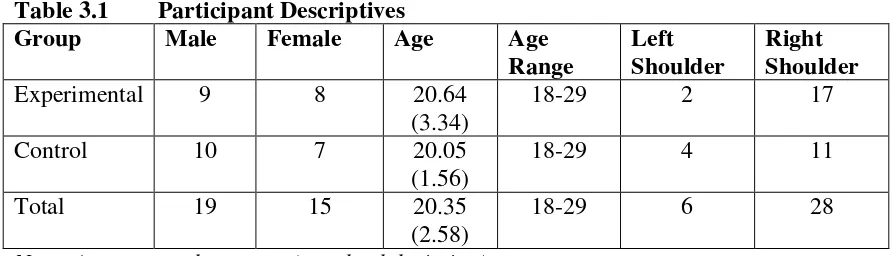
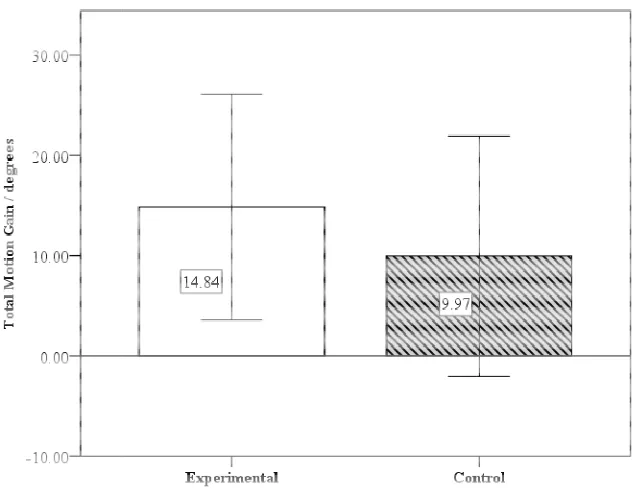
Total Motion of the Gleno
Similar analysis was done to compare differences for total motion between groups. This calculation is
ROM and ER ROM measurements. Observed m group (14.84° ± 11.27) were
Figure 3.7. Subjects in the more total motion than the
Figure
control group (165.60° ± 12.73
Total Motion of the Gleno-Humeral Joint
was done to compare differences for total motion between ulation is the sum between pre- and post-test dominant shoulder I ROM and ER ROM measurements. Observed mean gain scores for the experimental
° ± 11.27) were compared to the control group (9.97° ± 11.99°
the experimental group (170.22° ± 14.11°) exhibited slightly more total motion than the
Figure 3.7 Total Motion Gain Mean Values
± 12.73°) in pre-test measurements (Figure 3.8).
was done to compare differences for total motion between test dominant shoulder IR
experimental roup (9.97° ± 11.99°, p = 0.232); experimental group (170.22° ± 14.11°) exhibited slightly
Figure
test total motion for the experimental control group (175.57° ± 18.48
Figure
Figure 3.8 Total Motion Pre-Test Values
motion for the experimental group (185.05° ± 14.79°) as compared to ± 18.48°); Figure 3.9.
Figure 3.9 Total Motion Post-Test Values
Measurements and gain scores for all measurement pre- and post-tests, are categorized below in Tables 3.5 and 3.6, respectively. The level of significance for each measurement comparison is referenced in Table 3.6.
Table 3.5 Pre- and Post- Test measurements.
Measurement Test Experimental Control
Dom IR ROM Pre 60.36 (13.58) 58.42 (7.23) Post 73.60 (12.79) 66.89 (10.62) Non Dom IR ROM Pre 77.58 (11.76) 75.44 (7.40) Post 78.18 (15.03) 74.78 (9.29) Dom ER ROM Pre 109.86 (8.98) 107.18 (12.25) Post 111.45 (7.48) 108.67 (9.92) Non Dom ER ROM Pre 99.45 (9.10) 100.07 (6.92) Post 100.10 (7.42) 100.34 (5.99) IR Deficit ROM Pre 17.22 (6.76) 17.02 (3.63)
Post 4.58 (8.70) 7.89 (8.33)
Dominant Total Motion
Pre 170.22 (14.11) 165.60 (12.73) Post 185.05 (14.79) 175.57 (18.48)
Note: Means (Standard Deviation) in degrees
Dom = Dominant Shoulder
Non Dom = Non-Dominant Shoulder
Table 3.6 Gain Scores for All Measurements (Post-Test and Pre-Test).
Measurement Experimental Gain Control Gain Sig. level
p.
Effect Size *
Dominant IR 13.24 (7.78) 8.47 (8.78) 0.104 0.081
Non Dom. IR 0.60 (8.32) -0.65 (6.96) 0.636 0.007 Dominant ER 1.60 (7.45) 1.50 (7.52) 0.971 0.000 Non Dom. ER 0 .65 (4.17) 0.27 (5.23) 0.815 0.002 IR Deficit - 12.64 (11.49) -9.13 (8.33) 0.441 0.032
Total Motion 14.84 (11.27) 9.97 (11.99) 0.232 0.044
Note: Mean gain (Standard Deviation) in degrees
* Effect Size is estimated partial eta squared
Within groups analysis demonstrated the following results: 100% (n = 17) of the prone-passive participants increased IR ROM compared to 70% (n= 12) of the cross-body subjects; 100% (n = 34) of the participants in both groups decreased the amount of IR deficit; and, 95% (n = 16) of the prone-passive participants improved total degrees of motion compared to 82% (n = 14) in the cross-body treatment group.
IR ROM, IR Deficit, and Total Motion in Males and Females
Table 3.7 Gain Score for Dominant IR ROM, IR Deficit, and Total Motion, Males.
Measurement Experimental Gain Control Gain Sig. level
p.
IR ROM 14.39 (10.37) 6.71 (8.01) 0.087
IR Deficit -17.75 (11.58) -9.25 (7.16) 0.174
Total Motion 16.12 (12.63) 6.13 (11.34) 0.087
Note: Mean gain (Standard Deviation) in degrees
Table 3.8 references the mean gain score measurements for female participants in each group.
Table 3.8 Gain Score for Dominant IR ROM, IR Deficit, and Total Motion, Females.
Measurement Experimental Gain Control Gain Sig. level
p.
IR ROM 11.95 (3.47) 10.99 (9.84) 0.813
IR Deficit -6.89 (8.77) -8.95 (10.39) 0.852
Total Motion 13.39 (10.16) 15.46 (11.42) 0.719
Note: Mean gain (Standard Deviation) in degrees
IR and ER Correlation
Figure 3.10 Scatterplot with
Correlation Between Dominant IR and ER
Scatterplot with Linear Regression Line Demonstrating Correlation Between Dominant IR and ER Change
CHAPTER IV: DISCUSSION
The findings of this study have demonstrated the effects of the novel prone- passive stretching technique, and the cross-body technique on unilaterally restricted shoulder internal rotation in an overhead athlete population. We have introduced a previously untested treatment technique to the body of work in the area of conservative treatment for internal rotation deficit and/or posterior shoulder tightness.
Response to Intervention
At least two previous studies have demonstrated statistical significance for specific variables during the investigation of the cross-body technique (Manske et al., 2010; McClure et al., 2007). While our study did not achieve statistical significance between the prone-passive and cross-body stretching techniques, a determination of clinical significance could be inferred through the observed increase in IR ROM in the mean gain comparisons for the prone-passive subjects. Both the Manske et al. and McClure et al. studies refer to clinical significance and the application of the self-stretching technique as an appropriate method for improving IR restriction. While both of these studies demonstrated significance of an experimental group compared to a true control (no treatment applied), the purpose of this study was to determine if a novel stretching technique was more effective than a previously described clinical technique.
Gender Comparisons
Males responded more favorably than females to prone-passive treatment (Tables 3.7 and 3.8). Between groups, females did not respond differently depending on
treatment group, whereas males increased IR and decreased IR deficit more in the prone- passive group than the cross body. The comparisons observed between genders is not likely directly related to type of stretching technique, but more so a reflection of the prevalence of IR deficit in baseball (Table 3.4). In addition to the increased prevalence of IR deficit in males screened for inclusion in our study, males also demonstrated increased degrees of deficit compared to female participants. Thus, males had more room for improvement than females.
produce significant adaptive posterior shoulder muscle and capsule-ligamentous contracture, which likely leads to the observed higher prevalence of GIRD in males.
Differing Views on Total Motion in an IR Deficit Sample
Many have described a concomitant increase in external rotation (ER) when limitations to IR exist, with total motion remaining normal compared to the non-dominant shoulder. Others refer to this relationship as a shift toward ER (Borsa et al., 2005; Wilk et al., 2011). The assumption then may be that total motion remains the same even with a decrease in IR. Our findings do not concur with or support this assumption; rather, in our study, total motion was improved in the dominant shoulder after treatment for unilateral IR deficit. We observed increases to IR in the prone-passive group
compared to the cross-body participants, while ER remained unchanged regardless of treatment. The total motion concept states that an equal amount of IR and ER exists in the dominant and non-dominant shoulders, even in cases of GIRD (Wilk et al., 2002). Our results suggest there are differences in the degrees of total motion when IR ROM has been improved, producing an increase in total motion in an individual with IR deficit. We speculate that dominant shoulder total motion may be increased in healthy, non-GIRD overhead athletes compared to the non-dominant shoulder. These findings lead us to believe that soft tissue contracture, primarily in the posterior shoulder musculature, leads to GIRD and/or posterior shoulder tightness (PST).
Capsule or Muscle?
the exact reason why overhead throwers tend to gravitate toward unilateral internal rotation deficit, and probably from early on in their sport careers, but both prior and recent research is focusing on two soft tissue structures as the causative factor- the posterior gleno-humeral capsule and posterior shoulder musculature (Borstad &
Dashotter, 2011). This recent cadaveric study has re-visited the notion of the capsule as the primary cause of GIRD, but certainly has left the door open to suggest muscular tightness as another contributor.
about the differences between the two tissues is in the contractility properties of each. A capsule, to our knowledge, cannot be strengthened through therapeutic exercise like a muscle. Therefore, the musculotendinous units providing dynamic stabilization of the posterior shoulder may provide more stability to the gleno-humeral joint, especially in overhead dominant sports. This implies that a musculotendinous unit undergoes more tensile forces that lead to contracture than the capsule. Manske and colleagues (2010) demonstrated IR restrictions started to return as soon as four weeks after IR stretching and/or joint mobilization treatment stopped. This observation suggests that the visco-elastic property of muscle and the functional shortening thereof is the primary contributor of GIRD and/or PST in an otherwise healthy overhead athlete. Therefore, both soft tissues in the posterior shoulder girdle should be the focus of a conservative treatment protocol.
While we did not perform a follow-up measurement in this study, we did emphasize to participants the importance of continuing a maintenance internal rotation stretching program. Though IR deficits were decreased in this study, it would be illogical to think that ROM restrictions would not return in the absence of a maintenance
stretching program for overhead athletes.
Parameters of the Stretching Technique
Creep theory of collagenous tissue emphasizes the importance of repeated stretching or, more simply, movement within soft tissue in order to maintain elasticity (Rigby et al., 1959). This theory can be thought of as a change in length proportional to the amount of strain applied over time. An interesting observation was noted by the clinicians providing the prone-passive stretching technique in this study. Un-measured improvements to IR ROM were noticed after only 4-6 treatment sessions in the
experimental group. An obvious explanation to this occurrence would be the acute stretch response as demonstrated in many other studies investigating IR ROM restriction response to a single stretching treatment (Laudner et al., 2008; Moore et al., 2011). However, participants in the experimental group were not being treated on a daily basis; rather, treatments were administered three to four times per week. Even with multiple days in between sessions, we noticed a reduction to the previously encountered soft tissue limitations to IR ROM within the first 2 weeks of the program. While this observation was not measured or substantiated, this response to treatment may imply a shorter creep response, leading us to suggest a reduced number of treatments are needed to elicit improvements in IR ROM at the shoulder joint. This same phenomenon would also suggest a rather rapid reversal in tissue elongation in the absence of maintenance stretching.
Tensile Forces of the Stretching Techniques
torsion, is imparted on both capsular and musculotendinous tissues during the prone- passive technique. Torsional stress acts on the majority of the capsule surface area, which may allow for further stretching of the musculature when compared to the localized tensile forces in the posterior shoulder during the adduction movement of the cross-body stretch. The torsional stress may enhance elongation on multiple components of these tissues, facilitating a greater increase in ROM. A capsular twisting technique has previously been described as an effective treatment for adhesive capsulitis, a
multidirectional restriction to shoulder motion (Henry, 1995).
The positioning of the patient in the prone position of this novel stretch allows other advantages compared to the self-stretch techniques proposed in this study. Instead of flexing the shoulder as in the sleeper stretch (whereby the humerus is forward flexed to 90° and somewhat horizontally adducted due to the weight of the upper body in a side lying position), the abducted position in the prone-passive stretch allows for greater capsular twist because the motions occurring in the shoulder girdle are occurring in both the sagittal and frontal planes (Hertling & Kessler, 2006). Flexing the arm, as in the sleeper stretch, may result in less capsular twisting, and therefore less tissue elongation when compared to the prone-passive technique.
later. We would suggest this soreness may be a result of the breakdown of adhesions and/or contracture in significantly compromised posterior shoulder tissues since the soreness tended to dissipate rather quickly and did not compromise the treatment schedule.
Importance of Maintaining Normal Glenohumeral Internal Rotation in Overhead
Athletes
It is well accepted that the prevalence of IR deficit in overhead athletes increases the risk of shoulder and elbow injuries to those individuals. A notable study has
demonstrated this correlation, particularly in baseball athletes (Wilk et al., 2011). This type of study influences the preventative nature of strength and conditioning programs as well as athletic therapy protocols.
Common injuries observed in patients with GIRD tend to reflect the repetitive stresses the shoulder undergoes in overhead sports. For instance, rotator cuff tendonitis, sub-acromial impingement, and labrum injury are all reflective of the pathologic
mechanics that are caused by restrictions to IR ROM in activities such as the overhand- throwing motion.
arthrokinematics in the throwing shoulder, one may be reducing the risk of the repetitive stress types of injuries.
Conclusion
REFERENCES
Awan, R., Smith, J., & Boon, A. J. (2002). Measuring shoulder internal rotation range of motion: A comparison of 3 techniques. Archives of Physical Medicine and Rehabilitation, 83(9), 1229-1234.
Bach, H. G. (2006). Posterior capsular contracture of the shoulder. Journal of the American Academy of Orthopaedic Surgeons, 14(5), 265-277.
Beach, M. L., Whitney, S. L., & Dickoffhoffman, S. A. (1992). Relationship of shoulder flexibility, strength, and endurance to shoulder pain in competitive swimmers.
Journal of Orthopaedic & Sports Physical Therapy, 16(6), 262-268.
Bennett, G. E. (1941). Shoulder and elbow lesions of the professional baseball pitcher.
Journal of American Medical Association, 117(7), 510-514.
Bennett, G. E. (1959). Elbow and shoulder lesions of baseball players. American Journal of Surgery, 98(3), 484-492.
Bigliani, L. U., Codd, T. P., Conner, P. M., Levine, W. N., Littlefield, M. A., Hershon, S. J. (1997). Shoulder motion and laxity in the professional baseball player.
American Journal of Sports Medicine, 25(5), 609-613.
Blanch, P. (2004). Conservative management of shoulder pain in swimming. Physical Therapy in Sport, 5(3), 109-124.
Borsa, P. A.,Wilk, K. E., Jacobson, J. A., Scibek, J. S., Dover, G. C., Reinold, M. M., Andrews, J. R. (2005). Correlation of range of motion and glenohumeral translation in professional baseball pitchers. American Journal of Sports Medicine, 33(9), 1392-1399.
Borstad, J.D., Dashottar, A. (2011). Quantifying strain on posterior shoulder tissues during 5 simulated clinical tests: A cadaver study. Journal of Orthopaedic and Sports Physical Therapy, 41(2), 90-99.
Burkhart, S. S., Morgan, C. D., Kibler, W. B. (2003a). The disabled throwing shoulder: Spectrum of pathology- Part I: Pathoanatomy and biomechanics. Arthroscopy-the Journal of Arthroscopic and Related Surgery, 19(4), 404-420.
Burkhart, S. S., Morgan, C. D., Kibler, W. B. (2003b). The disabled throwing shoulder: Spectrum of pathology - Part II: Evaluation and treatment of SLAP lesions in throwers. Arthroscopy-the Journal of Arthroscopic and Related Surgery, 19(5), 531-539.
Campbell, D. T., Stanley, J. C., (1963). Experimental and Quasi-Experimental Designs for Research (pp. 22-24). Houghton Mifflin Co.: Boston, MA.
Cools, A. M., Declercq, G., Cagnie, B., Cambier, D., Witvrouw, E. (2008). Internal impingement in the tennis player: rehabilitation guidelines. British Journal of Sports Medicine, 42(3), 165-171.
Crockett, H. C., Gross, L. B., Wilk, K. E., Schwartz, M. L., Reed, J., O'Mara, J., Reilly, M.T., Dugas, J. R., Meister, K., Lyman, S., Andrews, J. R. (2002). Osseous adaptation and range of motion at the glenohumeral joint in professional baseball pitchers. American Journal of Sports Medicine, 30(1), 20-26.
Cyriax, J. (1975). Textbook of Orthopedic Medicine: Diagnosis of Soft Tissue Lesions, 6th ed,(1). Baltimore, MD: Williams and Wilkins.
de Winter, Heemskerk, M., Terwee, C., Jans, M., Schaardenburg, D., Scholten, R., Bouter, L. (2004). Inter-observer reproducibility of measurements of range of motion in patients with shoulder pain using a digital inclinometer. BMC Musculoskeletal Disorders, 5(18). doi: 10.1186/1471-2474-5-18
Dillman, C. J., Fleisig, G. S., Andrews, J. R. (1993). Biomechanics of pitching with emphasis upon shoulder kinematics. Journal of Orthopaedic & Sports Physical Therapy, 18(2), 402-408.
Ellenbecker, T. S., Roetert, E. P., Bailie, D. S., Davies, G. J., Brown, S. W. (2002). Glenohumeral joint total rotation range of motion in elite tennis players and baseball pitchers. Medicine and Science in Sports and Exercise, 34(12), 2052-2056.
Ellenbecker, T. S., Roetert, E. P., Piorkowski, P. A., Schulz, D. A. (1996). Glenohumeral joint internal and external rotation range of motion in elite junior tennis players.
Journal of Orthopaedic & Sports Physical Therapy, 24(6), 336-341.
Fleisig, G. S., Andrews, J. R., Dillman, C. J., Escamilla, R. F. (1995). Kinetics of baseball pitching with implications about injury mechanisms. American Journal of Sports Medicine, 23(2), 233-239.
Goldman, B., Sauers, EL. (2004). The acute effectiveness of PNF stretching and joint mobilizations for increasing posterior shoulder mobility of the professional baseball player. Journal of Athletic Training, 39(2), S-64.
Grossman, M. G., Tibone, J. E., McGarry, M. H., Schneider, D. J., Veneziani, S., Lee, T. Q. (2005). A cadaveric model of the throwing shoulder: A possible etiology of superior labrum anterior-to-posterior lesions. Journal of Bone and Joint Surgery-American Volume, 87A(4), 824-831.
Halbrecht, J. L., Tirman, P., Atkin, D. (1999). Internal impingement of the shoulder: Comparison of findings between the throwing and nonthrowing shoulders of college baseball players. Arthroscopy-the Journal of Arthroscopic and Related Surgery, 15(3), 253-258.
Harryman, D. T., Sidles, J. A., Clark, J. M., McQuade, K. J., Gibb, T. D., Matsen, F. A. (1990). Translation of the humeral head on the glenoid with passive glenohumeral motion. Journal of Bone and Joint Surgery-American Volume, 72A(9), 1334-1343.
Hertling, D., Kessler, R. M. (2006). Management of Common Musculoskeletal Disorders: Physical Therapy Principles and Methods, 4th ed, (288-289).
Henry, J. A. (1995). Manual therapy of the shoulder. In Kelly, M. J., Clark, W. A. (Eds.),
Orthopaedic Therapy of the Shoulder (285-336). Philadelphia: JB Lippincott. Hung, C. J., Hsieh, C. L., Yang, P. L., Lin, J. J. (2010). Relationship between posterior
shoulder muscle stiffness and rotation in patients with stiff shoulder. Journal of Rehabilitation Medicine, 42(3), 216-220.
Jobe, C. M. (1995). Posterior superior glenoid impingement- Expanded spectrum.
Arthroscopy, 11(5), 530-536.
Jobe, F. W., Giangarra, C. E., Kvitne, R. S., Glousman, R. E. (1991). Anterior capsulolabral reconstruction of the should in athletes in overhand sports.
American Journal of Sports Medicine, 19(5), 428-434.
Johansen, R. L., Callis, M., Potts, J., Shall, L. M. (1995). A modified internal -roation stretching technique for overhand and throwing athletes. Journal of Orthopaedic & Sports Physical Therapy, 21(4), 216-219.
Kibler, B. W., Chandler, T. J., Livingston, B. P., Roetert, E. P. (1996). Shoulder range of motion in elite tennis players - Effect of age and years of tournament play.
American Journal of Sports Medicine, 24(3), 279-285.