R E S E A R C H
Open Access
Different curcumin forms selectively bind
fibrillar amyloid beta in post mortem
Alzheimer
’
s disease brains:
Implications for
in-vivo
diagnostics
Jurre den Haan
1*, Tjado H. J. Morrema
2, Annemieke J. Rozemuller
2, Femke H. Bouwman
1and Jeroen J. M. Hoozemans
2Abstract
The combined fluorescent and Aβ-binding properties of the dietary spice curcumin could yield diagnostic purpose in the search for a non-invasive Aβ-biomarker for Alzheimer’s disease (AD). However, evidence on the binding properties of curcumin, its conjugates and clinically used bio-available formulations to AD neuropathological hallmarks is scarce. We therefore assessed the binding properties of different curcumin forms to different neuropathological deposits in post-mortem brain tissue of cases with AD, other neurodegenerative diseases, and controls. Post mortem brain tissue was histochemically assessed for the binding of curcumin, its isoforms,
conjugates and bio-available forms and compared to routinely used staining methods. For this study we included brains of early onset AD, late onset AD, primary age-related tauopathy (PART), cerebral amyloid angiopathy (CAA), frontotemporal lobar degeneration (FTLD) with tau or TAR DNA-binding protein 43 (TDP-43) inclusions, dementia with Lewy bodies (DLB), Parkinson’s disease (PD) and control cases without brain pathology. We found that curcumin binds to fibrillar amyloid beta (Aβ) in plaques and CAA. It does not specifically bind to inclusions of protein aggregates in FTLD-tau cases, TDP-43, or Lewy bodies. Curcumin isoforms, conjugates and bio-available forms show affinity for the same Aβstructures. Curcumin staining overlaps with immunohistochemical detection of Aβin fibrillar plaques and CAA, and to a lesser extent cored plaques. A weak staining of neurofibrillary tangles was observed, while other structures immunopositive for phosphorylated tau remained negative. In conclusion, curcumin, its isoforms, conjugates and bio-available forms selectively bind fibrillar Aβin plaques and CAA in post mortem AD brain tissue. Curcumin, being a food additive with fluorescent properties, is therefore an interesting candidate for in-vivo diagnostics in AD, for example in retinal fluorescent imaging.
Keywords:Curcumin, Amyloid-beta, Alzheimer’s disease, Cerebral amyloid angiopathy (CAA), Neurodegeneration, Biomarker, Immunohistochemistry
Introduction
Aggregation and extracellular deposition of the amyloid-beta (Aβ) peptide in amyloid plaques is a pathological hallmark and an early event in the pathophysiology of Alzheimer’s disease (AD) [4,18]. Currently AD diagnosis can be made with help of clinical criteria with the
support of measurements of Aβ1–42and tau−181 in
cere-brospinal fluid (CSF), amyloid-positron emission tomog-raphy (PET), cortical atrophy on magnetic resonance imaging (MRI), or hypo-metabolism by fluorodeoxyglu-cose–PET (FDG-PET) [35]. As these are all either invasive or time consuming investigations, a patient friendly and repeatable amyloid or Aβ biomarker is urgently needed for diagnosis as well as therapy monitoring in clinical trials [15]. Curcumin is a dietary spice that has been shown to have combined fluorescent and Aβ-binding properties [16, 37, 39]. Therefore, curcumin has the potential to * Correspondence:[email protected]
1Department of Neurology, Amsterdam Neuroscience, VU University Medical
Center Alzheimer Center, Mailbox 7057, 1007, MB, Amsterdam, the Netherlands
Full list of author information is available at the end of the article
be used as a tool in a non-invasive Aβ-directed diag-nostic assay for AD. A recently published paper by Koronyo et al. shows possible application of curcumin for in-vivo detection of retinal amyloid [22].
Curcumin is a polyphenolic compound derived from the root Curcuma longa or turmeric, traditionally used in Indian cuisine and Ayurvedic medicine. Turmeric naturally contains three curcuminoids namely; curcumin (≈77%), demethoxycurcumin (DMC)(≈17%) and bis- de-methoxycurcumin (BDMC)(≈3%) [10]. Curcumin is fluor-escent by nature [16, 37]. It has anti-carcinogenic, anti-inflammatory, anti-oxidant and anti-angiogenic prop-erties and binds to Aβand interferes with Aβaggregation [10, 28]. More specifically curcumin binds Aβ-oligomers and fibrils in vitro [41, 52], binds plaques in APPSWE/
PS1ΔE9, Tg2576 and 5xFAD transgenic mice [23,29,30,53]
and binds Aβ-plaques in post mortem human AD brain tissue [23, 39, 49, 53]. Two reports also described moderate binding of curcumin to neurofibrillary tan-gles in AD brain tissue [36, 39].
In order to apply curcumin as a tool for in-vivo detec-tion of Aβits poor bio-availability and in-vivo metabol-ism should be considered. The majority (35–89%) of the orally administered curcumin is present in the feces, as the intestinal mucosa and mucus form a physical barrier [14, 43]. Secondly, curcumin that does reach the circula-tion undergoes phase 1 (reduccircula-tion) and phase 2 metabol-ism (conjugation) in the liver. Reductases reduce curcumin to dihydrocurcumin, tetrahydrocurcumin, hexahydrocur-cumin etc. [31,32,42]. In addition, curcumin is conjugated to sulfates and glucuronides [5,17,31,32]. Therefore, the majority of circulating curcumin is conjugated. To over-come curcumin’s poor bio-availability, nano-particulate drug delivery systems have been developed for oral admin-istration including micelles, solid lipid nanoparticles and li-posomes (Theracurmin™[20,40,44], Novasol™[7,21,45], Longvida™[11,22]).
So far, there are no reports on the Aβ-binding proper-ties of conjugated curcumin or bio-available forms of curcumin. Insight into binding properties of circulating curcuminoids in the blood (e.g. DMC, BDMC, and cur-cumin conjugates) and specific bio-available formula-tions is needed for the design of biomarker assays using oral (bio-available) curcumin formulations. In this study, we assessed the binding properties of curcumin, its isoforms, its conjugates and clinically used bioavailable curcumin supplements to neuropathological hallmarks of AD and other neurodegenerative features in post mortem brain tissue.
Materials and methods
Post mortem brain tissue
Post mortem brain tissue was obtained from the Netherlands Brain Bank (NBB; Amsterdam, the
Netherlands). Prior to death, donors signed informed consent for brain autopsy and use of brain tissue and medical records for research purposes. For this study we selected 5 early onset AD (EOAD)-patients, 5 late onset AD (LOAD) patients, 5 AD cases with capillary cerebral amyloid angiopathy (CAA type-1), 5 controls, 5 cases with primary age related tauopathy (PART), 3 fronto-temporal lobar degeneration (FTLD) cases with tau pathology (FTLD-tau), 1 FTLD case with TDP-43 path-ology (FTLD-TDP), 2 Parkinson’s disease (PD) cases and 1 dementia with Lewy bodies (DLB) case who donated their brains to the NBB between 2000 and 2014. EOAD cases had a reported disease onset before 65 years of age (mean age of death 61.6 years), while LOAD cases had reported disease onset > 65 years. Neuropathological diagnosis of AD was made following the NIA-AA cri-teria including Thal-staging for Aβ stage, Braak-and-Braak-staging for NFTs and CERAD-staging for neuritic plaques [1, 38, 48]. Pathological diagnosis of CAA-type 1, FTLD (tau and TDP-43), DLB and PD were made fol-lowing Thal-, Mackenzie-, McKeith- and Braak-staging criteria respectively [2, 25–27, 33, 34, 47]. Two of the FTLD-Tau cases were P301L mutations (chromosome 17), while one was a sporadic Pick’s disease case. The FTLD-TDP43 case was a progranulin mutation. See Table1for cohort characteristics.
Immunohistochemistry (IHC)
For each case we assessed formalin-fixed paraffin-em-bedded tissue sections (5 μm thick) from mesenceph-alon, hippocampus, frontal or occipital lobes. Paraffin-embedded tissue sections were deparaffinized and rehy-drated using xylene, alcohol and phosphate-buffered saline (PBS; pH 7.4). Endogenous peroxidase activity was blocked by 0.3% H2O2-treatment of the sections. Heat
induced antigen retrieval was performed in 0.01 M citrate buffer pH 6.0 using an autoclave. Sections were incubated overnight with primary antibody and subse-quently washed with PBS (see details in Table 2). Pri-mary antibodies were detected using EnVision (Dako, Glostrup, Denmark) followed by washing in PBS. Anti-body complex binding was visualized using 3,3′-diami-nobenzidine (DAB) and sections were counterstained with hematoxylin. Stained sections were covered using Quick-D (Klinipath, Beek, the Netherlands).
Thioflavin-S staining
Table 1Cohort characteristics
# PM delay (h:min) Pathological diagnosis (mutation) Sex Age Braak Amyloid
1 6:35 Control F 92 III 0
2 7:10 Control F 78 I A
3 4:35 Control F 78 II A
4 7:15 Control M 95 II B
5 5:15 Control M 83 I A
6 5:00 EOAD M 61 VI C
7 5:05 EOAD M 59 VI C
8 4:40 EOAD M 62 V B
9 4:45 EOAD M 64 V C
10 5:15 EOAD M 62 VI C
11 5:30 LOAD M 88 VI C
12 7:00 LOAD F 92 V C
13 4:40 LOAD F 89 V C
14 6:25 LOAD F 91 IV C
15 3:05 LOAD M 74 VI C
16 6:05 CAA type 1 M 68 II C
17 4:20 CAA type 1 M 81 V C
18 4:20 CAA type 1 F 96 V C
19 3:25 CAA type 1 M 94 V C
20 6:00 CAA type 1 F 75 V C
21 5:30 PART F 81 II 0
22 3:52 PART F 89 III 0
23 5:00 PART F 87 II 0
24 5:50 PART F 93 II 0
25 6:35 PART F 103 IV 0
26 3:35 FTLD-TDP (Progranulin) F 76 n.a. A
27 5:23 FTLD-Tau (P301L) M 60 n.a. 0
28 6:25 FTLD-Tau (P301L) M 64 n.a. 0
29 6:15 FTLD-Tau (PiD) M 60 n.a. 0
30 24:00 PD M 57 I 0
31 33:00 LBD/AD F 78 VI B
32 14:00 PD M 68 I 0
Abbreviations:CAA type 1capillary cerebral amyloid angiopathy type 1,EOADearly onset Alzheimer’s disease,FTLD-taufrontotemporal lobar degeneration with
tau pathology,FTLD-TDP-43frontotemporal lobar degeneration with TDP-43 pathology,LOADlate onset Alzheimer’s disease,n.a.not applicable,PARTprimary
age-related tauopathy,PiDPick’s disease,PM delaypost-mortem delay andPDParkinson’s disease
Table 2Primary antibodies used for immunohistochemistry and fluorescent staining
Antibody Source Species Dilution
IC-16 Carsten Korth, Heinrich Heine University Dusseldorf, Düsseldorf, Germany [50] Mouse 1:400
4G8 Biolegend, San Diego, CA, USA Mouse 1:8000
AT8 Invitrogen, Thermo Fisher Scientific, MA, USA Mouse 1:800
pTDP-43 Cosmo Bio, Tokyo, Japan Mouse 1:8000
Curcuminoid staining
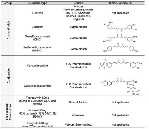
Curcuminoids, conjugates and bio-available forms of curcumin were dissolved in 0.5 M NaOH to a stock concentration of 20 mg/ml (see details in Table 3). Final concentrations of 0.2, 0.1 and 0.05 mg/ml were prepared by dilution in PBS. Deparaffinized and rehy-drated sections were incubated with curcumin solu-tions for 10 min, followed by washing in PBS. Stained sections were covered using TBS/glycerol mounting medium.
Combined curcumin staining and immunohistochemistry
Sections were deparaffinized and rehydrated, followed by blocking of endogenous peroxidase and antigen retrieval as described above. Subsequently, curcumin staining was
performed, sections were washed in PBS, and immuno-histochemistry was performed using anti-phosphorylated tau (anti-pTau, AT8,1:800) and anti-Aβ (IC-16, 1:400). Incubation was performed overnight, followed by wash-ing in PBS. Secondary fluorescently labeled antibody (Goat Anti-mouse 594, Life Tech, 1:250) was incubated for 1.5 h, followed by washing in PBS. Sections were counterstained with 4′6-diamidino-2-phenylindole (DAPI) and covered with TBS/Glycerol.
Imaging
IHC sections were imaged by an Olympus BX41 micro-scope using Leica Application Suite (LAS) AF lite soft-ware (Wetzlar, Germany). Fluorescently stained slides were imaged by a Leica DMi8 fluorescence microscope
using LAS AF lite and ImageJ software (National Institue of Health, USA).
Results
Curcumin binding in control and AD brain tissue
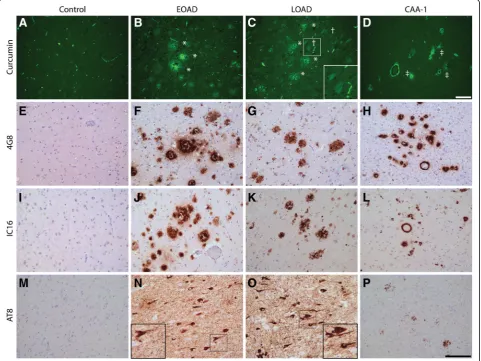
In general, it was observed that curcumin binding on human brain tissue presented no background staining and auto-fluorescent signals in neurons. In brain tissue of EOAD, LOAD and AD cases with CAA (CAA type 1), curcumin strongly bound to amyloid plaques and CAA (Fig.1). Curcumin staining revealed different types of plaques. We observed intense staining of primitive/ compact plaques and moderate staining of the cores in cored plaques. In AD cases, neurofibrillary tangles (NFTs) showed a weak detection by curcumin. Adjacent sections were assessed by immunohistochemistry for the presence of Aβand pTau in a corresponding area, which confirmed the presence of these pathological structures.
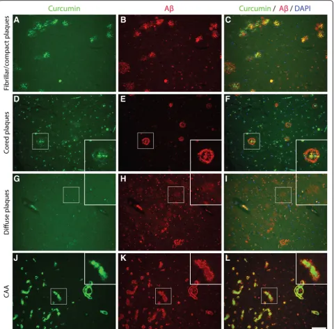
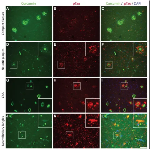
The binding of curcumin to these pathological struc-tures was investigated by co-stainings for curcumin and Aβ (IC-16) or pTau (AT8). Primitive/compact plaques showed almost a complete overlap of curcumin signal and immunodetection by Aβ(Fig. 2). Classic cored pla-ques presented co-labeling of the core, while the corona of these plaques were immuno-labelled by anti-Aβ and not by curcumin. No binding of curcumin was observed in diffuse Aβdeposits. CAA affected blood vessels were stained most intensively by curcumin and showed a complete overlap with the detection of CAA by Aβ. While compact plaques showed moderate neuritic changes, as presented by anti-pTau immuno-labeling, no co-labelling of anti-pTau and curcumin was present in these pla-ques (Fig. 3). Neuritic cored plaques showed a strong immunodetection of pTau around the core. The core of neuritic plaques showed a strong binding of curcumin while co-labelling with anti-pTau was absent. A strong
binding of curcumin was observed in CAA, however, no co-labelling with anti-pTau, observed around CAA af-fected capillaries, was detected. Co-labeling with anti-pTau confirmed that curcumin weakly detects NFTs.
Curcumin does not bind to non-AD pathology
We investigated whether curcumin showed binding to protein inclusions or aggregates in non-AD pathologies. PART is characterized by the presence of NFTs and tau
pathology in the absence of amyloid deposits [8, 19]. In cases with PART no specific binding of curcumin was observed. Co-labeling and single IHC with anti-pTau confirmed the presence of tau pathology and NFTs in the tissue section. In PART no binding of curcumin to NFTs was observed. Next, we assessed the binding of curcumin to tau inclusions in different FTLD-tau cases. Inclusions that were positive for pTau in cases with a MAPT P301L mutation, or in a case with sporadic
Pick’s disease, did not show binding of curcumin. Also inclusions positive for TDP-43 in FTLD-TDP, and α-synuclein containing inclusions in PD and DLB were not detected by curcumin (Fig. 4).
Staining of curcumin isoforms, conjugates and bio-available forms
In order to translate above findings to a possible in-vivo biomarker assay we assessed binding properties of differ-ent curcuminoids (DMC, BDMC), conjugates, and
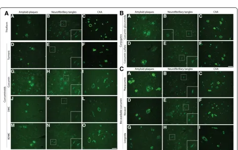
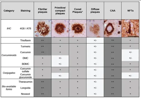
bio-available forms in AD cases. Given curcumin’s chemical similarities with Thioflavin, sharing an aromatic ring, we compared stainings to Thioflavin-S staining (Fig. 5). An overview of staining of different curcumin forms and Thioflavin-S to plaques, CAA and NFTs is given in Table 4.
First, as turmeric also contains two isoforms: DMC (≈17%) and BDMC (≈3%), we assessed their binding. Similar to Thioflavin-S and curcumin, both isoforms bound amyloid plaques and CAA and showed weak
Fig. 4Curcumin binding in non-AD pathologies. Curcumin staining of PART, FTLD-tau, FTLD-TDP-43, and PD cases (a-e). Fluorescent co-stainings for comparison of curcumin and tau (FTLD-tau), TDP-43 (FTD-TDP-43), andα-synuclein (PD) (f-j). Immunohistochemical DAB stainings for tau (AT8), TDP-43 (pTDP-43) andα-synuclein (LB-509) are shown for reference (k-o). Scale barsa-j50μm. Scale barsk-o100μm. Abbreviations: DAPI = 4′,6-diamidino-2-fenylindole, FTLD-tau = frontotemporal lobe degeneration with tau pathology, FTLD-TDP-43 = frontotemporal lobe degeneration with TDP-43 pathology, PART = primary age related tauopathy
staining of NFTs (Fig. 5a). Compared to Thioflavin-S, staining of fibrillar and primitive plaques by isoforms was slightly less apparent (Table4).
Next, we assessed binding properties of curcumin con-jugates (glucuronides, sulfates). As, curcumin is conju-gated by the liver to make it water soluble, conjugates represent the majority of circulating curcuminoids. Curcumin-glucuronide and curcumin-sulfate bound to amyloid plaques and CAA (Fig. 5b). Curcumin glucuro-nide showed slightly less apparent staining of fibrillar, primitive/compact plaques and NFTs compared to Thioflavin-S (Table 4).
Finally, in order to translate above findings to clinically used oral bio-available formulations of curcumin we compared three bio-available formulations, namely Theracurmin, Novasol and Longvida. All three forms showed staining of primitive/compact plaques, the core of cored plaques, CAA and faintly stain NFTs, similar to Thioflavin-S (Table4, Fig.5c).
Discussion
This study demonstrates that curcumin, curcuminoids, curcumin conjugates and bio-available formulations con-taining curcumin, bind to fibrillar amyloid in primitive/ compact plaques, cored plaques and CAA in AD brain tissue. Curcumin isoforms and conjugates show few se-lective binding to NFTs in AD brain tissue and no bind-ing to other neuropathological deposits, e.g. pTau and TDP-43 positive structures in FTLD and alpha-synuclein inclusions in PD and DLB. To the best of our knowledge we are the first to report that curcumin isoforms, conju-gates and bio-available forms have similar staining prop-erties as curcumin.
Comparing plaque types, binding of curcumin was more pronounced to amyloid deposits with a dense amyloid structure, as shown in Fig.2 and Table 4. This is supported by co detection of curcumin with amyloid-beta in compact plaques while in contrast, diffuse pla-ques showed a lack of co-detection (Fig. 3). This
Table 4Overview of binding of different curcumin forms to different pathological deposits
Staining of different pathological deposits by different curcumin forms was qualitatively compared for different plaque types, cerebral amyloid angiopathy (CAA)
and neurofibrillary tangles (NFTs). Immuno-histochemical (IHC) stainings for amyloid-beta (4G8, plaques and CAA) and phosphorylated tau (AT-8, NFT’s) are shown
for reference. Scalebar 50μm
++: clearly visible, +: visible, +/−: visible with appreciation of morphology. *The staining of the core of cored plaques rather than the corona was assessed here
indicates that curcumin binds preferably to dense amyl-oid structures, such as fibrillar plaques, rather than the amyloid beta peptide itself. The detection of amyloid by curcumin is comparable to the detection of amyloid by Thioflavin-S. This could be explained by the fact that curcumin and Thioflavin have chemical similarities, sharing a double aromatic ring [52].
Previous studies showed that curcumin binds Aβ-oligomers and fibrils in vitro [33, 44], Aβ-plaques in APPSWE/ PS1ΔE9, Tg2576 and 5xFAD transgenic mice [19,24,30,45] and in post mortem human AD brain tis-sue [19, 31, 41, 45]. The latter human studies however assessed relatively small patient cohorts and did not as-sess other neurodegenerative diseases. Our findings in a well-characterized and large cohort of cases confirm earlier findings of specific binding of curcumin to fibril-lar Aβ plaques, weak binding to NFT’s and rule out binding to pathological protein aggregates in other neu-rodegenerative diseases. Binding to fibrillary plaques might be of clinical relevance, as an increase of fibrillar plaques is associated with clinical progression to demen-tia [6]. In addition to curcumin binding to amyloid pla-ques, Mutsuga et al. described curcumin staining to CAA in larger vessels of an AD case as well as in various animals [39]. CAA can be subdivided in CAA type 1 (CAA-1) and CAA type 2(CAA-2) [47]. CAA-1 is characterized by Aβ presence in leptomeningeal and cortical arteries, arterioles, veins and venules as well as cortical capillaries, while CAA-2 on the other hand, affects leptomeningeal and cortical vessels with the exception of capillaries. In addition to earlier findings in CAA-2 by Mutsuga et al. we showed that curcumin also binds to CAA in capillaries in CAA-1 cases [39].
To the existing literature we added the comparison of the binding properties of different curcumin forms in human brain tissue, which is needed to translate findings for possible use in in-vivo diagnostic or therapeutic stud-ies. The majority of curcumin and its isoforms are con-jugated in-vivo and concon-jugated curcumin represents a large proportion of circulating curcumin [12]. To over-come poor bio-availability, delivery systems containing curcuminoids were developed. We showed that isoforms and conjugates and newly developed delivery systems of curcumin bind amyloid plaques and CAA and show comparable fluorescent properties as curcumin. For diagnostic studies this implies that all contents of newly developed delivery systems, including curcumin, DMC an BDMC and secondary formed curcumin conjugates harbor amyloid binding properties and could yield diag-nostic purpose. Newly developed delivery systems, e.g. micelles, solid lipid nanoparticles and liposomes, did not show to affect amyloid binding properties. Similar bind-ing properties could also indicate a therapeutic role of
isoforms and conjugates. Previous studies showed that curcumin reverses existing amyloid pathology and associated neurotoxicity in a mouse model [9, 53]. If conjugates harbor the same functional effects on amyloid formation and related neurotoxicity is un-known. More research is therefore needed to assess possible functional effects of isoforms and conjugates on AD pathology.
Strengths of our study are the well-described patient cohort including various neurodegenerative features, thorough assessment of different curcumin forms and comparison with commonly used neuropathological IHC. Our patient cohort consists of a fair amount of well described AD-cases, cases with other neurodegenera-tive conditions as well as healthy controls, while pre-vious studies assessed binding properties of curcumin mostly in animal models. Applying commonly used neuropathological IHC-staining we directly compared curcumin with established staining methods, which al-lows assessment of its specificity towards pathological structures. A limitation of our study is that the use of high concentrations curcumin in post mortem stain-ing might not reflect concentrations that could be reached in-vivo. Although turmeric, with a mass percentage of only ≈3% curcuminoids, showed similar staining patterns and intensity as pure curcumin it should be noted that clinically reached concentrations might be unable to effectively bind pathology. Future studies might therefore test the minimal concentra-tion needed to label pathology.
Secondly, we tested a wide variety of curcumin forms, however not synthesized forms of curcumin (e.g. CRANAD-28). CRANAD-28 might have amyloid pla-ques and CAA binding properties and beneficial blood brain barrier penetration, yet is not applicable for hu-man use [54]. Lastly, as we focused on isoforms and conjugates representing the majority of circulating cur-cumin we did not test reduced forms of curcur-cumin. Fu-ture studies might test binding properties of synthesized and reduced forms of curcumin.
curcuminoids might be used as labeling fluorophore for non-invasive fluorescent retinal imaging.
Conclusion
In conclusion, curcumin, its isoforms, conjugates and bio-available forms bind to fibrillar Aβ plaques and CAA, and faintly stain neurofibrillary tangles in post mortem AD brain tissue. They do not show binding in control brain or to specific structures observed in other neurodegenerative disease like FTLD and PD. As conju-gates and bio-available curcumin forms show comparable binding properties, curcumin might be an interesting candidate for in-vivo diagnostics in AD like retinal fluores-cent imaging.
Abbreviations
AD:Alzheimer’s disease; Aβ: Amyloid-beta; BDMC: Bis-demethoxycurcumin; BMC: Demethoxycurcumin; CAA: Cerebral amyloid angiopathy; CAA-1: Cerebral amyloid angiopathy type 1; CAA-2: Cerebral amyloid angiopathy type 2; CSF: Cerebrospinal fluid; DAB: 3,3′ –diaminobenzidine; DAPI: 4′ 6-diamidino-2-phenylindole; DLB: Dementia with Lewy bodies; EOAD: Early onset Alzheimer’s disease; FTLD: Frontotemporal lobar degeneration; IHC: Immunohistochemistry; LOAD: Late onset Alzheimer’s disease; MRI: Magnetic resonance imaging; NBB: The Netherlands Brain Bank; NFTs: Neurofibrillary tangles; PART: Primary age-related tauopathy; PBS: Phosphate-buffered saline; PD: Parkinson’s disease; PET: Positron emission tomography; PiD: Pick’s disease; PM delay: Post mortem delay; pTau: Phosphorylated tau; TBS: Tris-buffered saline; TDP-43: TAR DNA-binding protein 43
Acknowledgements
Anti-amyloid beta antibody (IC-16) was kindly provided by Carsten Korth, Heinrich Heine University, Dusseldorf, Germany.
Funding
This research was funded by Alzheimer Nederland.
Authors’contributions
JdH designed the study and experiments, performed analysis, prepared figures and drafted the manuscript. TM designed experiments, performed the experiments and performed analysis. JH performed analysis and supervised the study. FH designed and supervised the study. All authors read and approved the final manuscript.
Ethics approval and consent to participate
Prior to death, donors signed informed consent with the Netherlands Brain Bank (NBB) for brain autopsy and use of brain tissue and medical records for research purposes.
Consent for publication Not applicable
Competing interests
The authors declare that they have no competing interests.
Publisher’s Note
Springer Nature remains neutral with regard to jurisdictional claims in published maps and institutional affiliations.
Author details 1
Department of Neurology, Amsterdam Neuroscience, VU University Medical Center Alzheimer Center, Mailbox 7057, 1007, MB, Amsterdam, the Netherlands.2Department of Pathology, Amsterdam Neuroscience, VU University Medical Center, Amsterdam, the Netherlands.
Received: 21 June 2018 Accepted: 27 July 2018
References
1. Braak H, Alafuzoff I, Arzberger T, Kretzschmar H, Tredici K (2006) Staging of Alzheimer disease-associated neurofibrillary pathology using paraffin sections and immunocytochemistry. Acta Neuropathol 112:389–404.https:// doi.org/10.1007/s00401-006-0127-z
2. Braak H, Del Tredici K, Rub U, de Vos RA, Jansen Steur EN, Braak E (2003) Staging of brain pathology related to sporadic Parkinson's disease. Neurobiol Aging 24:197–211
3. Cordeiro MF, Normando EM, Cardoso MJ, Miodragovic S, Jeylani S, Davis BM, Guo L, Ourselin S, A'Hern R, Bloom PA (2017) Real-time imaging of single neuronal cell apoptosis in patients with glaucoma. Brain 140:1757– 1767.https://doi.org/10.1093/brain/awx088
4. Cummings JL (2004) Alzheimer’s disease. N Engl J Med 351:56–67.https:// doi.org/10.1056/NEJMra040223
5. Dhillon N, Aggarwal BB, Newman RA, Wolff RA, Kunnumakkara AB, Abbruzzese JL, Ng CS, Badmaev V, Kurzrock R (2008) Phase II trial of curcumin in patients with advanced pancreatic cancer. Clin Cancer Res 14: 4491–4499.https://doi.org/10.1158/1078-0432.CCR-08-0024
6. Dickson TC, Vickers JC (2001) The morphological phenotype of beta-amyloid plaques and associated neuritic changes in Alzheimer’s disease. Neuroscience 105:99–107
7. Dutzmann S, Schiborr C, Kocher A, Pilatus U, Hattingen E, Weissenberger J, Gessler F, Quick-Weller J, Franz K, Seifert V et al (2016) Intratumoral concentrations and effects of orally administered micellar curcuminoids in glioblastoma patients. Nutr Cancer 68:943–948.https://doi.org/10.1080/ 01635581.2016.1187281
8. Duyckaerts C, Braak H, Brion JP, Buee L, Del Tredici K, Goedert M, Halliday G, Neumann M, Spillantini MG, Tolnay M et al (2015) PART is part of Alzheimer disease. Acta Neuropathol 129:749–756.https://doi.org/10.1007/s00401-015-1390-7 9. Garcia-Alloza M, Borrelli LA, Rozkalne A, Hyman BT, Bacskai BJ (2007)
Curcumin labels amyloid pathology in vivo, disrupts existing plaques, and partially restores distorted neurites in an Alzheimer mouse model. J Neurochem 102:1095–1104.https://doi.org/10.1111/j.1471-4159.2007.04613.x 10. Goel A, Kunnumakkara AB, Aggarwal BB (2008) Curcumin as "Curecumin":
from kitchen to clinic. Biochem Pharmacol 75:787–809.https://doi.org/10. 1016/j.bcp.2007.08.016
11. Gota VS, Maru GB, Soni TG, Gandhi TR, Kochar N, Agarwal MG (2010) Safety and pharmacokinetics of a solid lipid curcumin particle formulation in osteosarcoma patients and healthy volunteers. J Agric Food Chem 58:2095– 2099.https://doi.org/10.1021/jf9024807
12. Heger M, van Golen RF, Broekgaarden M, Michel MC (2014) The molecular basis for the pharmacokinetics and pharmacodynamics of curcumin and its metabolites in relation to cancer. Pharmacol Rev 66:222–307.https://doi. org/10.1124/pr.110.004044
13. Ho CY, Troncoso JC, Knox D, Stark W, Eberhart CG (2014) Beta-amyloid, phospho-tau and alpha-synuclein deposits similar to those in the brain are not identified in the eyes of Alzheimer's and Parkinson's disease patients. Brain Pathol 24:25–32.https://doi.org/10.1111/bpa.12070
14. Holder GM, Plummer JL, Ryan AJ (1978) The metabolism and excretion of curcumin (1,7-bis-(4-hydroxy-3-methoxyphenyl)-1,6-heptadiene-3,5-dione) in the rat. Xenobiotica 8:761–768
15. Hooper C, Lovestone S, Sainz-Fuertes R (2008) Alzheimer’s disease, diagnosis and the need for biomarkers. Biomark Insights 3:317–323
16. Hun Bong P (2000) Spectral and Photophysical behaviors of curcumin and curcuminoids. Bull Korean Chem Soc 21(1):81–86
17. Ireson CR, Jones DJ, Orr S, Coughtrie MW, Boocock DJ, Williams ML, Farmer PB, Steward WP, Gescher AJ (2002) Metabolism of the cancer
chemopreventive agent curcumin in human and rat intestine. Cancer Epidemiol Biomark Prev 11:105–111
18. Jack CR, Holtzman DM (2013) Biomarker modeling of alzheimer’s disease. Neuron 80:1347–1358.https://doi.org/10.1016/j.neuron.2013.12.003 19. Jellinger KA, Alafuzoff I, Attems J, Beach TG, Cairns NJ, Crary JF, Dickson DW,
Hof PR, Hyman BT, Jack CR Jr et al (2015) PART, a distinct tauopathy, different from classical sporadic Alzheimer disease. Acta Neuropathol 129: 757–762.https://doi.org/10.1007/s00401-015-1407-2
agent with improved bioavailability, in healthy human volunteers. Cancer Chemother Pharmacol 69:65–70.https://doi.org/10.1007/s00280-011-1673-1 21. Kocher A, Bohnert L, Schiborr C, Frank J (2016) Highly bioavailable micellar
curcuminoids accumulate in blood, are safe and do not reduce blood lipids and inflammation markers in moderately hyperlipidaemic individuals. Mol Nutr Food Res.https://doi.org/10.1002/mnfr.201501034
22. Koronyo Y, Biggs D, Barron E, Boyer DS, Pearlman JA, Au WJ, Kile SJ, Blanco A, Fuchs DT, Ashfaq Aet al (2017) Retinal amyloid pathology and proof-of-concept imaging trial in Alzheimer's disease. JCI Insight 2: Doihttps://doi. org/10.1172/jci.insight.93621
23. Koronyo-Hamaoui M, Koronyo Y, Ljubimov AV, Ca M, Ko MK, Black KL, Schwartz M, Farkas DL (2011) Identification of amyloid plaques in retinas from Alzheimer’s patients and noninvasive in vivo optical imaging of retinal plaques in a mouse model. NeuroImage 54:S204–S217.https://doi.org/10. 1016/j.neuroimage.2010.06.020
24. La Morgia C, Ross-Cisneros FN, Koronyo Y, Hannibal J, Gallassi R, Cantalupo G, Sambati L, Pan BX, Tozer KR, Barboni P et al (2015) Melanopsin retinal ganglion cell loss in Alzheimer’s disease. Ann Neurol.https://doi.org/10. 1002/ana.24548
25. Love S, Chalmers K, Ince P, Esiri M, Attems J, Jellinger K, Yamada M, McCarron M, Minett T, Matthews F et al (2014) Development, appraisal, validation and implementation of a consensus protocol for the assessment of cerebral amyloid angiopathy in post-mortem brain tissue. Am J Neurodegenerative Dis 3:19–32
26. Mackenzie IR, Neumann M, Bigio EH, Cairns NJ, Alafuzoff I, Kril J, Kovacs GG, Ghetti B, Halliday G, Holm IE et al (2010) Nomenclature and nosology for neuropathologic subtypes of frontotemporal lobar degeneration: an update. Acta Neuropathol 119:1–4.https://doi.org/10.1007/s00401-009-0612-2 27. Mackenzie IRA, Neumann M, Bigio EH, Cairns NJ, Alafuzoff I, Kril J, Kovacs
GG, Ghetti B, Halliday G, Holm IE et al (2009) Nomenclature for
neuropathologic subtypes of frontotemporal lobar degeneration: consensus recommendations. Acta Neuropathol 117:15–18.https://doi.org/10.1007/ s00401-008-0460-5
28. Maiti P, Dunbar GL (2018) Use of curcumin, a natural polyphenol for targeting molecular pathways in treating age-related neurodegenerative diseases. Int J Mol Sci 19.https://doi.org/10.3390/ijms19061637 29. Maiti P, Hall TC, Paladugu L, Kolli N, Learman C, Rossignol J, Dunbar GL
(2016) A comparative study of dietary curcumin, nanocurcumin, and other classical amyloid-binding dyes for labeling and imaging of amyloid plaques in brain tissue of 5x-familial Alzheimer's disease mice. Histochem Cell Biol 146:609–625.https://doi.org/10.1007/s00418-016-1464-1
30. Maiti P, Paladugu L, Dunbar GL (2018) Solid lipid curcumin particles provide greater anti-amyloid, anti-inflammatory and neuroprotective effects than curcumin in the 5xFAD mouse model of Alzheimer’s disease. BMC Neurosci 19:7.https://doi.org/10.1186/s12868-018-0406-3
31. Marczylo TH, Steward WP, Gescher AJ (2009) Rapid analysis of curcumin and curcumin metabolites in rat biomatrices using a novel ultraperformance liquid chromatography (UPLC) method. J Agric Food Chem 57:797–803. https://doi.org/10.1021/jf803038f
32. Marczylo TH, Verschoyle RD, Cooke DN, Morazzoni P, Steward WP, Gescher AJ (2007) Comparison of systemic availability of curcumin with that of curcumin formulated with phosphatidylcholine. Cancer Chemother Pharmacol 60:171–177.https://doi.org/10.1007/s00280-006-0355-x 33. McKeith IG, Boeve BF, Dickson DW, Halliday G, Taylor JP, Weintraub D,
Aarsland D, Galvin J, Attems J, Ballard CG et al (2017) Diagnosis and management of dementia with Lewy bodies: fourth consensus report of the DLB consortium. Neurology 89:88–100.https://doi.org/10.1212/wnl. 0000000000004058
34. McKeith IG, Galasko D, Kosaka K, Perry EK, Dickson DW, Hansen LA, Salmon DP, Lowe J, Mirra SS, Byrne EJ et al (1996) Consensus guidelines for the clinical and pathologic diagnosis of dementia with Lewy bodies (DLB): report of the consortium on DLB international workshop. Neurology 47: 1113–1124
35. McKhann GM, Knopman DS, Chertkow H, Hyman BT, Jack CR, Kawas CH, Klunk WE, Koroshetz WJ, Manly JJ, Mayeux R et al (2011) The diagnosis of dementia due to Alzheimer’s disease: recommendations from the National Institute on Aging-Alzheimer’s Association workgroups on diagnostic guidelines for Alzheimer’s disease. Alzheimers Dement 7:263–269.https:// doi.org/10.1016/j.jalz.2011.03.005
36. Mohorko N, Repovs G, Popovic M, Kovacs GG, Bresjanac M (2010) Curcumin labeling of neuronal fibrillar tau inclusions in human brain samples. J
Neuropathol Exp Neurol 69:405–414.https://doi.org/10.1097/NEN. 0b013e3181d709eb
37. Mondal S, Ghosh S, Moulik SP (2016) Stability of curcumin in different solvent and solution media: UV-visible and steady-state fluorescence spectral study. J Photochem Photobiol B 158:212–218.https://doi.org/10. 1016/j.jphotobiol.2016.03.004
38. Montine TJ, Phelps CH, Beach TG, Bigio EH, Cairns NJ, Dickson DW, Duyckaerts C, Frosch MP, Masliah E, Mirra SS et al (2012) National Institute on Aging-Alzheimer’s Association guidelines for the neuropathologic assessment of Alzheimer’s disease: a practical approach. Acta Neuropathol 123:1–11.https://doi.org/10.1007/s00401-011-0910-3
39. Mutsuga M, Chambers JK, Uchida K, Tei M, Makibuchi T, Mizorogi T, Takashima A, Nakayama H (2012) Binding of curcumin to senile plaques and cerebral amyloid Angiopathy in the aged brain of various animals and to neurofibrillary tangles in Alzheimer’s brain. J Vet Med Sci 74:51–57.https:// doi.org/10.1292/jvms.11-0307
40. Nakagawa Y, Mukai S, Yamada S, Matsuoka M, Tarumi E, Hashimoto T, Tamura C, Imaizumi A, Nishihira J, Nakamura T (2014) Short-term effects of highly-bioavailable curcumin for treating knee osteoarthritis: a randomized, double-blind, placebo-controlled prospective study. J Orthop Sci 19:933– 939.https://doi.org/10.1007/s00776-014-0633-0
41. Ono K, Hasegawa K, Naiki H, Yamada M (2004) Curcumin has potent anti-amyloidogenic effects for Alzheimer’s beta-amyloid fibrils in vitro. J Neurosci Res 75:742–750.https://doi.org/10.1002/jnr.20025
42. Pan MH, Huang TM, Lin JK (1999) Biotransformation of curcumin through reduction and glucuronidation in mice. Drug Metab Dispos 27:486–494 43. Ravindranath V, Chandrasekhara N (1981) Metabolism of curcumin--studies
with [3H]curcumin. Toxicology 22:337–344
44. Sasaki H, Sunagawa Y, Takahashi K, Imaizumi A, Fukuda H, Hashimoto T, Wada H, Katanasaka Y, Kakeya H, Fujita M et al (2011) Innovative preparation of curcumin for improved oral bioavailability. Biol Pharm Bull 34:660–665. https://doi.org/10.1248/bpb.34.660
45. Schiborr C, Kocher A, Behnam D, Jandasek J, Toelstede S, Frank J (2014) The oral bioavailability of curcumin from micronized powder and liquid micelles is significantly increased in healthy humans and differs between sexes. Mol Nutr Food Res 58:516–527.https://doi.org/10.1002/mnfr.201300724 46. Schön C, Hoffmann NA, Ochs SM, Burgold S, Filser S, Steinbach S, Seeliger
MW, Arzberger T, Goedert M, Kretzschmar HA et al (2012) Long-term in vivo imaging of Fibrillar tau in the retina of P301S transgenic mice. PLoS One 7: 1–9.https://doi.org/10.1371/journal.pone.0053547
47. Thal DR, Ghebremedhin E, Rub U, Yamaguchi H, Del Tredici K, Braak H (2002) Two types of sporadic cerebral amyloid angiopathy. J Neuropathol Exp Neurol 61:282–293
48. Thal DR, Rub U, Orantes M, Braak H (2002) Phases of a beta-deposition in the human brain and its relevance for the development of AD. Neurology 58:1791–1800
49. Veldman ER, Jia Z, Halldin C, Svedberg MM (2016) Amyloid binding properties of curcumin analogues in Alzheimer’s disease postmortem brain tissue. Neurosci Lett 630:183–188.https://doi.org/10.1016/j.neulet.2016.07.045 50. Verwey NA, Hoozemans JJ, Korth C, van Royen MR, Prikulis I, Wouters D,
Twaalfhoven HA, van Haastert ES, Schenk D, Scheltens P et al (2013) Immunohistochemical characterization of novel monoclonal antibodies against the N-terminus of amyloid beta-peptide. Amyloid 20:179–187. https://doi.org/10.3109/13506129.2013.797389
51. Williams EA, McGuone D, Frosch MP, Hyman BT, Laver N, Stemmer-Rachamimov A (2017) Absence of Alzheimer disease Neuropathologic changes in eyes of subjects with Alzheimer disease. J Neuropathol Exp Neurol 76:376–383.https://doi.org/10.1093/jnen/nlx020
52. Yanagisawa D, Taguchi H, Yamamoto A, Shirai N, Hirao K, Tooyama I (2011) Curcuminoid binds to amyloid-beta1-42 oligomer and fibril. J Alzheimers Dis 24(Suppl 2):33–42.https://doi.org/10.3233/JAD-2011-102100 53. Yang F, Lim GP, Begum AN, Ubeda OJ, Simmons MR, Ambegaokar SS, Chen
PP, Kayed R, Glabe CG, Frautschy SA et al (2005) Curcumin inhibits formation of amyloid beta oligomers and fibrils, binds plaques, and reduces amyloid in vivo. J Biol Chem 280:5892–5901.https://doi.org/10.1074/jbc.M404751200 54. Zhang X, Tian Y, Yuan P, Li Y, Yaseen MA, Grutzendler J, Moore A, Ran C