Module 11
Energy Producing and Distributing Systems
What this module is about
You have just finished the module on the musculo-skeletal system. Did you discover concepts you did not know before? As the human body is a complex machine, there are more to learn about it. Let’s take a tour of the energy producing and distributing system.
This module has the following lessons: Lesson 1 – Digestive System
Lesson 2 – Respiratory System
Lesson 3 – Circulatory System
What you are expected to learn
After going through this module, you are expected to: 1. Explain why digestion of food is important.
2. Name the parts of the human digestive system and explain its role. 3. Explain the role of the human digestive system.
4. Describe how some accessory organs and glands help the body in the digestive process.
5. Define respiration and its two phases.
6. Trace the pathway of oxygen and carbon dioxide through the human respiratory system.
7. Draw the parts of the respiratory system. 8. Label the parts of the respiratory system. 9. Identify the functions of the circulatory system. 10. Identify the parts of the circulatory system. 11. Describe the parts of the circulatory system. 12. Illustrate the human heart.
How to learn from this module
In order to achieve the objectives of this module, you have to remember the following:
1. Read and follow instructions carefully. 2. Answer the pre test.
3. Take down notes and record points for clarification.
4. Take the posttest and check your answers against the answer key at the end of the module.
5. Try to get at least 75% level of proficiency in each test.
6. Work quietly and honestly.
What to do before (Pretest)
I. Multiple Choice. Choose the letter of the correct answer. Write the chosen letter on a separate sheet of paper.
1. What is the process of changing food into a simpler substance for use by the cells?
a. chewing c. eating
b. digestion d. excretion
2. What do you call the process of changing food into a simpler substance with the help of enzymes?
a. chemical digestion c. mastication
b. churning d. mechanical digestion 3. Where does the final digestion of food take place?
a. large intestine c. mouth
b. liver d. small intestine
4. Normally, an average meal will stay in the stomach for about how many hours?
a. 2 to 4 c. 4 to 5
b. 3 to 4 d. 5 to 6
5. What is the product of digestion in the mouth?
a. bile c. chyme
6. What do you call the wave -like contraction in the walls of the digestive tract?
a. chewing c. masticating
b. digesting d. peristalsis
7. Roughage foods are good for the digestive system because these help a. prevent diarrhea c. regulate body temperature b. prevent ulcers d. regulate bowel movement
8. Which system is responsible for the exchange of oxygen and carbon dioxide between the air and the cells?
a. circulatory c. excretory b. digestive d. respiratory 9. Which organ serves to filter and warm the air entering it?
a. air sac c. diaphragm
b. bronchus d. nasal cavity 10. Where does exchange of gases take place?
a. alveoli c. bronchioles
b. bronchi d. diaphragm
11. The chemical process in which oxygen and carbon dioxide are exchanged between the outside air and the cells is called
a. breathing c. exhaling
b. inhaling d. respiration
12. The life support system that feeds the cells with food and oxygen is the a. circulatory c. excretory
b. digestive d. respiratory
13. Which organ is NOT a part of the circulatory system?
a. blood c. esophagus
b. blood vessel d. heart
14. Which organ is referred to as the living pump?
a. heart c. lung
b. liver d. stomach
15. All of the following carry blood to and from all parts of the body EXCEPT: a. arteries c. large intestines
II. Write each organ in the column of the system to which it belongs.
nose blood stomach
air sacs heart capillaries
liver diaphragm mouth
blood vessels esophagus arteries
gall bladder bronchi small intestine
trachea larynx veins
Digestive Circulatory Respiratory
III. Identification: Identify the following systems
[image:4.612.165.456.359.665.2]
Figure 4. Respiration Process
[image:5.612.153.445.53.242.2]
Figure 5. The Heart
http//www4.tgpi.com.au/users/amcgann/body/circulatory.html B
[image:5.612.120.492.277.680.2]Lesson 1. The Digestive System
Why do we eat? Is eating necessary to stay alive? We need food to provide energy for moving about. Food also supplies the raw materials needed for growth and repair of body parts.
Body cells need food for energy, growth and repair. However, when food is eaten, it is not in a form that can be used by cells in the body. Food must be broken down into a form that cells can use. The body changes food into a usable form by means of a group of organs known as the digestive system.
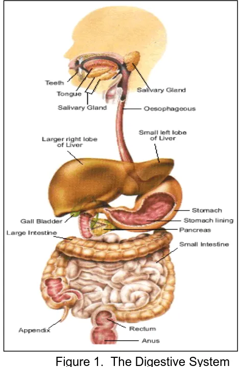
If you were told that your digestive system is like a factory, would you believe it? It is true. In a factory, raw materials are brought in and transported to different places in the factory. As the raw materials move through the factory, they change in various ways. Finally, they come out of the factory as several new products.
Your digestive system works in a similar way. Food enters the digestive system through your mouth. Food is the raw material. Once inside your body, it moves through the different organs of your digestive system. It is where the food is changed.
The function of the digestive system is digestion. Digestion is the breakdown of organic compounds into their simple forms for use by the cells. Digestion is the life support job of the digestive system. The digestive system breaks down food in two ways:
mechanically and chemically.
Organs of the digestive system that chew, tear, churn, squeeze and grind food help in mechanical digestion. On the other hand, the organs that make and use chemicals to break apart the food and reduce it to liquid help in chemical digestion.
There are two groups of organs in the digestive system. One group makes up the GASTROINTESTINAL TRACT, a food tube that is open at each end and includes the mouth, esophagus, stomach, and intestine.
The other group of organs makes and stores the chemicals that chemically break down the food. These are the liver, pancreas, gall bladder, and salivary glands.
Let’s take a trip through the human digestive system to see how it works. To make it a little more interesting, try to imagine what happens to a hamburger when eaten. Remember that ground meat is mostly protein, mayonnaise is mostly fat, and the bun is mostly carbohydrate. By the way, the trip takes about 18-20 hours.
Look at the figure on the next page. Trace the pathway of the hamburger that you ate in your body. Get set, go!
Humans and many other animals have a digestive system with two openings. Food enters the system by way of the mouth.
Mouth. The food tube, nine meters long, begins at the mouth. The teeth mechanically chew, chop and break the food apart. The breaking and gliding are physical changes. The SALIVARY GLANDS produce a chemical that starts the breakdown of carbohydrates. The product of digestion in the mouth is the bolus. Food moves from the mouth to the esophagus when you swallow.
Esophagus. The esophagus is a tube that connects the mouth to the stomach. Muscles of the esophagus push and transport foods and liquids to the stomach.
Stomach. The stomach is a bag-like muscular organ. The function of the stomach is to grind the food and mix it with the digestive juices. It can hold about one liter of liquid and food. The product of digestion in the stomach is chyme. The stomach has special cells in its walls that make gastric juice.
Gastric juice begins the chemical breakdown of proteins. After about four hours, the stomach pushes food into the small intestines.
Do you know?
[image:7.612.319.553.166.525.2]That the pH of the stomach is 2? It is more than enough to burn your dress!
Small Intestine. The small intestine is where most of the food is chemically digested. The small intestine itself makes several digestive juices. Some of these chemicals digest proteins into amino acids. Others digest carbohydrates into simple sugars.
Before describing what the small intestine does we must take a short detour. Let us pass by the three organs that are part of the digestive system. These organs are the liver, pancreas and gall bladder.
Liver. The liver is the largest organ in the body. It has a mass of about two kilograms. The liver makes bile, a green liquid that breaks up large fat droplets into small fat droplets and stores it in the gall bladder. When needed, bile enters the small intestine and aids in the digestion of fat. If bile is not needed, it is delivered to the gall bladder.
Pancreas. The pancreas is a small organ that makes three different kinds of enzymes and is found below the stomach. It makes about half of the liters of digestive juices made each day. These juices aid in the digestion of all three organic compounds.
Gall Bladder. The gall bladder is a small pear shape sac that can hold about 50ml of bile. It stores the bile until it is needed by the small intestine to emulsify fats.
Large Intestine. The job of the large intestine is to remove the useful liquids from the undigested food. The undigested food, called FECES, is solidified and pushed out to the anus. If the large intestine did not return two liters of liquids to the body a day, a person could die from lack of water. Aside from water, this organ also reabsorbs salt for further use by the body.
The small intestine receives digestive juices from the LIVER and PANCREAS. The liver contributes BILE, which digests fat. But, it is the small intestine that makes and receives many digestive chemicals that complete the digestion of food.
Our entire trip through the digestive system took about 20 hours. Look at the figure 1 again. It shows how the entire digestive system looks from one end to the other.
Do you know?
What you will do
Activity 1.1
This activity will help you estimate the length of the digestive system. Materials needed:
yarns of different colors (blue, green, red, yellow) scissors
ruler Procedure:
1. Cut pieces of yarns according to the measurements provided below, but have an extra length for each piece for tying.
2. Use different colors of yarn to represent the different organs.
3. After the yarns have been cut, tie the pieces together and measure again. Blue … 25 cm
Green … 20 cm Red … 700 cm Yellow … 150 cm 895 cm Answer the following questions:
1. What is the longest measurement? What does this represent? 2. What is the shortest measurement? What does this represent?
3. What is the second to the shortest measurement? What does this represent? 4. How will you compare the longest yarn and the 2nd to the longest yarn? 5. What do you think is the reason why food stays in the body for 8 hours?
What you will do
Self-Test 1.1
Rearrange the parts of the digestive system in the correct sequence. Write the letters of the correct answer on the blank.
Digestion Process
a. small intestine b. anus
c. stomach d. mouth
e. large intestine f. esophagus g. rectum
1.________ 2.________ 3.________ 4.________ 5.________ 6.________ 7.________
How Nutrients Get Into the Blood
Nutrients get into the blood system through the small intestine. The inside of the small intestine is not smooth like a garden hose. Instead, the inside of the small intestine is lined with millions of tiny hair-like projections called VILLI. (singular: villus)
Villi increase the surface area of the small intestine to about five times than that of a smooth surface. As the food moves slowly between, over and around the villi, nutrients are absorbed into the bloodstream inside each villus.
The wall of each villus is only one cell thick. Digested foods can easily pass through these layers of cell. Examine the cross section of the small intestine on the right and notice the finger–like folds called the rugae. These are lined with hair-like projections called the villi.
[image:10.612.308.551.460.648.2]Key to answers on page 26.
Figure 2. The Villi
microvilli epithelial cell
capillary (blood) lacteal (lymph)
What you will do
Activity 1.2
Procedure:
Draw two circles – Circle A and Circle B as shown below
A B
1. Place a piece of string around the inside of circle A. 2. Cut off any extra string so it fits exactly.
3. Measure the string in centimeters.
4. Place a piece of string around the inside of circle B. 5. Cut off any extra string so it fits exactly.
6. Measure the string in centimeters.
Which is longer circle A or circle B? You should have discovered that the inside of circle B is three times longer than the inside of circle A. The same kind of thing happens in the small intestines. Villi make the surface larger. The larger surface means digested food is absorbed better.
What you will do
Self Test 1.2
1. Where are most of the food nutrients absorbed? 2. Describe the inside of the small intestine.
3. How do the villi increase absorption?
4. What is the function of the digestive system? 5.
Lesson 2. Respiratory System
The Exchange of Gases
Do you know that you can survive for several days without water and survive for a month without food, but you cannot survive for more than five minutes without oxygen?
Oxygen is the part of the air that we breathe. Air is a mixture of different gases. The air you breathe is made up of
Oxygen … 21.0%
Nitrogen … 78.1% Carbon Dioxide … 0.03% Other gases … 0.87%
Life depends on breathing because the cells of the body need oxygen. You breathe in to bring fresh air into the lungs. The lungs must separate the oxygen from the air. Then you breathe out to get rid of the carbon dioxide that the body does not need.
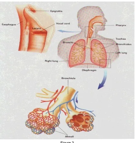
Breathing is a mechanical process. It is a process of pumping air into and out of the lungs. Breathing is done by a group of organs that make up the RESPIRATORY SYSTEM
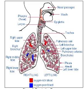
(Figure 3). The function of the respiratory system is to exchange oxygen and carbon dioxide between the air and the cells.
The respiratory organs filter particles from the incoming air. They help control the temperature and water content of air. They also aid in producing the sounds used in speech and play important roles in the sense of smell and the regulation of pH.
Now, let us take a tour of our respiratory system. The following are the parts and their functions.
Nose. The function of the nose is to filter and warm the entering air with the help of the cilia (hairs inside it).
Pharynx. The pharynx is commonly called the throat. It connects the nose with the windpipe.
Trachea. The trachea is commonly called the windpipe.
Bronchus. The trachea branches into two tubes, the BRONCHI, inside the lungs. Each bronchus continues to branch and rebranch until it is very small. Each tube finally ends in a tiny air sac called an ALVEOLUS (plural: alveoli).
The lungs are really two bags full of thousands and thousands of alveoli. It is at the alveoli inside the lungs that gases are exchanged.
Diaphragm. The diaphragm is a large muscle that lies flat at the bottom of the chest cavity. The diaphragm aids in breathing by moving up and down.
[image:13.612.93.538.214.681.2]What you will do
Activity 2.1
A. Demonstration of breathing Procedure:
a. Close your mouth, then press your nose
b. Do it for a few seconds or for as long as you can hold breathing. Answer the following questions:
a. How did you feel as you press your nose with your mouth closed? Why? b. What happens when the air cannot enter the body?
c. What air do you inhale? What air do you exhale? B. Respiration Process
Create a mental picture of the respiratory system. Using the words in column A write the correct sequence in column B.
Column A Column B
air sacs bronchioles nose trachea pharynx bronchi larynx
1.________ 2.________ 3.________ 4.________ 5.________ 6.________ 7.________
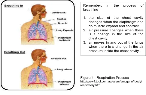
When you breathe in or inhale, the diaphragm contracts. Inhaling moves the diaphragm down and makes the size of the chest cavity larger. At the same time, the ribs move up and increase the size of the chest cavity. There is now more space and less air pressure inside the lungs. Air pushes in from the outside where there is a higher air pressure. It pushes into the lungs where there is a lower air pressure.
When you breathe out, or exhale, the diaphragm relaxes. The diaphragm and ribs return to their original place. The chest cavity returns to its original size. There is now less space and more air pressure inside the lungs. It pushes the air to the outside where there is a lower air pressure.
What you will do
Self-Test 2.1
1. What change causes the air to move into and out of the lungs? 2. What is the function of the respiratory system?
The Process of Respiration
Breathing and respiration are two different processes. BREATHING is a mechanical process of pumping air into and out of the lungs. The lungs are like two bags turned inside out, inside the body. The gases in the lungs must get to the cell, and the waste gases in the cells must get to the lungs. For this to happen, a chemical process is needed.
Key to answers on page 27.
Remember, in the process of breathing
1. the size of the chest cavity changes when the diaphragm and rib muscle expand and contract. 2. air pressure changes when there
is a change in the size of the chest cavity.
[image:15.612.66.552.59.377.2]3. air moves in and out of the lungs when there is a change in the air pressure inside the chest cavity.
Figure 4. Respiration Process
Respiration takes place in two stages. External respiration is the exchange of gases between the air and the blood. Internal respiration is the exchange of gases between the blood and the cells.
External respiration takes place at the alveoli. The surfaces of the alveoli are covered with a network of tiny blood vessels that are called CAPILLARIES.
The walls of the alveoli are one cell thick. The walls of the capillaries are also one cell thick. By diffusion, oxygen passes from the alveoli in the lungs into the capillaries across two rows of cells. At the same time, carbon dioxide passes from the capillaries to the alveoli across the same two rows of cells.
What you will do
Activity 2.3
Experimental data and statistics show that 90 percent of all lung cancer cases are the result of smoking. Write an advertising campaign to urge people especially the young (adolescents), to avoid smoking. Include information on the effect of smoking on the respiratory system.
What you will do
Self-Test 2.2
Study the illustration of the respiratory system very well. Identify the parts as numbered
{
2
1
7
6
4
3
Lesson 3. The Circulatory System
The circulatory system is the life support system that feeds your cells with food and oxygen. It also takes away the waste products. The circulatory system is like a network of highways, streets and alleys connecting all the cells together into a community. In turn the community of cells keeps the body alive. There are two divisions of this system, the
lymphatic, which helps to return tissue fluid to the blood, and the blood division, which is a closed circuit.
There are three main parts to the circulatory system. These are the heart, the blood vessels and the blood.
THE CIRCULATORY SYSTEM
Parts Function
Heart ………pumps the blood
Blood Vessels ……... carry the blood Arteries
Veins Capillaries
Blood ……….carries the materials
The Human Heart
Look at your fist. Note its size. Your HEART (Figure 5) is a bundle of muscles about the size of your fist. The heart is shaped like a cone. It is located in the center of your chest between the lungs. It is tilted to one side and points downward to the left.
[image:17.612.276.553.357.683.2]There is a VALVE between each atrium and ventricle to prevent the blood from flowing backwards. The valves are like one-way doors that keep the blood moving in only one direction.
What you will do
Self-Test 3.1
1. What is the size, shape and location of the heart? 2. Name the four chambers of the heart.
3. What is the function of the valves?
How the Heart Works
The function of the circulatory system is to carry materials to and from all parts of the body. The power for the circulatory system comes from the muscular action of the heart. The heart does its work by contracting and relaxing.
All of the muscle tissues of the heart do not contract at the same time. Different parts of the heart contract at different times. When the top contracts, the bottom relaxes. When the bottom contracts, the top relaxes. When a chamber contracts, it becomes smaller and the blood inside gets squeezed or pumped out. To have a better understanding of how the heart works, do this simple activity.
What you will do
Activity 3.1 The Pumping on the Heart
Materials: Plastic bottle (preferably white) Water
Dye Procedure:
1. Fill the plastic bottle with colored water.
2. Observe the content closely. No liquid is pushed out. The same is true with the heart. If the heart muscles don’t squeeze together, no blood is pumped.
3. Squeeze the bottle with both hands and observe what happens. When the heart muscles squeeze together, blood is pumped.
Answer the following questions:
1. What is the function of the circulatory system?
_________________________________________________________________ 2. Name the chambers through which blood flows in the right order.
_________________________________________________________________ 3. What does the blood pick up?
_________________________________________________________________ 4. What kind of blood goes to the body?
_________________________________________________________________
There are four valves in the heart. The valves are one of the most important parts of the heart. When the atria contract, the tricuspid (right) and bicuspid valve (left) open. When the ventricles contract, the pulmonary and aortic valves open. When these two phases are repeated, you have one heartbeat cycle.
The two phases of the heartbeat cycle can be heard. “Lub-dub” is the sound your heart makes. The heart beat sound is caused by the contraction of the muscles and the slamming shut of the valves. In the “lub” phase the ventricles contract and the tricuspid and bicuspid valves close. In the “dub” phase, the pulmonary and aortic valves close. A normal heart repeats the “lub- dub” sound over and over again in perfect rhythm.
What you will do
Activity 3.2 Tracing the Flow of Blood through the Heart
The path the blood takes through the heart is listed below. Write the letter for each step in the proper place in drawing of the heart.
a. Blood returning from the body travels through a blood vessel connected to the heart.
b. The blood enters the right atrium.
c. After the right atrium fills, the atria contract. This pushes the blood past a one-way valve.
d. The blood fills the right ventricle.
f. This blood vessel goes to the lungs where the blood picks up oxygen.
g. Blood from the lung travels through a blood vessel to the heart.
h. The blood enters the left atrium.
i. After the left atrium is filled, it contracts. This pushes the blood to a one-way valve. j. The blood fills the left
ventricle.
k. The ventricles contract and push the blood to a valve into a blood vessel.
l. This blood, rich in oxygen, goes to the body.
The Blood Vessels
The circulatory system is a closed system. This means that the blood stays inside the heart and a set of tubes as it circulates.
Your blood moves through your body in tubes called VESSELS. There are three kinds of blood vessels that make up the circulatory system: ARTERIES, VEINS, and CAPILLARIES.
The arteries carry blood away from the heart. The veins carry blood towards the heart. The capillaries are very small tubes that connect the arteries to the veins.
[image:20.612.104.558.53.310.2]The blood vessels form an intricate transportation network to service every cell. Transportation is the function of the circulatory system. The human body has 100,000 kilometers of blood vessels. If laid end to end, a person’s blood vessels would circle the earth twice!
Figure 6. Flow of Blood http//www4.tpgi.com.au/users/amcgann/body/ circulatory.html http://www4.tpgi.com.au/users/amcgann/body/circulatory.html
Arteries. Arteries have thick, muscular walls. They are elastic and expand every time the ventricles contract. The force of the heart pumping keeps the blood moving through the arteries.
Arteries carry blood away from the heart. The blood in the arteries is bright red because it contains much oxygen.
The large artery is the AORTA. This is the first artery leaving the heart to the body. The arteries branch into smaller and smaller vessels, which end at the capillaries.
Veins. Veins have muscular walls too, but they are thinner than the walls of the arteries. There are one-way valves inside the veins to prevent the blood from flowing backwards. Blood is moved along when you move your muscles. This squeezes the blood inside the veins and pushes the blood towards the heart.
Veins carry blood to the heart. The blood in the veins is blue in color because it lacks oxygen. You can see some of your veins because they are right under the surface of the skin. The veins begin at the capillaries and join into larger veins until the largest vein empties into the heart.
Capillaries. Capillaries are tiny vessels that connect arteries to veins. The capillaries are so small that the red blood cells must pass through them in single file. Also, the walls of the capillaries are only one-cell thick.
The work of the circulatory system takes place at the capillaries. It is here that the exchange of materials between the blood and the cells takes place. At the capillaries, food and oxygen pass from the blood to the cells. Also, carbon dioxide and waste products pass from the cells to the blood.
[image:21.612.323.550.332.645.2]The circulatory system is really two separate systems. One part of the system pumps blood to the lungs. Another part of the system pumps blood to the body. Blood must go to the lungs to pick up oxygen before it can go to the body.
What you will do
Self-Test 3.2
1. What kind of system is the circulatory system? Explain. 2. Name the kinds of blood vessels.
3. What is the purpose of the blood vessels? 4. How does blood move in the arteries? 5. How does blood move in the veins? 6. What and where are the capillaries?
7. What vital function takes place at the capillaries?
What you will do
Activity 3.3 Circulation process
Rearrange the flow of blood to the heart in the correct sequence. Write the letter of the correct answer in the blank.
A. Pulmonary veins 1. __________ B. Tricuspid valve 2. __________ C. Superior and inferior vena cava 3. __________
D. Aorta 4. __________
E. Lungs 5. __________
F. Pulmonary artery 6. __________ G. Bicuspid / Mitral valve 7. __________
H. Right auricle 8. __________
I. Left ventricle 9. __________
J. Right ventricle 10. __________
K. Left auricle 11. __________
The blood is a fluid that carries most of the materials necessary for life. The blood has two different parts. The nonliving, liquid part is called PLASMA. It is a yellowish fluid that makes up 55 percent of your blood. The remaining 45 percent is made up of three kinds of cells: red blood cells, white blood cells and platelets.
Plasma is the fluid part of the blood in which the blood cells coat is a yellowish substance composed of 92% water and 8% of dissolved nutrients, mineral salts, antibodies
and hormones.
Key to answers on page 28.
The proteins present in the plasma are:
1. Albumin – helps in keeping the blood pressure normal. It regulates the amount of water in the plasma.
2. Globulin – contains antibodies.
3. Chemical Substances – effective against specific diseases 4. Fibrinogen – works with platelets in the clotting process.
Red Blood Cells (RBC) are also called erythrocytes. They are the most numerous cells in the blood. Each liter of blood contains 4.5 to 6 trillion red blood cells. The main component of red blood cells is the pigment known as
hemoglobin. Hemoglobin in the RBC binds and carries oxygen.
White Blood Cells (WBC) are larger and less numerous than RBC (red blood cells). WBC (white blood cells), also called leukocytes, contain nuclei and other types of organelles. WBC may circulate in the blood for weeks before leaving the blood and entering other tissues. WBC help defend the body against infection.
Let’s Summarize
1. Digestion is the process that changes food so that it can be used by the body. 2. Cells need food for growth and energy.
3. The digestive system breaks down food mechanically and chemically.
4. The digestive system must digest the food, extract the nutrients and dispose the waste.
5. The salivary glands produce a chemical that begins the digestion of carbohydrates.
6. The stomach grinds the food and mixes it with the gastric juice.
7. The liver and pancreas produce digestive juices that aid digestion in the small intestine.
8. The small intestine makes and receives many digestive chemicals that complete digestion of food.
9. Life depends on breathing because the cells need oxygen. 10. The nose filters and warms the entering air.
14. Breathing is the mechanical process of pumping air into and out of the lungs. 15. The air that we take is oxygen and the air that we breathe out is carbon dioxide. 16. The circulatory system transports materials to and from all parts of the body. 17. The main parts of the circulatory system are the heart, blood vessels and blood. 18. The heart is a pumping organ.
19. The heart is a bundle of muscles.
20. The heart is two hearts in one, a left heart with two chambers and a right heart with two chambers.
21. The left heart pumps blood to the lungs, and the right heart pumps blood to the body.
22. The two phase of respiration are: inhalation, the taking in of oxygen; and exhalation, the removal of carbon dioxide from the lungs.
23. The different parts of the respiratory system are: nose, pharynx, larynx, trachea, bronchi, bronchioles, bronchial tubes and air sacs.
24. The circulatory system is made up of the heart, the blood vessels and the blood. 25. Physical exercise makes your heart stronger because your heart beats faster.
This enables the blood to circulate efficiently. Thus supplying oxygen to the tissues and forcing out carbon dioxide.
26. Eating a balance diet, not smoking and exercise can help maintain a normal and healthy heart.
Posttest
I. Multiple Choice. Choose the letter of the correct answer. Write the chosen letter on a separate sheet of paper.
1. The chemical process in which oxygen and carbon dioxide are exchanged between the outside air and the cells is called
m. breathing c. exhaling
n. inhaling d. respiration
2. The life support system that feeds the cells with food and oxygen is the o. circulatory c. excretory
p. digestive d. respiratory
3. Which organ is NOT a part of the circulatory system?
q. blood c. esophagus
r. blood vessel d. heart
4. Which organ is referred to as the living pump?
s. heart c. lung
5. All of the following carry blood to and from all parts of the body EXCEPT: u. arteries c. large intestines
v. capillaries d. veins
6. What is the process of changing food into a simpler substance for use by the cells?
w. chewing c. eating
x. digestion d. excretion
7. What do you call the process of changing food into a simpler substance with the help of enzymes?
y. chemical digestion c. mastication
z. churning d. mechanical digestion 8. Where does the final digestion of food take place?
aa. large intestine c. mouth
bb. liver d. small intestine
9. Normally, an average meal will stay in the stomach for about how many hours?
cc. 2 to 4 c. 4 to 5
dd. 3 to 4 d. 5 to 6
10. What is the product of digestion in the mouth?
ee. bile c. chyme
ff. bolus d. feces
11. What do you call the wave -like contraction in the walls of the digestive tract?
gg. chewing c. masticating
hh. digesting d. peristalsis
12. Roughage foods are good for the digestive system because these help ii. prevent diarrhea c. regulate body temperature jj. prevent ulcers d. regulate bowel movement
13. Which system is responsible for the exchange of oxygen and carbon dioxide between the air and the cells?
kk. circulatory c. excretory ll. digestive d. respiratory 14. Which organ serves to filter and warm the air entering it?
mm. air sac c. diaphragm
Key to Answers
Pretest
I.
1. b 6. d 11. d
2. a 7. d 12. b
3. d 8. d 13. c
4. b 9. d 14. a
5. b 10. a 15. c
II.
Digestive Circulatory Respiratory
Liver Blood vessels Nose
Esophagus Blood Air sacs
Gall bladder Heart Trachea
Stomach Arteries Diaphragm
Mouth Veins Bronchi
Small intestine Capillaries larynx
III.
A. Respiratory B. Heart C. Digestive
Lesson 1
Activity 1.1
1. 700 cm – small intestine 2. 20 cm – rectum and anus 3. 25 cm – esophagus
4. There is a difference of 550 cm between the longest yarn and the shortest yarn.
Self-Test 1.1
Self-Test 1.2
1. small intestines
2. lined with millions of tiny hair-like projections (villi) 3. the surface is so long so it makes absorption fast
4. breakdown of food into simple form and removal of undigested solid food waste.
Lesson 2
Activity 2.1
A. a. Uncomfortable because there was no air. b. Breathing is difficult
c. Oxygen, carbon dioxide B. 1. Nose
2. Pharynx 3. Larynx 4. Trachea 5. Bronchi 6. Bronchioles 7. Air sacs
Self-Test 2.1
1. change in air pressure inside the chest cavity
2. exchanges oxygen and carbon dioxide between the air and the cells
Self-Test 2.2
1. lungs 5. bronchioles 2. nose 6. larynx 3. bronchi 7. pharynx 4. trachea 8. air sacs
Lesson 3
Self-Test 3.1
1. fist size, cone-shaped center of the chest 2. right and left atrium
right and left ventricle
Activity 3.1
1. carry materials to and from all parts of the body 2. right atrium
right ventricle left atrium left ventricle
3. carry materials to and from all parts of the body 4. oxygen
Activity 3.2
Self-Test 3.2
1. Closed. Blood passes through vessels around the body. 2. Arteries, veins, capillaries
3. Transportation
4. Carry blood away from the heart 5. Carry blood towards the heart
6. Tiny vessels that connect arteries to veins
7. Exchange of materials between the blood and cells takes place here.
Activity 3.3
1. C 7. A
2. H 8. K
3. B 9. G
4. J 10. I
5. F 11. D
Posttest
1. d 6. b 11. d
2. b 7. a 12. d
3. c 8. d 13. d
4. a 9. b 14. d
5. c 10. b 15. a
References
Books:
Daniel, L. (1994). Life science. Westerville, OH: Merill Publishing Co., Mcmillan/McGraw-Hill.
Grabowski, T. (2003). Principles of anatomy and physiology. NY: John Wiley and Sons, Inc.
Holo, W. (1984). Human anatomy and physiology. (3rd Ed.) Dubuque, Iowa: W. C. Brown Publishers.
Hopson, J.L. & Wessells, N.K. (1990). Essentials of biology. USA: McGraw-Hill Publishing Company.
Kaskel, A., Hummer, P.J. & Daniel, L. (1981). Biology on everyday experience. USA: Merill Publishing Co.
Mariele, E.N. (1998). Essentials of human anatomy and physiology. (3rd Ed.) USA: Addison-Wesley Longman, Inc.
Payne, H. (1995). Understanding your health. St Louis, Missouiri: Mosby Publishing Company.
Pikering, W.H. (2000). Complete biology. New York: Oxford University Press.
Electronic Sources:
Retrieved January 10, 2005 htttp//www4.tpgi.com.au/users/amcgann/body/html Retrieved January 10, 2005 from http//www.innerbody.com
Retrieved January 10, 2005 from http/www.bartleby.com