The Genomic Standards Consortium
Non contiguous-finished genome sequence and
description of
Ajay Kumar Mishra1, Jean-Christophe Lagier1, Catherine Robert1, Didier Raoult1 and
Pierre-Edouard Fournier1*
1 Unité de Recherche sur les Maladies Infectieuses et Tropicales Emergentes, UMR,
Aix-Marseille Université, France
* Corresponding author: Pierre-Edouard Fournier ([email protected])
Keywords, genome
strain JC401T sp. nov. is the type strain of P. timonensis sp. nov., a
new species within the
was isolated from the fecal flora of a healthy patient. P. timonensis is an obligate Gram-positive anaerobic coccus. Here we describe the features of this organism, together with the complete genome sequence and annotation. The 1,758,598 bp long genome (1 chromosome, no plasmid) contains 1,922 protein-coding and 22 RNA genes, including 5 rRNA genes.
Introduction
strain JC401T (= CSUR
P165= DSM 25367) is the type strain of P.
timonensis sp. nov. This bacterium is a
Gram-positive, anaerobic, indole-positive coccus that was isolated from the stool of a healthy Senegalese patient as part of a “culturomics” study aiming at cultivating individually all species within human feces.
Since the early days of bacterial taxonomy, defin-ing a bacterial species has been a matter of debate. Currently, the availability of a wide array of mo-lecular methods, notably 16S rRNA and full ge-nome sequencing, offers a possibility to base the description of new species on other methods than the “gold standard” of DNA-DNA hybridization [1]. In particular, sequence similarity of the 16S rRNA, although neither uniform across taxa nor neces-sarily predictive, enabled the taxonomic classifica-tion or reclassificaclassifica-tion of many taxa [2], and ge-nome sequencing has provided access to the com-plete genetic information of bacteria [3]. As a
con-sequence, we based our description of P.
timonensis sp. nov. on a polyphasic approach [4]
including their genome sequence and main pheno-typic characteristics (habitat, Gram-stain reaction, culture and metabolic characteristics, MALDI-TOF spectrum, and when applicable, pathogenicity).
Here we present a summary classification and a set of features for P. timonensis sp. nov. strain JC401T together with the description of the com-plete genomic sequencing and annotation. These characteristics support the creation of the P.
timonensis species.
The genuset al. 2001) was
created in 2001 [5] and consist of species that are non-saccharolytic, butyrate-producing, non-motile gram-positive anaerobic cocci and use peptones and oligopeptide as major energy source [6]. To
date, the genus
cies namel
genus
from various human clinical specimens such as vaginal discharges, ovarian, peritoneal, sacral and
lachrymal gland abscesses [5].
summer mastitis in cattle [5].
Organism information
consent, and the agreement of the National Ethics Committee of Senegal and the local ethics commit-tee of the IFR48 (Marseille, France) were obtained under agreement (09-022 and 11-017). The fecal specimen was preserved at -80°C after collection and sent to Marseille. Strain JC401T was isolated in June 2011 by cultivation on 5% sheep blood-enriched Brain Heart Infusion agar (Becton Dick-inson, Heidelberg, Germany). This strain exhibited
a 98% nucleotide sequence similarity with
validated
sification, see Table 1). This value was lower than the 98.7% 16S rRNA gene sequence threshold recommended by Stackebrandt and Ebers to de-lineate a new species without carrying out DNA-DNA hybridization [18].
Table 1. Classification and general features o strain JC401T according to the
MIGS recommendation [8]
MIGS ID Property Term Evidence codea
Domai TAS [9]
Phylum TAS [10-12]
Cla TAS [13,14]
Current classification Orde TAS [15,16]
Family XI Incertae sedis TAS [15,16]
Genu TAS [5]
Species IDA
Type strain JC401T IDA
Gram stain Positive IDA
Cell shape Coccoid IDA
Motility Nonmotile IDA
Sporulation Nonsporulating IDA
Temperature range Mesophile IDA
Optimum temperature 37°C IDA
MIGS-6.3 Salinity Growth in BHI medium + 1% NaCl IDA
MIGS-22 Oxygen requirement Anaerobic IDA
Carbon source Unknown NAS
Energy source Unknown NAS
MIGS-6 Habitat Human gut IDA
MIGS-15 Biotic relationship Free living IDA
Pathogenicity Unknown Biosafety level 2
MIGS-14 Isolation Human feces NAS
MIGS-4 Geographic location Senegal IDA
MIGS-5 Sample collection time September 2010 IDA
MIGS-4.1 Latitude 13.7167 IDA
MIGS-4.1 Longitude -16.4167 IDA
MIGS-4.3 Depth Surface IDA
MIGS-4.4 Altitude 51 m above sea level IDA
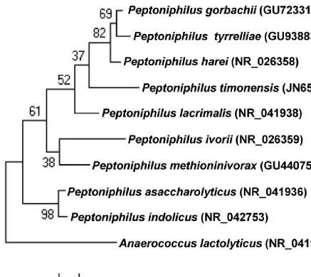
Figure 1. Phylogenetic tree highlighting the position of strain JC401T relative to other
type strains within the
quences were aligned using CLUSTALW, and phylogenetic inferences obtained using the maximum-likelihood method within the MEGA software. Numbers at the nodes are percentages of bootstrap values obtained by re-peating the analysis 500 times to generate a majority consensus tree outgroup. The scale bar represents a 2% nucleotide sequence divergence.
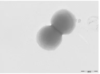
Different growth temperatures (25, 30, 37, 45°C) were tested. Growth was not observed at 25°C and 45°C, but optimal growth occurred between 30 and 37°C. Colonies were 0.5 mm in diameter on blood-enriched BHI agar. Growth of the strain was tested under anaerobic and microaerophilic con-ditions using GENbag anaer and GENbag microaer systems, respectively (BioMérieux), and in aerobic conditions, with or without 5% CO2. Growth was not achieved in aerobic (with and without CO2) conditions. The growth was observed in anaerobic conditions. Gram staining showed Gram-positive cocci (Figure 2). A motility test was negative. Cells
grown on agar are sporulated and have a mean diameter of 0.91 µm (Figure 3).
Strain JC401T exhibited a catalase activity but no ox-idase activity. Using API Rapid ID 32A, positive
reac-tions were obtained for α galactosidase, arginine
arylimidase, tyrosine arylamidase, histidine arylamidase, serine arylamidase and indole produc-tion. Weak reactions were observed for leucine arylamidase and phenylalanine arylamidase. P.
timonensis is susceptible to penicillin G, imipeneme,
Figure 2. Gram staining of P. timonensis strain JC401T
Figure 3. Transmission electron microscopy of P. timonensis strain JC401T, using a
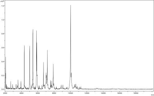
Matrix-assisted laser-desorption/ionization time-of-flight (MALDI-TOF) MS protein analysis was carried out as previously described [19]. Briefly, a pipette tip was used to pick one isolated bacterial colony from a culture agar plate, and to spread it as a thin film on an MTP 384 MALDI-TOF target plate (Bruker Daltonics, Leipzig, Germany). Twelve distinct deposits were done for strain JC401T from twelve isolated colonies. Each smear was overlaid with 2µL of matrix solution (saturat-ed solution of alpha-cyano-4-hydroxycinnamic acid) in 50% acetonitrile, 2.5% tri-fluoracetic-acid, and allowed to dry for five minutes. Meas-urements were performed with a Microflex spec-trometer (Bruker). Spectra were recorded in the positive linear mode for the mass range of 2,000 to 20,000 Da (parameter settings: ion source 1 (IS1), 20 kV; IS2, 18.5 kV; lens, 7 kV). A spectrum was obtained after 675 shots at a variable laser power. The time of acquisition was between 30 seconds and 1 minute per spot. The twelve JC401T spectra were imported into the MALDI BioTyper
software (version 2.0, Bruker) and analyzed by standard pattern matching (with default parame-ter settings) against the main spectra of 3,769
bac-teria, including 12 spectra from
species, which were used as reference data, in the BioTyper database. The method of identification included the m/z from 3,000 to 15,000 Da. For every spectrum, 100 peaks at most were taken into account and compared with spectra in the database. A score enabled the identification, or not, from the tested species: a score > 2 with a val-idated species enabled the identification at the species level, a score > 1.7 but < 2 enabled the identification at the genus level; and a score < 1.7 did not enable any identification. For strain JC401T, the obtained score was 1.2, thus suggest-ing that our isolate was not a member of a known species. We incremented our database with the spectrum from strain JC401T (Figure 4). The spec-trum was made available online in our free-access URMS database [20].
0.0 0.2 0.4 0.6 0.8 1.0 1.2 4 x10
In
te
ns
. [a
.u
.]
2000 4000 6000 8000 10000 12000 14000 16000 18000
m/z Figure 4: Reference mass spectrum from P. timonensis strain JC401T. Spectra from 12 individual colonies were
Genome sequencing information
Genome project history
The organism was selected for sequencing on the basis of its phylogenetic position and 16S rRNA
simi-larity to other members of th
and is part of a “culturomics” study of the human digestive flora aiming at isolating all bacterial spe-cies within human feces. It was the seventh genome
of a
Peptoniphilus timonensis sp. nov. The Genbank
ac-cession number is CAEL00000000 and consists of 97 large contigs. Table 2 shows the project infor-mation and its association with MIGS version 2.0 compliance.
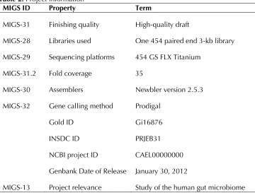
Table 2. Project information
MIGS ID Property Term
MIGS-31 Finishing quality High-quality draft
MIGS-28 Libraries used One 454 paired end 3-kb library MIGS-29 Sequencing platforms 454 GS FLX Titanium
MIGS-31.2 Fold coverage 35
MIGS-30 Assemblers Newbler version 2.5.3 MIGS-32 Gene calling method Prodigal
Gold ID Gi16876
INSDC ID PRJEB31
NCBI project ID CAEL00000000 Genbank Date of Release January 30, 2012
MIGS-13 Project relevance Study of the human gut microbiome
Growth conditions and DNA isolation
P. timonensis sp. nov. strain JC401T (CSUR P165,
DSM 25367) was grown anaerobically on 5% sheep blood-enriched Columbia agar at 37°C. Six petri dishes were spread and resuspended in 6x100µl of G2 buffer (EZ1 DNA Tissue kit, Qiagen). A first mechanical lysis was performed by glass powder on the Fastprep-24 device (Sample Prepa-ration system, MP Biomedicals, USA) during 2x20 seconds. DNA was then treated with 2.5µg/µL ly-sozyme (30 minutes at 37°C) and extracted using the BioRobot EZ1 Advanced XL (Qiagen). The DNA was then concentrated and purified using the Qiamp kit (Qiagen). The yield and the concentra-tion was measured by the Quant-it Picogreen kit
(Invitrogen) on the Genios Tecan fluorometer at 123.3ng/µl.
Genome sequencing and assembly
Labchip with an optimal at 568bp. Then the library was quantified on the Quant-it Ribogreen kit (Invi-trogen) on the Genios_Tecan fluorometer at 890 pg/µL. The library concentration equivalence was calculated as 2.87E+09 molecules/µL. The library was stored at -20°C until further use.
The shotgun library was clonal amplified with 0.25 and 0.5cpb in 2 emPCR reactions per conditions with the GS Titanium SV emPCR Kit (Lib-L) v2.The yields of the emPCR were 2.79% and 10.79% re-spectively in the range of 5 to 20% from the Roche procedure.
Approximately 790,000 beads for a ¼ region and 340000 beads for a 1/8 region were loaded on the GS Titanium PicoTiterPlate PTP Kit 70×75 and se-quenced with the GS FLX Titanium Sequencing Kit XLR70 (Roche). The run was performed overnight and then analyzed on the cluster through the gsRunBrowser and Newbler assembler (Roche). For the shotgun sequencing, 193,186 passed filter wells were obtained and generated 37.47Mb with a length average of 190 bp. The passed filter se-quences were assembled Using Newbler with 90% identity and 40 bp as overlap. The final assembly identified 7 scaffolds and 97 large contigs (>1500bp) generating a genome size of 1.76 Mb.
Genome annotation
Open Reading Frames (ORFs) were predicted using Prodigal [21] with default parameters but the pre-dicted ORFs were excluded if they were spanning a sequencing gap region. The predicted bacterial pro-tein sequences were searched against the GenBank database [22] and the Clusters of Orthologous Groups (COG) databases using BLASTP. The tRNAScanSE tool [23] was used to find tRNA genes, whereas ribosomal RNAs were found by using RNAmmer [24] and BLASTn against the GenBank database. ORFans were identified if their BLASTP E-value was lower than 1e-03 for alignment length greater than 80 amino acids. If alignment lengths were smaller than 80 amino acids, we used an E -value of 1e-05. Such parameter thresholds have already been used in previous works to define ORFans.
To estimate the mean level of nucleotide sequence similarity at the genome level between
only using BLASTN and the following parameters:
query coverage of ≥ 70% and a minimum
nucleo-tide length of 100 bp.
Genome properties
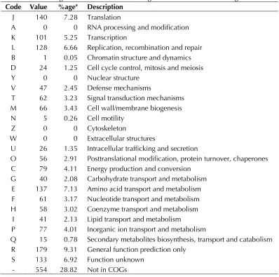
The genome is 1,758,598 bp long (1 chromosome, but no plasmid) with a 30.70% GC content (Figure 5 and Table 3). Of the 1,944 predicted genes, 1,922 were protein-coding genes and 22 were RNAs. A total of 1,368 genes (70.37%) were as-signed a putative function. A total of 186 genes were identified as ORFans (9.6%). The remaining genes were annotated as hypothetical proteins. The distribution of genes into COGs functional cat-egories is presented in Table 4. The properties and the statistics of the genome are summarized in Tables 3 and 4.
Comparison with the genomes from
oth
Draft genome sequences are currently available for six species. Here we compared the genome sequence of P. timonensis strain JC401T with
those of
strain 315-B.
The draft genome sequence of P. timonensis is
larger than
respectively) and smaller than
content of P. timonensis is comparable t
smaller than those of
(32.29 and 34.44% respectively). Additionally, P.
timonensis has more predicted genes than
respectively) and lesser than
The genes assigned to COGs of P. timonensis are
comparable t
tively) greater tha
lesser tha
tribution of genes into COG categories (Table 4) was almost similar in all the four genomes.
In addition, P. timonensis shared a mean 86.49% (range 77.75 to 99.15%), 85.54% (range 77.36 to 99.13) and 82.80% (range 77.43 to 95.39)
se-quence similarity with
Figure 5. Graphical circular map of the chromosome. From outside to the center: Genes on the forward strand (colored by COG categories), genes on the reverse strand (colored by COG categories), RNA genes (tRNAs green, rRNAs red), GC content, and GC skew.
Conclusion
On the basis of phenotypic, phylogenetic and ge-nomic analyses, we formally propose the creation of
sp. nov. that contains the
strain JC401T. This strain has been found in Senegal.
Description of
(tim.on.en’sis. L. gen.
masc. n. timonensis, of Timone, the name of the hospital where strain JC401T was cultivated. Isolat-ed from stool from an asymptomatic Senegalese patient. P. timonensis is an anaerobic Gram-positive bacterium. Grows on axenic medium at 37°C in an
anaerobic atmosphere. Strain JC401T exhibited a catalase activity but no oxidase activity. Positive
reactions were obtained for α galactosidase, arg i-nine arylimidase, tyrosine arylamidase, histidine arylamidase, serine arylamidase and indole pro-duction. Weak reactions were observed for leucine arylamidase and phenylalanine arylamidase. Posi-tive for indole. P. timonensis is susceptible to peni-cillin G, imipeneme, amoxipeni-cillin + clavulanic acid, vancomycin, clindamycin and metronidazole. Non-motile. The G+C content of the genome is 30.7%.
The type strain is JC401T (= CSUR P165= DSM
Table 3. Nucleotide content and gene count levels of the genome
Attribute Value % of totala
Genome size (bp) 1,758,598
DNA coding region (bp) 1,566,468 89.07 DNA G+C content (bp) 5,398,89 30.7
Total genes 1,944 100
RNA genes 22 1.13
Protein-coding genes 1,922 98.87 Genes with function prediction 1,343 69.08 Genes assigned to COGs 1,368 70.37 Genes with peptide signals 119 6.12 Genes with transmembrane helices 450 23.15
aThe total is based on either the size of the genome in base pairs or
the total number of protein coding genes in the annotated genome
Table 4. Number of genes associated with the 25 general COG functional categories Code Value %agea Description
J 140 7.28 Translation
A 0 0 RNA processing and modification K 101 5.25 Transcription
L 128 6.66 Replication, recombination and repair B 1 0.05 Chromatin structure and dynamics D 24 1.25 Cell cycle control, mitosis and meiosis Y 0 0 Nuclear structure
V 47 2.45 Defense mechanisms
T 62 3.23 Signal transduction mechanisms M 66 3.43 Cell wall/membrane biogenesis N 5 0.26 Cell motility
Z 0 0 Cytoskeleton
W 0 0 Extracellular structures
U 26 1.35 Intracellular trafficking and secretion
O 56 2.91 Posttranslational modification, protein turnover, chaperones C 79 4.11 Energy production and conversion
G 40 2.08 Carbohydrate transport and metabolism E 137 7.13 Amino acid transport and metabolism F 61 3.17 Nucleotide transport and metabolism H 58 3.02 Coenzyme transport and metabolism
I 41 2.13 Lipid transport and metabolism
P 77 4.01 Inorganic ion transport and metabolism
Q 15 0.78 Secondary metabolites biosynthesis, transport and catabolism R 179 9.31 General function prediction only
S 133 6.92 Function unknown - 554 28.82 Not in COGs
References
1. Rossello-Mora R. DNA-DNA Reassociation Methods Applied to Microbial Taxonomy and Their Critical Evaluation. In: Stackebrandt E (ed), Molecular Identification, Systematics, and popu-lation Structure of Prokaryotes. Springer, Berlin, 2006, p. 23-50.
2. Stackebrandt E, Ebers J. Taxonomic parameters revisited: tarnished gold standards. Microbiol To-day 2006; 33:152-155.
3. Welker M, Moore ER. Applications of whole-cell matrix-assisted laser-desorption/ionization time-of-flight mass spectrometry in systematic microbi-ology. Syst Appl Microbiol 2011; 34:2-11
4. Tindall BJ, Rosselló-Móra R, Busse HJ, Ludwig W,
Kämpfer P. Notes on the characterization of pro-karyote strains for taxonomic purposes. Int J Syst Evol Microbiol 2010; 60:249-266
5. Ezaki T, Kawamura Y, Li N, Li ZY, Zhao L, Shu S.
Proposal of the genera
for members of the genus Int J Syst Evol Microbiol 2001; 51:1521-1528
6. Song Y, Liu C, Finegold SM.
a
from clinical specimens of human origin. J Clin Microbiol 2007; 45:1746-1752 7. Rooney AP, Swezey JL, Pukall R, Schumann P,
Spring S
a Gram-positive anaerobic coccus isolated from retail ground beef. Int J Syst Evol Microbiol 2011; 61:1962-1967
8. Field D, Garrity G, Gray T, Morrison N, Selengut J, Sterk P, Tatusova T, Thomson N, Allen MJ, Angiuoli SV, et al. The minimum information about a genome sequence (MIGS) specification. Nat Biotechnol 2008; 26:541-547
9. Woese CR, Kandler O, Wheelis ML. Towards a natural system of organisms: proposal for the do-mains ArchProc Natl Acad Sci USA 1990; 87:4576-4579
10. Gibbons NE, Murray RGE. Proposals Concerning the Higher Taxa ofInt J Syst Bacteriol 1978; 28:1-6.
11. Garrity GM, Holt JG. The Road Map to the
Man-ual. In: Garrity GM, Boone DR, Castenholz RW (eds), Bergey's Manual of Systematic Bacteriolo-gy, Second Edition, Volume 1, Springer, New York, 2001, p. 119-169.
12. Murray RGE. The Higher Taxa, or, a Place for Everything...? In: Holt JG (ed), Bergey's Manual of Systematic Bacteriology, First Edition, Volume 1, The Williams and Wilkins Co., Baltimore, 1984, p. 31-34.
13. List Editor. List of new names and new combina-tions previously effectively, but not validly, pub-lished. List no. 132. Int J Syst Evol Microbiol 2010; 60:469-472.
14. Rainey FA. Class
Vos P, Garrity G, Jones D, Krieg NR, Ludwig W, Rainey FA, Schleifer KH, Whitman WB (eds), Bergey's Manual of Systematic Bacteriology, Se-cond Edition, Volume 3, Springer-Verlag, New York, 2009, p. 736.
15. Skerman VBD, Sneath PHA. Approved list of bac-terial names. Int J Syst Bact 1980; 30:225-420. 16. Prevot AR. Dictionnaire des bactéries pathogens.
In: Hauduroy P, Ehringer G, Guillot G, Magrou J, Prevot AR, Rosset, Urbain A (eds). Paris, Masson, 1953, p.1-692.
17. Ashburner M, Ball CA, Blake JA, Botstein D, But-ler H, Cherry JM, Davis AP, Dolinski K, Dwight SS, Eppig JT, et al. Gene ontology: tool for the unification of biology. The Gene Ontology Con-sortium. Nat Genet 2000; 25:25-29
18. Stackebrandt E, Ebers J. Taxonomic parameters revisited: tarnished gold standards. Microbiol To-day 2006; 33:152-155.
19. Seng P, Drancourt M, Gouriet F, La Scola B, Fournier PE, Rolain JM, Raoult D. Ongoing revo-lution in bacteriology: routine identification of bacteria by matrix-assisted laser desorption ioni-zation time-of-flight mass spectrometry. Clin In-fect Dis 2009; 49:543-551
20. URMS database
21. Prodiga
22. Benson DA, Karsch-Mizrachi I, Clark K, Lipman DJ, Ostell J, Sayers EW. GenBank. Nucleic Acids Res 2012; 40:D48-D53
23. Lowe TM, Eddy SR. t-RNAscan-SE: a program for imroved detection of transfer RNA gene in ge-nomic sequence. Nucleic Acids Res 1997;
25:955-964
![Table 1. Classification and general features of MIGS recommendation [8]Peptoniphilus timonensis strain JC401T according to the a](https://thumb-us.123doks.com/thumbv2/123dok_us/695948.2067561/2.612.79.536.218.677/table-classification-general-features-recommendation-peptoniphilus-timonensis-according.webp)