O R I G I N A L A R T I C L E
Open Access
Imaging features in post-mortem x-ray
dark-field chest radiographs and correlation
with conventional x-ray and CT
Alexander A. Fingerle
1*†, Fabio De Marco
2†, Jana Andrejewski
2, Konstantin Willer
2, Lukas B. Gromann
2,
Wolfgang Noichl
2, Fabian Kriner
3, Florian Fischer
3, Christian Braun
3, Hanns-Ingo Maack
4, Thomas Pralow
4,
Thomas Koehler
5,6, Peter B. Noël
1, Felix Meurer
1, Dominik Deniffel
1, Andreas P. Sauter
1, Bernhard Haller
7,
Daniela Pfeiffer
1, Ernst J. Rummeny
1, Julia Herzen
2and Franz Pfeiffer
1,2,6Abstract
Background:Although x-ray dark-field imaging has been intensively investigated for lung imaging in different animal models, there is very limited data about imaging features in the human lungs. Therefore, in this work, a reader study on nine post-mortem human chest x-ray dark-field radiographs was performed to evaluate dark-field signal strength in the lungs, intraobserver and interobserver agreement, and image quality and to correlate with findings of conventional x-ray and CT. Methods:In this prospective work, chest x-ray dark-field radiography with a tube voltage of 70 kVp was performed post-mortem on nine humans (3 females, 6 males, age range 52–88 years). Visual quantification of dark-field and transmission signals in the lungs was performed by three radiologists. Results were compared to findings on conventional x-rays and 256-slice computed tomography. Image quality was evaluated. For ordinal data, median, range, and dot plots with medians and 95% confidence intervals are presented; intraobserver and interobserver agreement were determined using weighted Cohenκ.
Results:Dark-field signal grading showed significant differences between upper and middle (p= 0.004–0.016, readers 1– 3) as well as upper and lower zones (p= 0.004–0.016, readers 1–2). Median transmission grading was indifferent between all lung regions. Intraobserver and interobserver agreements were substantial to almost perfect for grading of both dark-field (κ= 0.793–0.971 andκ= 0.828–0.893) and transmission images (κ= 0.790–0.918 andκ= 0.700–0.772). Pulmonary infiltrates correlated with areas of reduced dark-field signal. Image quality was rated good for dark-field images. Conclusions:Chest x-ray dark-field images provide information of the lungs complementary to conventional x-ray and allow reliable visual quantification of dark-field signal strength.
Keywords:Lung, Observer variation, Radiography (thoracic), Tomography (x-ray computed), X-ray dark-field imaging
Key points
X-ray dark-field chest radiographs provide
informa-tion complementary to conveninforma-tional chest x-ray
Dark-field signal shows apicobasal gradient in the
human lungs
Dark-field signal can be reliably quantified by visual
assessment
Pulmonary infiltrates, cardiomegaly, and
haemopericardium can reduce dark-field signal
Background
The discovery of xradiation marks the birth of diagnostic radiology and its use remains indispensable in daily clinical practice. However, even modern computed tomography (CT) imaging exploits only part of the physical interactions between x-rays and matter for contrast formation in x-ray-based images. Similar to visible light, x-rays can not only be
© The Author(s). 2019Open AccessThis article is distributed under the terms of the Creative Commons Attribution 4.0 International License (http://creativecommons.org/licenses/by/4.0/), which permits unrestricted use, distribution, and reproduction in any medium, provided you give appropriate credit to the original author(s) and the source, provide a link to the Creative Commons license, and indicate if changes were made.
* Correspondence:[email protected]
†Alexander A. Fingerle and Fabio De Marco contributed equally to this work.
1Department of Diagnostic and Interventional Radiology, School of Medicine & Klinikum rechts der Isar, Technical University of Munich, 81675 Munich, Germany
interpreted as particles, but also show wave-like properties, such as refraction, that can be utilised for contrast forma-tion [1,2]. A grating-based approach has been intensively investigated for its application in biomedical imaging [3,4]. Grating-based x-ray dark-field imaging allows detection, quantification, and visualisation of small-angle x-ray scattering [5], which is not possible with conventional x-ray imaging devices. This technique has been translated to the use of conventional x-ray sources [6].
Small-angle x-ray scattering occurs at interfaces between structures of different electron density, e.g., air-tissue inter-faces in the lungs or bone-fat interinter-faces in the spongious bone. In the field x-ray image, the strength of the dark-field signal represents the amount of small-angle x-ray scat-tering. Due to its specific histologic anatomy, with numerous air-tissue interfaces at the microscopic level of the alveoli, the lungs are of special interest for x-ray dark-field imaging. In 2013, it was already shown in a mouse model that normal lungs generate a high signal on x-ray dark-field radiography [7]. Further small animal studies have demonstrated the capability of x-ray dark-field imaging to detect and quantify pulmonary emphysema with significantly higher sensitivities compared to conventional radiography [8–12].
In animal studies, x-ray dark-field radiography has also shown better diagnostic performance than conventional x-ray for the detection of pulmonary fibrosis [13, 14], lung cancer [15, 16], pneumothorax [17, 18], neonatal lung injury [19], and acute lung inflammation [20]. As early imaging setups were optimised for small animals, the technology was further developed to enable imaging of human-sized animals. This has been demonstrated in a large animal model [18,21] and, finally, in the first x-ray dark-field chest radiograph of a human body [22].
So far, however, imaging features of x-ray dark-field radiography have not been described from the perspective of clinical radiology. To allow x-ray dark-field imaging to become a clinical imaging modality, imaging find-ings have to be reported in a consistent manner and correlated with established imaging modalities to fa-cilitate correct interpretation.
Therefore, the purpose of this study on post-mortem human chest x-ray dark-field radiographs was to address dark-field signal strength in the lungs, inter- and intraob-server agreement, and image quality and to correlate find-ings with conventional x-ray and CT.
Methods Human bodies
This prospective study was approved by the Institutional Re-view Board and was conducted between November 2015 and July 2018. Human bodies were transferred to the Insti-tute of Forensic Medicine at coroner’s inquest. Due to the mode of inclusion, no preselection of the human bodies ac-cording to certain criteria,e.g., the presence of specific lung
diseases, was possible. Externally visible conditions causing a significant impairment of the normal thoracic anatomy and signs of advanced decomposition were exclusion criteria. Imaging was performed before autopsy no longer than 36 h after death with bodies cooled to slow decomposition. The imaging was not part of the forensic analysis. Altogether, nine bodies (3 females, age range 52–88 years; 6 males, age range 60–83 years) were imaged. Airway pressure was kept constant (20–25 mbar) during x-ray dark-field imaging by endotracheal intubation and mechanical ventilation.
X-ray dark-field imaging
The setup was previously described [18,21]. The employed three-grating arrangement is asymmetric [periodicity of G0, G1, and G2 is 68.72μm, 8.73μm, and 10μm, respectively; inter-grating distances: d(G0–G1) = 1.60 m, d(G1–G2) = 0.25 m]. Gold heights for all gratings range between 150 and 200μm. As already described [23], the shadow of G1 is directly projected onto G2. G1 and G2 are tiled to each cover an area of 40 × 2.5 cm2. The tiling procedure has been already described [24]. All gratings are mounted on a swing pivoting around the focal spot. Acquisition is performed via fringe-scanning, yielding a field of view of 32 × 35 cm2. The source (MRC 200 0310 ROT-GS 1004, Philips Medical Sys-tems, Hamburg, Germany) is an actively cooled tungsten rotating anode and was operated at 70 kVp, where a mean visibility of 31% was achieved. A flat panel detector (Pixium RF 4343, Trixell, Moirans, France) was used. Source and detector remain stationary during acquisition. Imaging was performed in supine position with anterior-posterior beam setup. Acquisition time was 40 s. For additional informa-tion, see Additional file1: Figure S1.
CT imaging
Human bodies were imaged in supine position on a 256-slice CT unit (Brilliance iCT, Philips, Amsterdam, Netherlands). High-resolution chest CT was performed in craniocaudal direction with 128 × 0.625 mm collima-tion and 0.383 pitch. Tube voltage was 120 kVp. Mean tube current was 537 mA. CT images were recon-structed with iDose4, a hybrid iterative reconstruction technique (Philips, Amsterdam, Netherlands), at level 2 in axial, coronal, and sagittal view with a slice thickness of 3 mm, 1024 × 1024 matrix, and 350-mm field of view.
Data acquisition and processing of x-ray dark-field imaging The fringe-scanning method [25] was used for data ac-quisition: a fringe pattern is induced on the detector by detuning inter-grating distances. Acquisition while mov-ing the pattern across the sample produces images of the same features at multiple relative grating shifts.
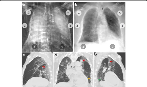
Fig. 1Visual evaluation scheme and grading scale for dark-field chest radiographs and conventional x-rays. In dark-field (a) and conventional (b) chest x-ray, lungs are divided into six regions: right lung-upper zone (RL-UZ), right lung-middle zone (RL-MZ), right lung-lower zone (RL-LZ), left lung-upper zone (LL-UZ), left lung-middle zone (LL-MZ), and left lung-lower zone (LL-LZ). In each region, dark-field (a) and transmission (b) signals are visually graded using a (c) 6-point ordinate scale
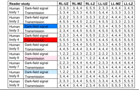
Table 1Dark-field and transmission signal grading of all nine human bodies
due to beam-hardening, a correction algorithm compar-able to the method presented in [27] was used.
The dark-field radiographs were low-pass-filtered (2D Gaussian filter kernel, σ= 3.2 pixels). This reduces noise levels by a factor of ~ 11.3 for white noise, leading to a vis-ual impression more similar to conventional radiography. Although low-pass filtering obscures small features, these are nearly undetectable in the unfiltered images due to the high noise levels. The used kernel size was found to be an acceptable trade-off between image impression and resolution. No filtering was applied to conventional radiographs.
Reader study
Visual image analysis was independently performed by three residents with 3 (F. M.), 5 (A. S.), and 5 (D. D.) years of ex-perience in chest imaging on a clinical Picture Archiving
and Communication System workstation. For training pur-poses, the dark-field radiograph of human body 4 was pre-sented before the reading session to demonstrate low and high dark-field signal intensity. In the first reading session, window settings were fixed to allow optimal comparison of low and high dark-field signal intensities in dark-field radio-graphs and opacification in conventional chest x-rays to avoid influence of individual windowing. A linear mapping between grey values and logarithmic visibility reduction ra-tios,−ln(V/V0), was used. Window level and width were set to 8,500 and 5,000, respectively. Converting these numbers back to physical quantities, this means that a logarithmic visibility reduction ratio of -0.268 corresponds to “black,” and a value of 0.343 corresponds to“white.”
The nine x-ray dark-field radiographs had to be graded separately one after the other without the possibility to compare or change gradings. Next, the conventional x-rays
were presented. On each image, the left and right lung were divided into three regions of equal height, upper, middle, and lower zones, using the apex and the costodiaphrag-matic recess as anatomical landmarks. Dark-field signal intensity and degree of transmission (or opacification) of the upper, middle, and lower zones of the left and right lung were graded on a 6-point (0–5) ordinal scale (Fig.1). For the dark-field signal intensity grading,“0”represents no (dark area in the radiograph) and“5”a high (bright area in the radiograph) dark-field signal. “1–4” represent inter-mediate dark-field signal intensities (Fig.4for comparison). For the transmission grading, “0” represents no transmis-sion or hyperattenuation like in the clinical case of a pleural effusion where no ventilated lung parenchyma is visible.“5” represents a normal, healthy lung with high transmission or hypoattenuation. “1–4” represents intermediate transmis-sion grades (Fig.7for comparison).
The reading session was repeated after 4 weeks. In a separate reading session, the readers independently graded image quality for right and left lung on a 6-point ordinate scale: 1 = not diagnostic, 2 = sufficient, 3 = satis-factory, 4 = good, 5 = very good, and 6 = excellent. As stan-dardised image quality criteria for dark-field radiographs do not exist, the readers were instructed to evaluate the following aspects: symmetrical reproduction of the thorax, reproduction of the whole lung, and presence of artefacts interfering with the grading of pulmonary dark-field signal intensity (e.g., vertical streaking artefacts, dark-field signal from bony structures). For transmission images, the “European guidelines on quality criteria for diagnostic radiographic images”[28] were applied wherever possible
considering imaging of a human body in supine position. In this setting, readers were free to change window/level values to optimise individual image impression.
Correlation of dark-field and transmission radiography with CT findings
As there exists no data on x-ray dark-field imaging fea-tures of human lung pathologies, we performed a CT scan of each human body to correlate findings in chest CT images with signal changes in dark-field and trans-mission radiographs. CT images were reviewed by an at-tending radiologist with 10 years of experience in chest radiology (A. A. F.) using axial, sagittal, and coronal re-constructions. Pulmonary findings and extrapulmonary findings with a potential effect on dark-field signal
Table 2Intra- and interobserver agreement of dark-field and transmission signal grading
Reader 1 Reader 2 Reader 3
Intraobserver agreement (κvalues)
Dark-field signal 0.959 0.971 0.793
Transmission 0.907 0.918 0.790
Interobserver agreement (κvalues)
Dark-field signal
Reader 1 – 0.848 0.828
Reader 2 0.848 – 0.893
Reader 3 0.828 0.893 –
Transmission
Reader 1 – 0.772 0.744
Reader 2 0.772 – 0.700
Reader 3 0.744 0.700 –
Visual grading of dark-field signal and transmission shows substantial (weighted Cohenκ= 0.61–0.80) to almost perfect (κ= 0.81–1.00) intraobserver and interobserver agreement, according to Landis and Koch [29]. Time difference between reading sessions for assessment of intraobserver agreement was 4 weeks
intensity were recorded. Apart from septal thickening, the extent of pulmonary findings was visually quantified for every lobe in 10% intervals. For pleural effusions, the maximum width in anterior-posterior direction was measured in centimeters. Other findings were qualita-tively recorded. CT findings were correlated with the vis-ual assessment of dark-field signal strength in a descriptive model.
Statistical analysis
Statistical analysis was performed using GraphPad Prism 7 for Mac OS X (Version 7.0d, GraphPad Soft-ware Inc., USA) and R version 3.4.4 (R Foundation for Statistical Computing, Vienna, Austria). For or-dinal data, median and range are presented and dot plots with medians and 95% confidence interval (based on the Hodges-Lehmann method) are shown. Intraobserver and interobserver agreement of dark-field signal and transmission grading were evaluated using weighted Cohen κ with squared weights. Cohen
κ coefficients with values < 0 were regarded as poor, 0–0.20 as slight, 0.21–0.40 as fair, 0.41–0.60 as mod-erate, 0.61–0.80 as substantial, and 0.81–1.00 as (al-most) perfect agreement, according to Landis et al. [29]. Differences in distributions of dark-field signal and transmission grading for the upper, middle, and lower zones were tested separately for the right and left lung and each reader using the Friedman test. If
Friedman test indicated a significant (p< 0.050) asso-ciation between region and dark-field signal, Wil-coxon matched-pairs signed-rank test was performed for pairwise comparisons of regions of each lung for each reader. The correlation of dark-field signal with transmission grading was tested with Spearman’s rank correlation coefficient for each region of lungs and each reader. Differences in the grading of image qual-ity between the left and right lung for dark-field and transmission radiographs, respectively, and between dark-field and transmission radiographs for left and right lung, respectively, were tested with Wilcoxon matched-pairs signed-rank test.
Results
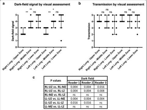
Visual grading of dark-field and transmission signals Statistical analysis of the dark-field and transmission gradings (Table 1) indicated a significant (right lung, reader 1–3, p< 0.0001; left lung, reader 1, p= 0.001, reader 2, p= 0.001, reader 3, p= 0.001) association between the lung zones and dark-field signal, which was further investigated by pairwise comparisons of regions of each lung for each reader (Fig. 2). The me-dian dark-field signal showed significant differences between the lung upper zones and lung middle zones. The median dark-field signal was also significantly different between the lung upper zones and lung lower zones, except for reader 3 in the left lung.
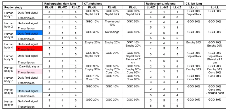
Table 3Correlation of dark-field and transmission radiography with CT findings
For all nine human bodies, median dark-field signal and transmission gradings for different lung regions are presented next to computed tomography (CT) findings in pulmonary lobes. Colour indicates cases with highest (blue, red) and lowest (light blue, pink) overall median dark-field signal or transmission grading that are shown as individual figures (Figs.3,4,5, and6)
For transmission radiographs, statistical analysis did not demonstrate significant associations with lung zones (right lung, reader 1, p= 0.535, reader 2, p= 0.482, reader 3, p= 0.312; left lung, reader 1, p= 0.568, reader 2,p> 0.9999, reader 3,p= 0.315).
These results indicate a correlation between the quan-tity of pulmonary tissue in the beam path (which is lower in lung apex than in middle and lower zones) and dark-field signal magnitude, whereas transmission chest x-rays are indifferent to this aspect.
Dark-field signal and transmission grading in each re-gion of the right and left lung for each reader showed significant correlations only in the right lung lower zone for reader 2 (p= 0.0079) and 3 (p= 0.0278) x-rays. All other tests did not show significant correlations (reader 1, RL-UZ, p> 0.9999, RL-MZ, p= 0.437, RL-LZ, p= 0.329, LL-UZ, p> 0.9999, LL-MZ, p= 0.385, LL-LZ, p= 0.100; reader 2, RL-UZ,p= 0.980, RL-MZ,p= 0.929, LL-UZ,p> 0.394, LL-MZ, p= 0.143, LL-LZ, p= 0.077; reader 3, RL-UZ, p= 0.827, RL-MZ, p= 0.603, LL-UZ, p= 0.921, LL-MZ,p= 0.185, LL-LZ,p= 0.333).
Intra- and interobserver agreement
The intraobserver agreement was from substantial to al-most perfect (κ= 0.793–0.971 for dark-field signal and
κ= 0.790–0.918 for transmission) for visual grading of dark-field and transmission signals (Table2).
Comparable results were obtained for interobserver agreement (Table2) that was almost perfect (κ= 0.828– 0.893) for visual grading of dark-field signal and sub-stantial to almost perfect correlation (κ= 0.700–0.772) for visual grading of transmission between all three readers.
Image quality
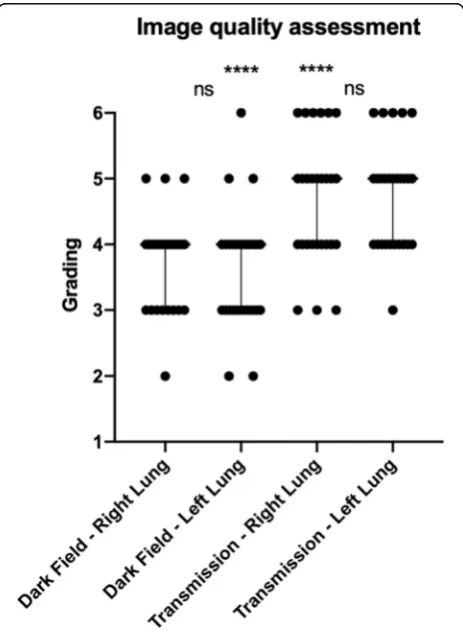
Median image quality grading (Fig.3) assessed by visual evaluation of all nine radiographs and all readers was good (4, interquartile range 1) for left and right lung of dark-field images and very good (5, interquartile range 1) for left and right lung of transmission images. Median image quality grading did not show significant differ-ences between the left and right lung for dark-field (p= 0.511) and transmission (p= 0.688) radiographs. Median
image quality grading was significantly different between dark-field and transmission radiographs for the left (p< 0.0001) and right (p< 0.0001) lung.
Correlation of dark-field and transmission radiography with CT findings
In the majority of CT images, ground-glass opacities were present in the lungs to a variable extent (Table 3, Figs.4,5,6, and7). Further pulmonary findings included emphysematous changes, consolidations, tree-in-bud sign, and (interlobular) septal thickening. Extrapulmon-ary findings were pleural effusions and an enlargement of the heart with haemopericardium.
In human body 4 (Fig. 4), we observed only minor (20% of total lobe) emphysematous changes in the parenchyma of all lobes. In the dark-field radiograph, a similar signal increase from the apex to the base of the lung is present in both the left and right lung, whereas transmission radiography shows no differ-ences. The lowest dark-field signal was reported in the lower zone of the left lung in human body 8 (Fig. 5). The corresponding CT revealed moderate
ground-glass opacities affecting 50% of the lower lobe and minor (10%) consolidations. Additionally, an en-larged heart with haemopericardium was extending into the left hemithorax. The highest dark-field signal over all zones of the lungs was present in human body 3 (Fig. 6), although a difference between the apex and the middle and lower zones was still visible. In the corresponding CT images, minor to moderate (20– 40%) ground-glass opacities were observed in all but the middle lobe. In human body 6 (Fig. 7), the transmission was lowest in the lower zone of the right lung correspond-ing to widespread (60%) consolidations in the lower lobe and moderate to extensive (50%/90%) ground-glass opaci-ties in the middle or upper lobe, respectively. In addition, extensive emphysematous changes in all lobes were present. Dark-field signal was also low in all zones of the right lung.
Discussion
X-ray dark-field radiography is a novel imaging mo-dality with high potential for lung imaging that has been translated from an experimental method to
clinical applicability in recent years. However, so far, its feasibility for imaging of the human lungs has only been demonstrated in a single post-mortem chest radiograph. Therefore, the purpose of our study was to assess the imaging features of nine post-mortem dark-field chest radiographs from a clinical point of view as a final step before evaluation of dark-field radiography in clinical studies.
In the visual assessment of post-mortem dark-field chest radiographs, we observed a gradient of dark-field signal strength from the apex to the base of the lungs. This can be attributed to an increasing amount of lung parenchyma in the x-ray beam path, as small-angle x-ray scattering increases with the amount of scattering material. However, in conventional chest x-rays, the transmission did not correlate with the dark-field signal, demonstrating that this presents a unique imaging feature of dark-field radiography. This finding is in accordance with results of animal studies [12,
13, 21]. In a clinical context, this would have to be considered when investigating pathologies that de-crease dark-field signal. In centrilobular emphysema, a form of chronic obstructive pulmonary disease that
primarily affects the upper lobes, knowledge of this
finding will be essential for correct image
interpretation.
X-ray dark-field radiography is an imaging modality which we believe will primarily be evaluated by visual assessment. Hence, for its clinical application, it is of major importance that imaging findings are consist-ently reported. We could demonstrate that visual grading of x-ray dark-field signal in the lungs shows substantial to almost perfect intra- and interobserver agreement, comparable to visual assessment of trans-mission in conventional chest x-rays. These results confirm outcomes of reader studies performed on x-ray dark-field radiographs of different lung patholo-gies in small animals [12, 14] and underline clinical applicability.
sufficient image quality for its application and evaluation in clinical trials.
Numerous preclinical animal studies have demon-strated the effect of specific lung pathologies on the dark-field signal. For example, in an animal model of idiopathic pulmonary fibrosis [13, 14], x-ray dark-field imaging allowed visualisation of early fibrotic changes in the lungs with dark-field images showing circum-scribed areas of markedly reduced dark-field signal in lung parenchyma affected by the pathologic process next to normal areas with high dark-field signal. In our study, the correlation of pathological findings in conventional x-ray and CT images of individual hu-man bodies with dark-field signal intensity showed comparable results in the human lungs. Areas of pulmonary consolidation may contribute to a major reduction of dark-field signal, whereas ground-glass opacities, representing interstitial and alveolar infil-trates, showed an inconsistent effect on the reduction of dark-field signal.
In one human body, an enlargement of the heart correlated with a strong decrease of the dark-field sig-nal in the left lung lower zone, probably by reducing the amount of lung parenchyma in the x-ray beam path. This is a relevant finding as it shows that extra-pulmonary pathologies can also influence the dark-field signal intensity and would have to be considered when interpreting clinical dark-field radiographs.
the lungs was free of pathologies and could have served as a reference. Furthermore, due to lung anatomy, it is difficult to correlate a CT finding in a pulmonary lobe to dark-field signal changes in a lung zone of a radio-graph. However, we gained insight into findings that may have a more pronounced effect on dark-field signal reduction, e.g., pulmonary consolidation, that has not been addressed in animal studies. X-ray dark-field im-aging was performed in supine position, which leads to dystelectasis in the dorsal basal parts of the lungs and may influence dark-field signal. To at least partially compensate for this, we performed endotracheal intub-ation and kept airway pressure constant during x-ray dark-field imaging. Since x-ray dark-field radiography is a novel imaging modality, the possibility to train the readers for the visual evaluation of the dark-field images was limited. The number of human bodies included in our study is relatively small. Still, our results are in accordance with previous animal studies and demonstrate clinical applicability.
In conclusion, our study on post-mortem human x-ray dark-field chest radiography demonstrates that x-ray dark-field images provide complementary information of the lungs to conventional x-ray, allow reliable visual quantification of dark-field signal strength, and have reached an image quality warranting an evaluation in clinical trials.
Additional file
Additional file 1: Figure S1.Overview of the dark-field imaging setup, qualitative explanation of dark-field contrast formation, and summary of grating parameters and distances. (DOCX 6811 kb)
Abbreviation
CT:Computed tomography
Authors’contributions
AAF, FDM, KW, PBN, DP, JH, and FP conceived of and designed the study. AAF, FDM, JA, KW, LBG, WN, FK, FK, FF, CB, FM, DD, and AS performed the experiments. AAF and BH performed the statistical analysis. All authors contributed to the literature research and edited the manuscript. AAF, FDM, and FP are guarantors of the integrity of the entire study. All authors reviewed and approved the final manuscript.
Funding
We acknowledge financial support through the DFG (Gottfried Wilhelm Leibniz program) and the European Research Council (AdG 695045). This work was carried out with the support of the Karlsruhe Nano Micro Facility (KNMF,www.kit.edu/knmf), a Helmholtz Research Infrastructure at Karlsruhe Institute of Technology (KIT).
Availability of data and materials
The datasets generated and/or analysed during the current study are available from the corresponding author on reasonable request.
Ethics approval and consent to participate
Institutional review board was obtained (ethics committee at the Faculty of Medicine of the Ludwig-Maximilians-University Munich, project number 14-13). Written informed consent was waived by the review board. The study was conducted according to the Declaration of Helsinki.
Consent for publication
Not applicable.
Competing interests
The authors declare that they have no competing interests.
Author details
1Department of Diagnostic and Interventional Radiology, School of Medicine & Klinikum rechts der Isar, Technical University of Munich, 81675 Munich, Germany.2Chair of Biomedical Physics, Department of Physics and Munich School of BioEngineering, Technical University of Munich, 85748 Garching, Germany.3Institute of Forensic Medicine, Ludwig-Maximilians-University Munich, 80336 Munich, Germany.4Philips Medical Systems DMC GmbH, 22335 Hamburg, Germany.5Philips GmbH Innovative Technologies, Research Laboratories, 22335 Hamburg, Germany.6Institute for Advanced Study, Technical University of Munich, 85748 Garching, Germany.7Institute of Medical Informatics, Statistics and Epidemiology, Technical University of Munich, 81675 Munich, Germany.
Received: 11 February 2019 Accepted: 29 May 2019
References
1. Bonse U, Hart M (1965) An x-ray interferometer. Appl Phys Lett 6:155–156. https://doi.org/10.1063/1.1754212
2. Momose A (2005) Recent advances in x-ray phase imaging. Jpn J Appl Phys 44:6355–6367.https://doi.org/10.1143/JJAP.44.6355
3. Pfeiffer F (2012) Milestones and basic principles of grating-based x-ray and neutron phase-contrast imaging. AIP Conf Proc 1466:2–11.https://doi.org/ 10.1063/1.4742261
4. Pfeiffer F, Herzen J, Willner M et al (2013) Grating-based x-ray phase contrast for biomedical imaging applications. Z Med Phys 23:176–185. https://doi.org/10.1016/j.zemedi.2013.02.002
5. Pfeiffer F, Bech M, Bunk O et al (2008) Hard-x-ray dark-field imaging using a grating interferometer. Nat Mater 7:134–137.https://doi.org/10.1038/nmat2096 6. Pfeiffer F, Weitkamp T, Bunk O, David C (2006) Phase retrieval and
differential phase-contrast imaging with low-brilliance x-ray sources. Nat Phys 2:258–261.https://doi.org/10.1038/nphys265
7. Bech M, Tapfer A, Velroyen A et al (2013) In-vivo dark-field and phase-contrast x-ray imaging. Sci Rep 3:3209.https://doi.org/10.1038/srep03209 8. Schleede S, Meinel FG, Bech M et al (2012) Emphysema diagnosis using
x-ray dark-field imaging at a laser-driven compact synchrotron light source. Proc Natl Acad Sci U S A 109:17880–17885.https://doi.org/10.1073/pnas. 1206684109
9. Meinel FG, Schwab F, Schleede S et al (2013) Diagnosing and mapping pulmonary emphysema on x-ray projection images: incremental value of grating-based x-ray dark-field imaging. PLoS One 8:e59526.https://doi.org/ 10.1371/journal.pone.0059526
10. Yaroshenko A, Meinel FG, Bech M et al (2013) Pulmonary emphysema diagnosis with a preclinical small-animal x-ray dark-field scatter-contrast scanner. Radiology 269:427–433.https://doi.org/10.1148/radiol.13122413 11. Meinel FG, Yaroshenko A, Hellbach K et al (2014) Improved diagnosis of pulmonary emphysema using in vivo dark-field radiography. Invest Radiol 49:653–658.https://doi.org/10.1097/RLI.0000000000000067
12. Hellbach K, Yaroshenko A, Meinel FG et al (2015) In vivo dark-field radiography for early diagnosis and staging of pulmonary emphysema. Invest Radiol 50:430–435.https://doi.org/10.1097/RLI.0000000000000147 13. Yaroshenko A, Hellbach K, Yildirim AÖ et al (2015) Improved in vivo
assessment of pulmonary fibrosis in mice using x-ray dark-field radiography. Sci Rep 5:17492.https://doi.org/10.1038/srep17492
14. Hellbach K, Yaroshenko A, Willer K et al (2017) X-ray dark-field radiography facilitates the diagnosis of pulmonary fibrosis in a mouse model. Sci Rep 7: 340.https://doi.org/10.1038/s41598-017-00475-3
15. Meinel FG, Schwab F, Yaroshenko A et al (2014) Lung tumors on multimodal radiographs derived from grating-based x-ray imaging–a feasibility study. Phys Med 30:352–357.https://doi.org/10.1016/j.ejmp.2013.11.001 16. Scherer K, Yaroshenko A, Bölükbas DA et al (2017) X-ray dark-field
17. Hellbach K, Yaroshenko A, Willer K et al (2016) Facilitated diagnosis of pneumothoraces in newborn mice using x-ray dark-field radiography. Invest Radiol 51:597–601.https://doi.org/10.1097/RLI.0000000000000285 18. Hellbach K, Baehr A, De Marco F et al (2018) Depiction of pneumothoraces
in a large animal model using x-ray dark-field radiography. Sci Rep 8:2602. https://doi.org/10.1038/s41598-018-20985-y
19. Yaroshenko A, Pritzke T, Koschlig M et al (2016) Visualization of neonatal lung injury associated with mechanical ventilation using x-ray dark-field radiography. Sci Rep 6:24269.https://doi.org/10.1038/srep24269 20. Hellbach K, Meinel FG, Conlon TM et al (2018) X-ray dark-field imaging to
depict acute lung inflammation in mice. Sci Rep 8:2096.https://doi.org/10. 1038/s41598-018-20193-8
21. Gromann L, De Marco F, Willer K et al (2017) In-vivo x-ray dark-field chest radiography of a pig. Sci Rep 7:4807.https://doi.org/10.1038/s41598-017-05101-w 22. Willer K, Fingerle AA, Gromann LB et al (2018) X-ray dark-field imaging of
the human lung - a feasibility study on a deceased body. PLoS One 13: e0204565.https://doi.org/10.1371/journal.pone.0204565
23. Huang Z-F, Kang K-J, Zhang L et al (2009) Alternative method for differential phase-contrast imaging with weakly coherent hard x rays. Phys Rev A 79: 013815.https://doi.org/10.1103/PhysRevA.79.013815
24. Schröter TJ, Koch FJ, Meyer P et al (2017) Large field-of-view tiled grating structures for x-ray phase-contrast imaging. Rev Sci Instrum 88.https://doi. org/10.1063/1.4973632
25. Kottler C, Pfeiffer F, Bunk O, Grünzweig C, David C (2007) Grating interferometer based scanning setup for hard x-ray phase contrast imaging. Rev Sci Instrum 78:043710.https://doi.org/10.1063/1.2723064
26. Koehler T, Daerr H, Martens G et al (2015) Slit-scanning differential x-ray phase-contrast mammography: proof-of-concept experimental studies. Med Phys 42:1959–1965.https://doi.org/10.1118/1.4914420
27. Pelzer G, Anton G, Horn F et al (2016) A beam hardening and dispersion correction for x-ray dark-field radiography. Med Phys 43:2774–2779.https:// doi.org/10.1118/1.4948671
28. Carmichael JHE, Maccia C, Moores BM et al. (1996) European guidelines on quality criteria for diagnostic radiographic images. Study Group on Quality Criteria Development of the European Commission. European Commission. Directorate-General XII: Science, Research and Development Available via. https://publications.europa.eu/s/lKRX. Accessed 20 May 2019
29. Landis JR, Koch GG (1977) The measurement of observer agreement for categorical data. Biometrics 33:159–174.https://doi.org/10.2307/2529310 30. Zhu H, Zhang L, Wang Y et al (2017) Improved image quality and
diagnostic potential using ultra-high-resolution computed tomography of the lung with small scan FOV: a prospective study. PLoS One 12:e0172688. https://doi.org/10.1371/journal.pone.0172688
Publisher’s Note