Introduction
A “red eye” is a common complaint encountered by general practitioners, pharmacists and health care workers in everyday practice. It can however be a challenge to accurately diagnose and manage this condition – a sentiment that is shared by specialist ophthalmologists.1 Allergic conjunctivitis is therefore often
underdiagnosed and consequently undertreated.
Several non-infective inflammatory conditions presenting as hypersensitivity reactions may be associated with clinical manifestations of ocular allergy.2 Typically the anterior segment
of the eye is affected, and all four types of hypersensitivity reactions may be involved; either as a single cause, or as an overlap between the different types. Specific IgE antibodies are almost always present in all types of allergic conjunctivitis whereby the inflammatory response towards allergens is a result of degranulation of the sensitized mast cells resulting in the clinical ocular allergic expression. 3 Recent findings indicate that
conjunctival symptoms are present in 30-70% of adults suffering from acute allergic rhinitis, while 30% are associated with chronic and persistent episodes.4
Environmental factors, especially notable in our South African setting, are important. For instance, there is a significant association between communities residing in close proximities to mine dumps and the occurrence of rhinoconjunctivitis, wheezing and asthma.5 The frequency of patients presenting
with ophthalmological complaints seems to be on the increase, and several causes have been proposed. Some of these causes include air pollution, early childhood exposure to allergens, genetic predisposition and several immunological conditions.6 During physical examination all forms of
non-severe allergic conjunctivitis presents in a very similar and often indistinguishable manner, therefore history taking and onset of symptoms plays an important role in the diagnosis.7 It
is important for general practitioners to be able to distinguish between bacterial, viral and allergic causes of conjunctivitis on clinical assessment.
Types of allergic conjunctivitis
Allergic conjunctivitis can be classified as either severe or non-severe. Non-severe allergic conjunctivitis includes Acute Allergic Conjunctivitis (AAC), Seasonal Allergic Conjunctivitis (SAC) and Perennial Allergic Conjunctivitis (PAC). Severe allergic conjunctivitis comprises Vernal keratoconjunctivitis (VKC), giant papillary conjunctivitis (GPC) and atopic keratoconjunctivitis (AKC).8
Non-severe allergic conjunctivitis:
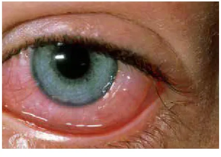
Environmental exposure to various allergens results in a sudden onset hypersensitivity reaction. Characteristically, symptoms develop rapidly and usually resolve spontaneously within 24 hours after removal of the causative factor. Acute allergic conjunctivitis is characterized by eyelid edema, tearing, severe itching, hyperemia and chemosis. The symptoms are frequently bilateral, however one eye may be affected more than the other. Mild crusting with a non-purulent discharge is not uncommon. In the presence of nasal symptoms (rhinoconjunctivitis), it is commonly attributed to exposure to outdoor airborne pollens and occurs at the same time each year, therefore termed Seasonal allergic conjunctivitis.9 The development of symptoms in seasonal
allergic conjunctivitis is less severe compared to typical sudden onset allergic conjunctivitis, but symptom resolution may take a few weeks to subside, and varies with the pollen count. Perennial
The allergic eye
Andre Marais, BPharm, MBChB, MSC.Pharm, Senior lecturer and Clinical Pharmacologist
Department of Pharmacology, School of Medicine, Faculty of Health Sciences, University of Pretoria, South Africa
Correspondence to: [email protected] / [email protected]
Keywords: Allergic conjunctivitis, atopic keratoconjunctivitis, giant papillary conjunctivitis, treatment, vernal keratoconjunctivitis
Abstract
Allergic conjunctivitis is a common condition encountered by the healthcare professional. Several factors have been implicated as possible causes and almost all involve an IgE mediated immune response. Symptoms may vary from mild to severe and if misdiagnosed, or incorrect treatment is given, the result could lead to permanent vision loss. It is important for the general practitioner to differentiate between allergic, bacterial and viral causes of conjunctivitis, and to treat and refer appropriately.
allergic conjunctivitis is the most common type of eye allergy and symptoms may persist throughout the year. It is estimated that around 20% of the world’s population is affected by PAC.10
The potential chronicity of this disease makes the classification between acute and chronic somewhat debatable. Perennial allergic conjunctivitis is frequently associated with known indoor allergens such as house dust mites, molds and animal dander. Symptoms tend to be more severe in the mornings and may be accompanied by ritualistic sneezing episodes.
Allergic eye disease primarily affects the conjunctiva resulting in various degrees of redness. This may involve the bulbar (globe), palpebral or tarsal (mucus membrane lining the inner surface of the eyelids) conjunctiva. 10
In all instances of non-severe allergic conjunctivitis the condition is painless, does not affect vision and causes minimal sensitivity to light. Alternative diagnosis or specialist referral should be considered to rule out more serious conditions including scleritis, iritis, episcleritis or angle closure glaucoma if ocular pain, vision disturbances or intense redness around the iris are present.
Severe allergic conjunctivitis
Atopic keratoconjunctivitis (AKC) is an uncommon condition affecting patients between the ages of 30 and 50 years. It is closely associated with atopic dermatitis and eczema. Affected patients present with severe pruritus and eyelid swelling which may lead to blindness, corneal scarring and cataract formation.11
Diagnosis is based on clinical features and associated atopic findings. Treatment includes long term application of topical antihistamines and mast cell stabilizers. Corticosteroid drops may be used for a period not exceeding 2 weeks after which the likelihood of raising the intraocular pressure increases dramatically. The accompanying eyelid dermatitis should be treated with application of a low potency corticosteroid cream two to four times daily. Non-responders warrant specialist ophthalmological referral where topical calcineurin inhibitors (i.e cyclosporine) could be used for symptom improvement.12
Vernal keratoconjunctivitis is a complex chronic allergic inflammatory disease mainly mediated by Th2 lymphocytes resulting in the over-expression of mast cells, eosinophils and certain cytokines and growth factors. Specific IgE testing (serum and lacrimal fluid) in these patients is positive in only approximately half of affected individuals. 13 Boys are mostly
affected, and the diagnosis is made during the first decade of life. The clinical presentation of VKC is similar to atopic keratoconjunctivitis in respect to the severe ocular pruritus, but the associated corneal complications are significantly lower. Characteristic findings include >1 mm tarsal giant conjunctival papillae and limbal gelatinous changes (Trantas dots), ptosis and corneal ulcers. (Figure 3) Diagnosis is based on typical epidemiology and clinical features. The disease is self-limiting and may disappear spontaneously in the second decade. First line therapy includes topical antihistamines and mast cell stabilizers. Immunomodulatory agents (cyclosporine A and tacrolimus) have been used in the long term management of resistant cases. The overuse of prescribed corticosteroids may lead to iatrogenic corneal damage and cataract formation, justifying that these patients be referred to an ophthalmologist for management.14
Figure 1: Allergic conjunctivitis
Image available from http://www.newhealthguide.org/images/10420176/Allergic-Conjunctivitis.jpg
Figure 2: Atopic keratoconjunctivitis
Image available from http://www.reviewofoptometry.com/ CMSImagesContent/2005/4/2_1407_0.jpg
Figure 3: Vernal keratoconjunctivitis
Giant papillary conjunctivitis is a disease mostly affecting young adults wearing contact lenses. The pathogenesis is therefore not exclusively a result of immunological mechanisms, but includes a mechanical component.15 Debris precipitates on the surface of
hard contact lenses in particular, which triggers an immunological IgE response. This could be an immediate type I hypersensitivity reaction, or in some instances a delayed type IV reaction, only presenting after several months of wear.16 Initial symptoms
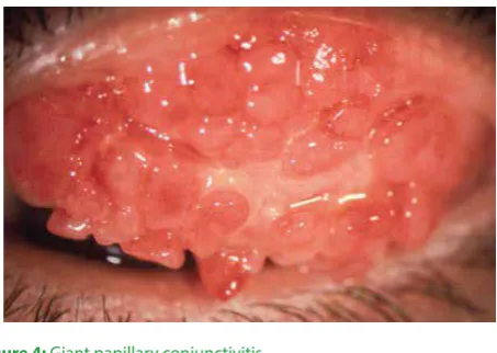
are mild irritation, itching and slight mucus production. If left untreated progression to intense discomfort, blurry vision, severe discharge and complete intolerance to wearing contact lenses may develop. In late stages the development of enlarged (giant) upper tarsal papillae (cobblestone appearance), conjunctival hyperemia, ptosis and eyelid thickening is present (Figure 4). Management consists of contact lens “holidays” for two to four weeks allowing for the inflammation to subside. Old lenses should be discarded and alternative contact lens preservatives should be explored. Topical antihistamines and mast cell stabilizers are effective first line therapy. Non responders may be given topical corticosteroids for a period of 2 weeks not exceeding 6 weeks of treatment.
Pharmacological therapy
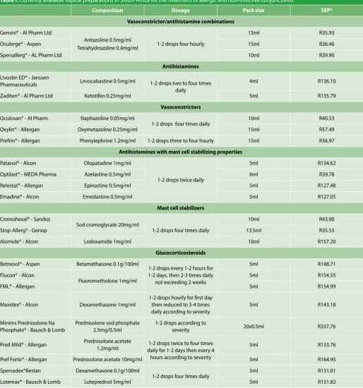
Various pharmacological agents are available to treat allergic conjunctivitis. Many of these agents, excluding the glucocorticoids, are available in pharmacies, without a prescription. It however remains imperative not to treat any condition resembling a “red eye” before an accurate diagnosis has been made. Table 1 illustrates the currently available topical ophthalmological preparations indicated for use in allergic and non-infective conjunctivitis.
Antihistamines, Vasoconstrictors and combinations (Decongestants):
Topical eye drops containing a H1 antihistamine (antazoline, levocabastine, ketotifen), a α1 vasoconstrictor (tetrahydrozoline, naphazoline, oxymetazoline, phenylephrine) or a combination of both are available without a prescription. Antihistamines inhibit the action of the primary mast cell mediator by reversibly antagonizing/blocking the H1 receptor in the conjunctiva
and eyelids. Vasoconstrictors decrease conjunctival edema by activating post ganglionic alpha-1-adrenergic receptors in the arterial blood vessels. Alpha agonists in addition decrease the conjunctival hyperemia. These agents should only be used for short term symptom relief as prolonged use may cause rebound hyperemia.17 Dosing with combination vasoconstrictor/
antihistamines is up to 4 times per day for a maximum of 7 days.
Antihistamines with mast cell stabilizing properties:
Antihistamines with mast cell stabilizing properties are also available without prescription. In addition to the antagonistic effect on the histamine receptors, these agents inhibit mast cell degranulation thereby preventing histamine, tryptase and prostaglandins from being released. Onset of action is within a few minutes. Dosing is twice daily and treatment should be continued for 2 weeks at least allowing for the inflammation to subside. Drugs in this group include olopatadine (with additional anticholinergic effects), azelastine, epinastine and emedastine.
Side effects of these drugs are uncommon but may include headache, blurred vision, foreign body sensation and ocular discomfort. In addition, these agents are frequently available as nasal sprays, which may contribute to a bitter taste and nasal burning.
Mast cell stabilizers:
In contrast to the antihistamines with mast cell stabilizing properties, pure mast cell stabilizers (sodium cromoglycate and lodoxamide) are less effective and display a longer onset of action (5-14 days) thereby making these agents unsuitable for acute immediate symptom relief. These drugs are useful in prophylactic use and provide a good option for SAC. Dosing is 4 times per day and may be continued for prolonged periods.
Glucocorticosteroids:
Due to the documented serious side effects of topical glucocorticosteroids, these agents are not advocated for routine use and are only indicated for refractory cases of allergic conjunctivitis. Use should be avoided if any viral infection is suspected. Glucocorticosteroids induce the formation of lipocortin, which acts as the body’s natural anti-inflammatory agent; thereby reducing all inflammatory products of the arachidonic acid cascade. 18 Treatment should ideally be initiated
under the care of an ophthalmologist, and only be used for short periods. Traditional topical glucocorticosteroids (prednisolone, fluorometholone, betamethasone and dexamethasone) have been associated with severe ocular side effects including raised intra ocular pressure, cataract formation, glaucoma and increased risk for secondary infections. The development of modified “soft” steroids (loteprednol and rimexolone) are formulated specifically to undergo rapid inactivation after corneal penetration; thereby limiting the incidence of raising the intraocular pressure. 19
Rimexolone is not yet available in South Africa. In general, glucocorticosteroids are administered every one to two hours during the first 24-48 hours and then 4 times daily for 2 weeks. Figure 4: Giant papillary conjunctivitis
Conclusion
Acute allergic conjunctivitis is a common condition and usually settles without treatment. Immediate relief may be achieved with the use of topical vasoconstrictor/antihistamine/mast cell stabilizers, either alone or in combination. Severe cases warrant treatment with corticosteroids and should preferably be done under specialist ophthalmological supervision. Chronic forms of allergic conjunctivitis may be difficult to treat and pose the risk of
permanent eye damage. Patients must be educated concerning their condition and general measures including instructions not to rub their eyes, avoidance of known triggers and proper contact lens care should be done at each encounter.
References
1. Granet D. Allergic rhinoconjunctivitis and differential diagnosis of the red eye. Allergy Asthma Proc. 2008;29(6):565-74.
2. Rosario N, Bielory L. Epidemiology of allergic conjunctivitis. Current opinion in allergy and clinical immunology. 2011;11(5):471-6.
3. Bonini S. Atopic keratoconjunctivitis. Allergy. 2004;59.
Table I: Currently available topical preparations in South Africa for the treatment of allergic and non-infective conjunctivitis
Composition Dosage Pack size SEP*
Vasoconstrictor/antihistamine combinations
Gemini® - Al Pharm Ltd
Antazoline 0.5mg/ml
Tetrahydrozoline 0.4mg/ml 1-2 drops four hourly
15ml R35.93
Oculerge® - Aspen 15ml R26.46
Spersallerg® - AL Pharm Ltd 10ml R39.90
Antihistamines
Livostin ED® - Janssen
Pharmaceuticals Levocabastine 0.5mg/ml 1-2 drops two to four times daily
4ml R136.10
Zaditen® - Al Pharm Ltd Ketotifen 0.25mg/ml 5ml R135.79
Vasoconstrictors
Oculosan® - Al Pharm Naphazoline 0.05mg/ml
1-2 drops four times daily 10ml R40.53 Oxylin® - Allergan Oxymetazoline 0.25mg/ml 15ml R57.49 Prefrin® - Allergan Phenylephrine 1.2mg/ml 1-2 drops three to four hourly 15ml R36.97
Antihistamines with mast cell stabilizing properties
Patanol® - Alcon Olopatadine 1mg/ml
1-2 drops twice daily
5ml R134.62 Optilast® - MEDA Pharma Azelastine 0.5mg/ml 6ml R39.78 Relestat® - Allergan Epinastine 0.5mg/ml 5ml R127.48 Emadine® - Alcon Emedastine 0.5mg/ml 5ml R127.05
Mast cell stabilizers
Cromohexal® - Sandoz
Sod cromoglycate 20mg/ml
1-2 drops four times daily
10ml R43.90
Stop-Allerg® - Genop 13.5ml R35.53
Alomide® - Alcon Lodoxamide 1mg/ml 10ml R157.20
Glucocorticosteroids
Betnesol® - Aspen Betamethasone 0.1g/100ml
1-2 drops every 1-2 hours for 1-2 days, then 2-3 times daily
not exceeding 2 weeks
5ml R148.71 Flucon® - Alcon
Fluorometholone 1mg/ml 5ml R154.55
FML® - Allergan 5ml R154.99
Maxidex® - Alcon Dexamethasone 1mg/ml
1-2 drops hourly for first day then reduced to 3-4 times daily according to severity
5ml R143.18
Minims Prednisolone Na Phosphate® - Bausch & Lomb
Prednisolone sod phosphate 2.5mg/0.5ml
1-2 drops according to
severity 20x0.5ml R337.76
Pred Mild® - Allergan Prednisolone acetate 1.2mg/ml 1-2 drops twice to four times daily for 1-2 days then every 4
hours according to severity
5ml R133.76
Pref Forte® - Allergan Prednisolone acetate 10mg/ml 5ml R164.95 Spersadex®Restan Dexamethasone 0.1g/100ml
4. Leonardi A, Castegnaro A, Valerio AL, Lazzarini D. Epidemiology of allergic conjunctivitis: clinical appearance and treatment patterns in a population-based study. Current opinion in allergy and clinical immunology. 2015;15(5):482-8.
5. Nkosi V, Wichmann J, Voyi K. Mine dumps, wheeze, asthma, and rhinoconjunctivitis among adolescents in South Africa: any association? International Journal of Environmental Health Research. 2015;25(6):583-600.
6. Leonardi S, del Giudice Miraglia M, La Rosa M, Bellanti JA. Atopic disease, immune system, and the environment. Allergy Asthma Proc. 2007;28.
7. Mantelli F, Lambiase A, Bonini S. A simple and rapid diagnostic algorithm for the detec-tion of ocular allergic diseases. Current opinion in allergy and clinical immunology. 2009;9(5):471-6.
8. Ono SJ, Abelson MB. Allergic conjunctivitis: Update on pathophysiology and prospects for future treatment. Journal of Allergy and Clinical Immunology. 2005;115(1):118-22. 9. Bousquet J, Khaltaev N, Cruz AA, Denburg J, Fokkens WJ, Togias A, et al. Allergic Rhinitis
and its Impact on Asthma (ARIA) 2008 update (in collaboration with the World Health Or-ganization, GA(2)LEN and AllerGen). Allergy. 2008;63 Suppl 86:8-160.
10. La Rosa M, Lionetti E, Reibaldi M, Russo A, Longo A, Leonardi S, et al. Allergic conjunctivitis: a comprehensive review of the literature. Italian Journal of Pediatrics. 2013;39(1):1-8. 11. Hu Y, Matsumoto Y, Adan ES, Dogru M, Fukagawa K, Tsubota K, et al. Corneal in vivo
confo-cal scanning laser microscopy in patients with atopic keratoconjunctivitis. Ophthalmology.
2008;115(11):2004-12.
12. Hingorani M, Moodaley L, Calder VL, Buckley RJ, Lightman S. A randomized, placebo-controlled trial of topical cyclosporin A in steroid-dependent atopic keratoconjunctivitis. Ophthalmology. 1998;105(9):1715-20.
13. Pattnaik L, Acharya L. A comprehensive review on vernal keratoconjunctivitis with empha-sis on proteomics. Life Sciences. 2015;128:47-54.
14. Pleyer U, Leonardi A. Vernal keratoconjunctivitis. Ophthalmologe. 2015;112(2):177-89. 15. Leonardi A, De Dominicis C, Motterle L. Immunopathogenesis of ocular allergy: a
sche-matic approach to different clinical entities. Current opinion in allergy and clinical immu-nology. 2007;7.
16. Donshik PC. Contact lens chemistry and giant papillary conjunctivitis. Eye & contact lens. 2003;29(1 Suppl):S37-9; discussion S57-9, S192-4.
17. Spector SL, Raizman MB. Conjunctivitis medicamentosa. J Allergy Clin Immunol. 1994;94(1):134-6.
18. Sibilia J. Corticosteroids and inflammation. La Revue du praticien. 2003;53(5):495-501. 19. Druzgala P, Wu WM, Bodor N. Ocular absorption and distribution of loteprednol etabonate,